Show PDB file:   
         Plain Text   HTML   (compressed file size)
QuickSearch:   
by PDB,NDB,UniProt,PROSITE Code or Search Term(s)  
(-)Asymmetric Unit
(-)Asym. Unit - sites
(-)Biological Unit 1
(-)Biol. Unit 1 - sites
(-)Biological Unit 2
collapse expand < >
Image Asymmetric Unit
Asymmetric Unit  (Jmol Viewer)
Image Asym. Unit - sites
Asym. Unit - sites  (Jmol Viewer)
Image Biological Unit 1
Biological Unit 1  (Jmol Viewer)
Image Biol. Unit 1 - sites
Biol. Unit 1 - sites  (Jmol Viewer)
Image Biological Unit 2
Biological Unit 2  (Jmol Viewer)

(-) Description

Title :  THE TYPE III CRYSTAL STRUCTURE OF STREPTOCOCCUS AGALACTIAE SORTASE C1
 
Authors :  B. Khare, S. V. L. Narayana
Date :  29 Mar 11  (Deposition) - 07 Sep 11  (Release) - 28 Mar 12  (Revision)
Method :  X-RAY DIFFRACTION
Resolution :  3.00
Chains :  Asym. Unit :  A,B
Biol. Unit 1:  A  (1x)
Biol. Unit 2:  B  (1x)
Keywords :  Sortase Fold, Beta-Barrel, Class C Sortase, Pili Biogenesis, Hydrolase (Keyword Search: [Gene Ontology, PubMed, Web (Google))
 
Reference :  B. Khare, V. Krishnan, K. R. Rajashankar, H. I-Hsiu, M. Xin, H. Ton-That, S. V. Narayana
Structural Differences Between The Streptococcus Agalactiae Housekeeping And Pilus-Specific Sortases: Srta And Srtc1.
Plos One V. 6 22995 2011
PubMed-ID: 21912586  |  Reference-DOI: 10.1371/JOURNAL.PONE.0022995

(-) Compounds

Molecule 1 - SORTASE FAMILY PROTEIN
    ChainsA, B
    EngineeredYES
    Expression SystemESCHERICHIA COLI
    Expression System StrainXL1BLUE
    Expression System Taxid562
    Expression System Vector TypePQE30
    FragmentUNP RESIDUES 43-260
    GeneSAG0647, SRTC1 (SAG0647)
    Organism ScientificSTREPTOCOCCUS AGALACTIAE SEROGROUP V
    Organism Taxid216466
    StrainSAG 2603V/R
    SynonymSORTASE C1

 Structural Features

(-) Chains, Units

  12
Asymmetric Unit AB
Biological Unit 1 (1x)A 
Biological Unit 2 (1x) B

Summary Information (see also Sequences/Alignments below)

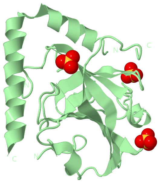
(-) Ligands, Modified Residues, Ions  (1, 6)

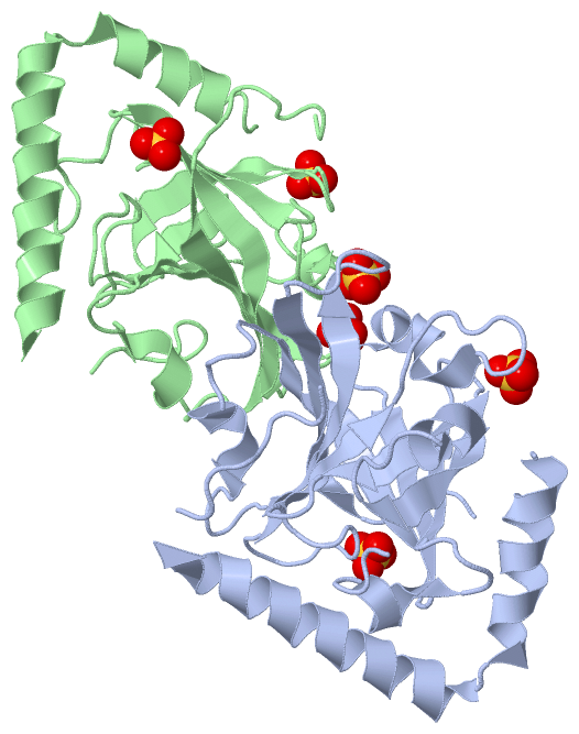
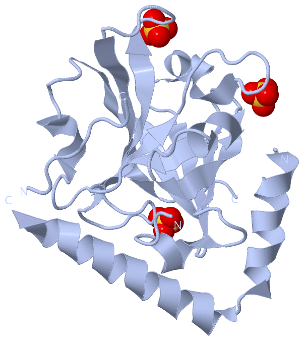
Asymmetric Unit (1, 6)
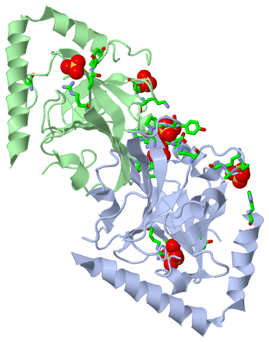
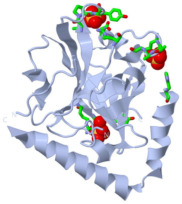
No.NameCountTypeFull Name
1SO46Ligand/IonSULFATE ION
Biological Unit 1 (1, 3)
No.NameCountTypeFull Name
1SO43Ligand/IonSULFATE ION
Biological Unit 2 (1, 3)
No.NameCountTypeFull Name
1SO43Ligand/IonSULFATE ION

(-) Sites  (6, 6)

Asymmetric Unit (6, 6)
No.NameEvidenceResiduesDescription
1AC1SOFTWAREARG A:200 , PRO A:202 , TYR A:203 , HOH A:318BINDING SITE FOR RESIDUE SO4 A 220
2AC2SOFTWAREARG A:123 , ASP A:135 , THR A:185 , PRO A:186 , TYR A:187BINDING SITE FOR RESIDUE SO4 A 221
3AC3SOFTWAREHIS A:3 , THR A:127 , ALA A:128 , LYS A:129BINDING SITE FOR RESIDUE SO4 A 222
4AC4SOFTWAREARG B:200 , PRO B:202 , TYR B:203BINDING SITE FOR RESIDUE SO4 B 220
5AC5SOFTWAREGLN B:83 , LYS B:129BINDING SITE FOR RESIDUE SO4 B 221
6AC6SOFTWAREARG B:65 , ARG B:123 , ASP B:135 , THR B:185 , PRO B:186 , TYR B:187BINDING SITE FOR RESIDUE SO4 B 222

(-) SS Bonds  (0, 0)

(no "SS Bond" information available for 3RBI)

(-) Cis Peptide Bonds  (2, 2)

Asymmetric Unit
No.Residues
1Thr A:185 -Pro A:186
2Thr B:185 -Pro B:186

 Sequence-Structure Mapping

(-) SAPs(SNPs)/Variants  (0, 0)

(no "SAP(SNP)/Variant" information available for 3RBI)

(-) PROSITE Motifs  (0, 0)

(no "PROSITE Motif" information available for 3RBI)

(-) Exons   (0, 0)

(no "Exon" information available for 3RBI)

(-) Sequences/Alignments

Asymmetric Unit
   Reformat: Number of residues per line =  ('0' or empty: single-line sequence representation)
  Number of residues per labelling interval =   
  UniProt sequence: complete  aligned part    
   Show mapping: SCOP domains CATH domains Pfam domains Secondary structure (by author)
SAPs(SNPs) PROSITE motifs Exons
(details for a mapped element are shown in a popup box when the mouse pointer rests over it)
Chain A from PDB  Type:PROTEIN  Length:193
 aligned with Q8E0S7_STRA5 | Q8E0S7 from UniProtKB/TrEMBL  Length:305

    Alignment length:205
                                    52        62        72        82        92       102       112       122       132       142       152       162       172       182       192       202       212       222       232       242     
         Q8E0S7_STRA5    43 SHANINAFKEAVTKIDRVEINRRLELAYAYNASIAGAKTNGEYPALKDPYSAEQKQAGVVEYARMLEVKEQIGHVIIPRINQDIPIYAGSAEENLQRGVGHLEGTSLPVGGESTHAVLTAHRGLPTAKLFTNLDKVTVGDRFYIEHIGGKIAYQVDQIKVIAPDQLEDLYVIQGEDHVTLLTCTPYMINSHRLLVRGKRIPYVEK 247
               SCOP domains ------------------------------------------------------------------------------------------------------------------------------------------------------------------------------------------------------------- SCOP domains
               CATH domains ------------------------------------------------------------------------------------------------------------------------------------------------------------------------------------------------------------- CATH domains
               Pfam domains ------------------------------------------------------------------------------------------------------------------------------------------------------------------------------------------------------------- Pfam domains
         Sec.struct. author .hhhhhhhhhhhhh.hhhhhhhhhhhhhhhhhhhhhh.----........--------....hhhhh.....eeeee....eeeeeee....hhhhh.eee............eeeeee........hhhhhhhhh....eeeeee..eeeeeeeeeeeee....hhhhh.....eeeeeeeee......eeeeeeeeee..... Sec.struct. author
                 SAPs(SNPs) ------------------------------------------------------------------------------------------------------------------------------------------------------------------------------------------------------------- SAPs(SNPs)
                    PROSITE ------------------------------------------------------------------------------------------------------------------------------------------------------------------------------------------------------------- PROSITE
                 Transcript ------------------------------------------------------------------------------------------------------------------------------------------------------------------------------------------------------------- Transcript
                 3rbi A   2 SHANINAFKEAVTKIDRVEINRRLELAYAYNASIAGAK----YPALKDPY--------VVEYARMLEVKEQIGHVIIPRINQDIPIYAGSAEENLQRGVGHLEGTSLPVGGESTHAVLTAHRGLPTAKLFTNLDKVTVGDRFYIEHIGGKIAYQVDQIKVIAPDQLEDLYVIQGEDHVTLLTCTPYMINSHRLLVRGKRIPYVEK 206
                                    11        21        31       | -  |     51        61        71        81        91       101       111       121       131       141       151       161       171       181       191       201     
                                                                39   44     51       60                                                                                                                                                  

Chain B from PDB  Type:PROTEIN  Length:194
 aligned with Q8E0S7_STRA5 | Q8E0S7 from UniProtKB/TrEMBL  Length:305

    Alignment length:209
                                    52        62        72        82        92       102       112       122       132       142       152       162       172       182       192       202       212       222       232       242         
         Q8E0S7_STRA5    43 SHANINAFKEAVTKIDRVEINRRLELAYAYNASIAGAKTNGEYPALKDPYSAEQKQAGVVEYARMLEVKEQIGHVIIPRINQDIPIYAGSAEENLQRGVGHLEGTSLPVGGESTHAVLTAHRGLPTAKLFTNLDKVTVGDRFYIEHIGGKIAYQVDQIKVIAPDQLEDLYVIQGEDHVTLLTCTPYMINSHRLLVRGKRIPYVEKTVQK 251
               SCOP domains ----------------------------------------------------------------------------------------------------------------------------------------------------------------------------------------------------------------- SCOP domains
               CATH domains ----------------------------------------------------------------------------------------------------------------------------------------------------------------------------------------------------------------- CATH domains
               Pfam domains ----------------------------------------------------------------------------------------------------------------------------------------------------------------------------------------------------------------- Pfam domains
         Sec.struct. author ...hhhhhhhhhhh.hhhhhhhhhhhhhhhhhhhhhh-------........--------............eeeee....eeeeeee..........eee............eeeeee............hhhhh....eeeeee..eeeeeeeeeeeee....hhhhh.....eeeeeeeee......eeeeeeeeee......... Sec.struct. author
                 SAPs(SNPs) ----------------------------------------------------------------------------------------------------------------------------------------------------------------------------------------------------------------- SAPs(SNPs)
                    PROSITE ----------------------------------------------------------------------------------------------------------------------------------------------------------------------------------------------------------------- PROSITE
                 Transcript ----------------------------------------------------------------------------------------------------------------------------------------------------------------------------------------------------------------- Transcript
                 3rbi B   2 SHANINAFKEAVTKIDRVEINRRLELAYAYNASIAGA-------ALKDPYSA--------EYARMLEVKEQIGHVIIPRINQDIPIYAGSAEENLQRGVGHLEGTSLPVGGESTHAVLTAHRGLPTAKLFTNLDKVTVGDRFYIEHIGGKIAYQVDQIKVIAPDQLEDLYVIQGEDHVTLLTCTPYMINSHRLLVRGKRIPYVEKTVQK 210
                                    11        21        31      |  -    |   51 |       -|       71        81        91       101       111       121       131       141       151       161       171       181       191       201         
                                                               38      46     53       62                                                                                                                                                    

   Legend:   → Mismatch (orange background)
  - → Gap (green background, '-', border residues have a numbering label)
    → Modified Residue (blue background, lower-case, 'x' indicates undefined single-letter code, labelled with number + name)
  x → Chemical Group (purple background, 'x', labelled with number + name, e.g. ACE or NH2)
  extra numbering lines below/above indicate numbering irregularities and modified residue names etc., number ends below/above '|'

 Classification and Annotation

(-) SCOP Domains  (0, 0)

(no "SCOP Domain" information available for 3RBI)

(-) CATH Domains  (0, 0)

(no "CATH Domain" information available for 3RBI)

(-) Pfam Domains  (0, 0)

(no "Pfam Domain" information available for 3RBI)

(-) Gene Ontology  (2, 2)

Asymmetric Unit(hide GO term definitions)
Chain A,B   (Q8E0S7_STRA5 | Q8E0S7)
cellular component
    GO:0016021    integral component of membrane    The component of a membrane consisting of the gene products and protein complexes having at least some part of their peptide sequence embedded in the hydrophobic region of the membrane.
    GO:0016020    membrane    A lipid bilayer along with all the proteins and protein complexes embedded in it an attached to it.

 Visualization

(-) Interactive Views

Asymmetric Unit
  Complete Structure
    Jena3D(integrated viewing of ligand, site, SAP, PROSITE, SCOP information)
    WebMol | AstexViewer[tm]@PDBe
(Java Applets, require no local installation except for Java; loading may be slow)
    STRAP
(Java WebStart application, automatic local installation, requires Java; full application with system access!)
    RasMol
(require local installation)
    Molscript (VRML)
(requires installation of a VRML viewer; select preferred view via VRML and generate a mono or stereo PDF format file)
 
  Ligands, Modified Residues, Ions
    SO4  [ RasMol | Jena3D ]  +environment [ RasMol | Jena3D ]
 
  Sites
    AC1  [ RasMol ]  +environment [ RasMol ]
    AC2  [ RasMol ]  +environment [ RasMol ]
    AC3  [ RasMol ]  +environment [ RasMol ]
    AC4  [ RasMol ]  +environment [ RasMol ]
    AC5  [ RasMol ]  +environment [ RasMol ]
    AC6  [ RasMol ]  +environment [ RasMol ]
 
  Cis Peptide Bonds
    Thr A:185 - Pro A:186   [ RasMol ]  
    Thr B:185 - Pro B:186   [ RasMol ]  
 
Biological Units
  Complete Structure
    Biological Unit 1  [ Jena3D ]
    Biological Unit 2  [ Jena3D ]

(-) Still Images

Jmol
  protein: cartoon or spacefill or dots and stick; nucleic acid: cartoon and stick; ligands: spacefill; active site: stick
Molscript
  protein, nucleic acid: cartoon; ligands: spacefill; active site: ball and stick

 Databases and Analysis Tools

(-) Databases

Access by PDB/NDB ID
  3rbi
    Family and Domain InformationProDom | SYSTERS
    General Structural InformationGlycoscienceDB | MMDB | NDB | OCA | PDB | PDBe | PDBj | PDBsum | PDBWiki | PQS | PROTEOPEDIA
    Orientation in MembranesOPM
    Protein SurfaceSURFACE
    Secondary StructureDSSP (structure derived) | HSSP (homology derived)
    Structural GenomicsGeneCensus
    Structural NeighboursCE | VAST
    Structure ClassificationCATH | Dali | SCOP
    Validation and Original DataBMRB Data View | BMRB Restraints Grid | EDS | PROCHECK | RECOORD | WHAT_CHECK
 
Access by UniProt ID/Accession number
  Q8E0S7_STRA5 | Q8E0S7
    Comparative Protein Structure ModelsModBase
    Genomic InformationEnsembl
    Protein-protein InteractionDIP
    Sequence, Family and Domain InformationInterPro | Pfam | SMART | UniProtKB/TrEMBL
 
Access by Enzyme Classificator   (EC Number)
  (no 'Enzyme Classificator' available)
    General Enzyme InformationBRENDA | EC-PDB | Enzyme | IntEnz
    PathwayKEGG | MetaCyc
 
Access by Disease Identifier   (MIM ID)
  (no 'MIM ID' available)
    Disease InformationOMIM
 
Access by GenAge ID
  (no 'GenAge ID' available)
    Age Related InformationGenAge

(-) Analysis Tools

Access by PDB/NDB ID
    Domain InformationXDom
    Interatomic Contacts of Structural UnitsCSU
    Ligand-protein ContactsLPC
    Protein CavitiescastP
    Sequence and Secondary StructurePDBCartoon
    Structure AlignmentSTRAP(Java WebStart application, automatic local installation, requires Java; full application with system access!)
    Structure and Sequence BrowserSTING
 
Access by UniProt ID/Accession number
  Q8E0S7_STRA5 | Q8E0S7
    Protein Disorder PredictionDisEMBL | FoldIndex | GLOBPLOT (for more information see DisProt)

 Related Entries

(-) Entries Sharing at Least One Protein Chain (UniProt ID)

UniProtKB/TrEMBL
        Q8E0S7_STRA5 | Q8E0S73rbj 3rbk 3tb7 3tbe 4g1j

(-) Related Entries Specified in the PDB File

3rbj CRYSTAL STRUCTURE OF THE LID-MUTANT OF STREPTOCOCCUS AGALACTIAE SORTASE C1
3rbk THE TYPE II CRYSTAL STRUCTURE OF STREPTOCOCCUS AGALACTIAE SORTASE C1
3rcc CRYSTAL STRUCTURE OF THE STREPTOCOCCUS AGALACTIAE SORTASE A