|
|
|
|
 Description
|
|
Compounds
|
||||||||||||||||||||||||||||||||||||||||||||||||||||||||||||||||||||||||||||||
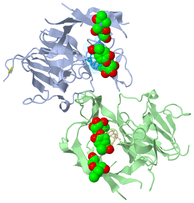
Chains, Units
Summary Information (see also Sequences/Alignments below) |
Ligands, Modified Residues, Ions (5, 10)
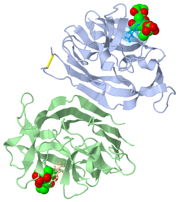
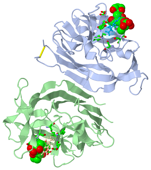
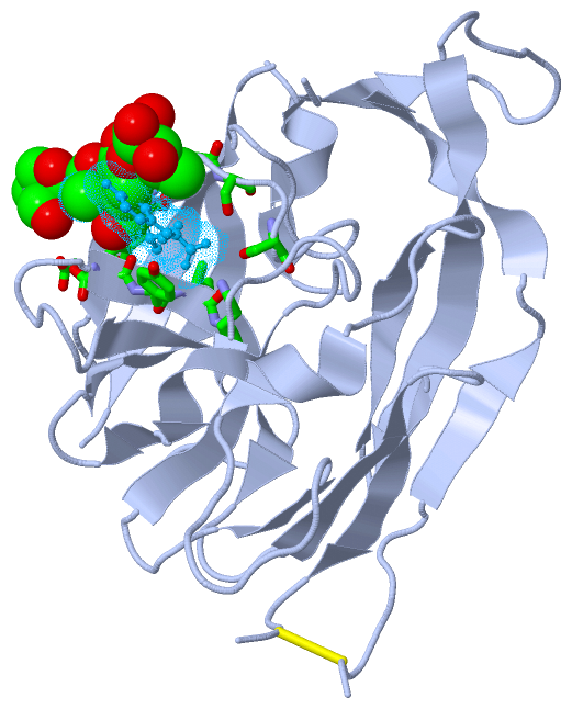
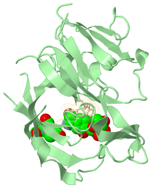
Asymmetric Unit (5, 10)
|
Sites (2, 2)
Asymmetric Unit (2, 2)
|
SS Bonds (1, 1)
Asymmetric Unit
|
||||||||
Cis Peptide Bonds (2, 2)
Asymmetric Unit
|
||||||||||||
SAPs(SNPs)/Variants (0, 0)| (no "SAP(SNP)/Variant" information available for 3S04) |
PROSITE Motifs (3, 6)
Asymmetric Unit (3, 6)
|
||||||||||||||||||||||||||||||||||||||||||||||||||||||||||||||||||||||||||||||||||||||||||||||||||||||||||||||||||||||||||||||||||||||||||||||||||||||||||||||||
Exons (0, 0)| (no "Exon" information available for 3S04) |
Sequences/Alignments
Asymmetric UnitChain A from PDB Type:PROTEIN Length:209 aligned with LEP_ECOLI | P00803 from UniProtKB/Swiss-Prot Length:324 Alignment length:245 89 99 109 119 129 139 149 159 169 179 189 199 209 219 229 239 249 259 269 279 289 299 309 319 LEP_ECOLI 80 FIYEPFQIPSGSMMPTLLIGDFILVEKFAYGIKDPIYQKTLIETGHPKRGDIVVFKYPEDPKLDYIKRAVGLPGDKVTYDPVSKELTIQPGCSSGQACENALPVTYSNVEPSDFVQTFSRRNGGEATSGFFEVPKNETKENGIRLSERKETLGDVTHRILTVPIAQDQVGMYYQQPGQQLATWIVPPGQYFMMGDNRDNSADSRYWGFVPEANLVGRATAIWMSFDKQEGEWPTGLRLSRIGGIH 324 SCOP domains d3s04a_ A: Type 1 signal pept idase SCOP domains CATH domains ----------------------------------------------------------------------------------------------------------------------------------------------------------------------------------------------------------------------------------------------------- CATH domains Pfam domains ----------------------------------------------------------------------------------------------------------------------------------------------------------------------------------------------------------------------------------------------------- Pfam domains SAPs(SNPs) ----------------------------------------------------------------------------------------------------------------------------------------------------------------------------------------------------------------------------------------------------- SAPs(SNPs) PROSITE ---------SPASE_I_-------------------------------------------------SPASE_I_2 --------------------------------------------------------------------------------------------------------------SPASE_I_3 ------------------------------------------ PROSITE Transcript ----------------------------------------------------------------------------------------------------------------------------------------------------------------------------------------------------------------------------------------------------- Transcript 3s04 A 80 FIYEPFQIPSGSMMPTLLIGDFILVEKFA----------------HPKRGDIVVFKYPEDPKLDYIKRAVGLPGDKVTYDPVSKELTIQPGCS---ACENALPVTYSNVEPSDFVQTF--------TSGFFEVPKNETKENGIRLSERKETLGDVTHRILTVPIAQDQVGMYYQQPGQQLATWIVPPGQYFMMGDNRDNSADSRYWGFVPEANLVGRATAIWMSFD---------LRLSRIGGIH 324 89 99 |- - | 129 139 149 159 169 | |179 189 | - |209 219 229 239 249 259 269 279 289 299 | - | 319 108 125 172 176 197 206 305 315 Chain B from PDB Type:PROTEIN Length:223 aligned with LEP_ECOLI | P00803 from UniProtKB/Swiss-Prot Length:324 Alignment length:245 89 99 109 119 129 139 149 159 169 179 189 199 209 219 229 239 249 259 269 279 289 299 309 319 LEP_ECOLI 80 FIYEPFQIPSGSMMPTLLIGDFILVEKFAYGIKDPIYQKTLIETGHPKRGDIVVFKYPEDPKLDYIKRAVGLPGDKVTYDPVSKELTIQPGCSSGQACENALPVTYSNVEPSDFVQTFSRRNGGEATSGFFEVPKNETKENGIRLSERKETLGDVTHRILTVPIAQDQVGMYYQQPGQQLATWIVPPGQYFMMGDNRDNSADSRYWGFVPEANLVGRATAIWMSFDKQEGEWPTGLRLSRIGGIH 324 SCOP domains d3s04b_ B: Type 1 signal pept idase SCOP domains CATH domains ----------------------------------------------------------------------------------------------------------------------------------------------------------------------------------------------------------------------------------------------------- CATH domains Pfam domains ----------------------------------------------------------------------------------------------------------------------------------------------------------------------------------------------------------------------------------------------------- Pfam domains SAPs(SNPs) ----------------------------------------------------------------------------------------------------------------------------------------------------------------------------------------------------------------------------------------------------- SAPs(SNPs) PROSITE ---------SPASE_I_-------------------------------------------------SPASE_I_2 --------------------------------------------------------------------------------------------------------------SPASE_I_3 ------------------------------------------ PROSITE Transcript ----------------------------------------------------------------------------------------------------------------------------------------------------------------------------------------------------------------------------------------------------- Transcript 3s04 B 80 FIYEPFQIPSGSMMPTLLIGDFILVEKFA--------------TGHPKRGDIVVFKYPEDPKLDYIKRAVGLPGDKVTYDPVSKELTIQPGC--------ALPVTYSNVEPSDFVQTFSRRNGGEATSGFFEVPKNETKENGIRLSERKETLGDVTHRILTVPIAQDQVGMYYQQPGQQLATWIVPPGQYFMMGDNRDNSADSRYWGFVPEANLVGRATAIWMSFDKQEGEWPTGLRLSRIGGIH 324 89 99 |- - | 129 139 149 159 169 | -| 189 199 209 219 229 239 249 259 269 279 289 299 309 319 108 123 171 180
Chain I from PDB Type:PROTEIN Length:6
SCOP domains ------ SCOP domains
CATH domains ------ CATH domains
Pfam domains ------ Pfam domains
SAPs(SNPs) ------ SAPs(SNPs)
PROSITE ------ PROSITE
Transcript ------ Transcript
3s04 I 2 xxGxAY 7
|| |
2-DSE
3-DAL
5-02V
Chain J from PDB Type:PROTEIN Length:6
SCOP domains ------ SCOP domains
CATH domains ------ CATH domains
Pfam domains ------ Pfam domains
SAPs(SNPs) ------ SAPs(SNPs)
PROSITE ------ PROSITE
Transcript ------ Transcript
3s04 J 2 xxGxAY 7
|| |
|| |
2-DSE
3-DAL
5-02V
|
||||||||||||||||||||
SCOP Domains (1, 2)
Asymmetric Unit
 |
CATH Domains (0, 0)| (no "CATH Domain" information available for 3S04) |
Pfam Domains (0, 0)| (no "Pfam Domain" information available for 3S04) |
Gene Ontology (13, 13)|
Asymmetric Unit(hide GO term definitions) Chain A,B (LEP_ECOLI | P00803)
|
||||||||||||||||||||||||||||||||||||||||||||||||||||||||||||||||||||||||||||||||||||||||||||||||
Interactive Views
|
|||||||||||||||||||||||||||||||||||||||||||||||||||||||||||||||||||||||||||||||||||||||||||||||||||||||||||||||||||||||||||||||||||||||||||||||||||||||||||||||||||||||||||||||||||||||||||||
Still Images
|
||||||||||||||||
Databases
|
||||||||||||||||||||||||||||||||||||||||||||||||||||||||||||||||||||||||||||||||||||||||||||||||||||||||||||||||||||||||||||||||||||||||||||||||||||||||||||||||
Analysis Tools
|
|||||||||||||||||||||||||||||||||||||||||||||||||||||||||||||
Entries Sharing at Least One Protein Chain (UniProt ID)
Related Entries Specified in the PDB File
|
|