|
|
|
|
 Description
|
|
Compounds
|
||||||||||||||||||||||||||||||||||||||||||||||||||||||||||||||||||||||||||||||
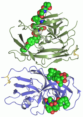
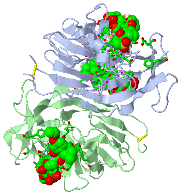
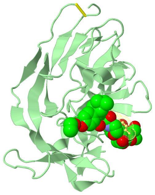
Chains, Units
Summary Information (see also Sequences/Alignments below) |
Ligands, Modified Residues, Ions (8, 18)
Asymmetric Unit (8, 18)
|
Sites (12, 12)
Asymmetric Unit (12, 12)
|
SS Bonds (2, 2)
Asymmetric Unit
|
||||||||||||
Cis Peptide Bonds (4, 4)
Asymmetric Unit
|
||||||||||||||||||||
SAPs(SNPs)/Variants (0, 0)| (no "SAP(SNP)/Variant" information available for 3IIQ) |
PROSITE Motifs (3, 6)
Asymmetric Unit (3, 6)
|
||||||||||||||||||||||||||||||||||||||||||||||||||||||||||||||||||||||||||||||||||||||||||||||||||||||||||||||||||||||||
Exons (0, 0)| (no "Exon" information available for 3IIQ) |
Sequences/Alignments
Asymmetric UnitChain A from PDB Type:PROTEIN Length:217 aligned with LEP_ECOLI | P00803 from UniProtKB/Swiss-Prot Length:324 Alignment length:245 89 99 109 119 129 139 149 159 169 179 189 199 209 219 229 239 249 259 269 279 289 299 309 319 LEP_ECOLI 80 FIYEPFQIPSGSMMPTLLIGDFILVEKFAYGIKDPIYQKTLIETGHPKRGDIVVFKYPEDPKLDYIKRAVGLPGDKVTYDPVSKELTIQPGCSSGQACENALPVTYSNVEPSDFVQTFSRRNGGEATSGFFEVPKNETKENGIRLSERKETLGDVTHRILTVPIAQDQVGMYYQQPGQQLATWIVPPGQYFMMGDNRDNSADSRYWGFVPEANLVGRATAIWMSFDKQEGEWPTGLRLSRIGGIH 324 SCOP domains d3iiqa_ A: Type 1 signal pep tidase SCOP domains CATH domains 3iiqA01 A:79-153,A:263-319 U mud Fragment, subunit A 3iiqA02 A:154-188, A:224-262 --------- ------------------3iiqA02 A:154-188,A:224-262 3iiqA01 A:79-153,A:263-319 Umud Fragment, subunit A ---- CATH domains Pfam domains ----------------------------------------------------------------------------------------------------------------------------------------------------------------------------------------------------------------------------------------------------- Pfam domains SAPs(SNPs) ----------------------------------------------------------------------------------------------------------------------------------------------------------------------------------------------------------------------------------------------------- SAPs(SNPs) PROSITE ---------SPASE_I_-------------------------------------------------SPASE_I_2 --------------------------------------------------------------------------------------------------------------SPASE_I_3 ------------------------------------------ PROSITE Transcript ----------------------------------------------------------------------------------------------------------------------------------------------------------------------------------------------------------------------------------------------------- Transcript 3iiq A 79 FIYEPFQIPSGSMMPTLLIGDFILVEKF-----------------HPKRGDIVVFKYPEDPKLDYIKRAVGLPGDKVTYDPVSKELTIQPGCS---ACENALPVTYSNVEPSDFVQTFS--------SGFFEVPKNETKENGIRLSERKETLGDVTHRILTVPIAQDQVGMYYQQPGQQLATWIVPPGQYFMMGDNRDNSADSRYWGFVPEANLVGRATAIWMSFDKQEGEWPTGLRLSRIGGIH 323 88 98 | - - | 128 138 148 158 168 | |178 188 |- 208 218 228 238 248 258 268 278 288 298 308 318 106 124 171 175 197 206 Chain B from PDB Type:PROTEIN Length:224 aligned with LEP_ECOLI | P00803 from UniProtKB/Swiss-Prot Length:324 Alignment length:244 90 100 110 120 130 140 150 160 170 180 190 200 210 220 230 240 250 260 270 280 290 300 310 320 LEP_ECOLI 81 IYEPFQIPSGSMMPTLLIGDFILVEKFAYGIKDPIYQKTLIETGHPKRGDIVVFKYPEDPKLDYIKRAVGLPGDKVTYDPVSKELTIQPGCSSGQACENALPVTYSNVEPSDFVQTFSRRNGGEATSGFFEVPKNETKENGIRLSERKETLGDVTHRILTVPIAQDQVGMYYQQPGQQLATWIVPPGQYFMMGDNRDNSADSRYWGFVPEANLVGRATAIWMSFDKQEGEWPTGLRLSRIGGIH 324 SCOP domains d3iiqb_ B: Type 1 signal pe ptidase SCOP domains CATH domains 3iiqB01 B:80-153,B:263-323 Umud Fragment, subunit A 3iiqB02 B:154-188,B: 224-262 ------------ --------------------3iiqB02 B:154-188,B:224-262 3iiqB01 B:80-153,B:263-323 Umud Fragment, subunit A CATH domains Pfam domains ---------------------------------------------------------------------------------------------------------------------------------------------------------------------------------------------------------------------------------------------------- Pfam domains SAPs(SNPs) ---------------------------------------------------------------------------------------------------------------------------------------------------------------------------------------------------------------------------------------------------- SAPs(SNPs) PROSITE --------SPASE_I_-------------------------------------------------SPASE_I_2 --------------------------------------------------------------------------------------------------------------SPASE_I_3 ------------------------------------------ PROSITE Transcript ---------------------------------------------------------------------------------------------------------------------------------------------------------------------------------------------------------------------------------------------------- Transcript 3iiq B 80 IYEPFQIPSGSMMPTLLIGDFILVEKF---------------TGHPKRGDIVVFKYPEDPKLDYIKRAVGLPGDKVTYDPVSKELTIQPGCSSG--CENALPVTYSNVEPSDFVQTFSRRN---ATSGFFEVPKNETKENGIRLSERKETLGDVTHRILTVPIAQDQVGMYYQQPGQQLATWIVPPGQYFMMGDNRDNSADSRYWGFVPEANLVGRATAIWMSFDKQEGEWPTGLRLSRIGGIH 323 89 99 | - - | 129 139 149 159 169 | |179 189 199| | 209 219 229 239 249 259 269 279 289 299 309 319 106 122 173 | 200 204 176
Chain C from PDB Type:PROTEIN Length:6
SCOP domains ------ SCOP domains
CATH domains ------ CATH domains
Pfam domains ------ Pfam domains
SAPs(SNPs) ------ SAPs(SNPs)
PROSITE ------ PROSITE
Transcript ------ Transcript
3iiq C 1 xxGxAY 6
|| |
1-DSE
2-DAL
4-5PG
Chain D from PDB Type:PROTEIN Length:6
SCOP domains ------ SCOP domains
CATH domains ------ CATH domains
Pfam domains ------ Pfam domains
SAPs(SNPs) ------ SAPs(SNPs)
PROSITE ------ PROSITE
Transcript ------ Transcript
3iiq D 1 xxGxAY 6
|| |
|| |
1-DSE
2-DAL
4-5PG
|
||||||||||||||||||||
SCOP Domains (1, 2)
Asymmetric Unit
 |
CATH Domains (2, 4)
Asymmetric Unit
|
Pfam Domains (0, 0)| (no "Pfam Domain" information available for 3IIQ) |
Gene Ontology (13, 13)|
Asymmetric Unit(hide GO term definitions) Chain A,B (LEP_ECOLI | P00803)
|
||||||||||||||||||||||||||||||||||||||||||||||||||||||||||||||||||||||||||||||||||||||||||||||||
Interactive Views
|
|||||||||||||||||||||||||||||||||||||||||||||||||||||||||||||||||||||||||||||||||||||||||||||||||||||||||||||||||||||||||||||||||||||||||||||||||||||||||||||||||||||||||||||||||||||||||||||||||||||||||||||||||||||||||||||||||||||||||||||||||||||||||||||||||||||||||||||||||||||||||||||||||
Still Images
|
||||||||||||||||
Databases
|
||||||||||||||||||||||||||||||||||||||||||||||||||||||||||||||||||||||||||||||||||||||||||||||||||||||||||||||||||||||||||||||||||||||||||||||||||||||||||||||||
Analysis Tools
|
|||||||||||||||||||||||||||||||||||||||||||||||||||||||||||||
Entries Sharing at Least One Protein Chain (UniProt ID)
Related Entries Specified in the PDB File
|
|