| molecular function |
|---|
| | GO:0001016 | | RNA polymerase III regulatory region DNA binding | | Interacting selectively and non-covalently with a DNA region that controls the transcription of a gene by RNA polymerase III. Binding may occur as a sequence specific interaction or as an interaction observed only once a factor has been recruited to the DNA by other factors. |
| | GO:0016209 | | antioxidant activity | | Inhibition of the reactions brought about by dioxygen (O2) or peroxides. Usually the antioxidant is effective because it can itself be more easily oxidized than the substance protected. The term is often applied to components that can trap free radicals, thereby breaking the chain reaction that normally leads to extensive biological damage. |
| | GO:0043027 | | cysteine-type endopeptidase inhibitor activity involved in apoptotic process | | Stops, prevents or reduces the activity of a cysteine-type endopeptidase involved in the apoptotic process. |
| | GO:0016491 | | oxidoreductase activity | | Catalysis of an oxidation-reduction (redox) reaction, a reversible chemical reaction in which the oxidation state of an atom or atoms within a molecule is altered. One substrate acts as a hydrogen or electron donor and becomes oxidized, while the other acts as hydrogen or electron acceptor and becomes reduced. |
| | GO:0004601 | | peroxidase activity | | Catalysis of the reaction: donor + hydrogen peroxide = oxidized donor + 2 H2O. |
| | GO:0051920 | | peroxiredoxin activity | | Catalysis of the reaction: 2 R'-SH + ROOH = R'-S-S-R' + H2O + ROH. |
| | GO:0072541 | | peroxynitrite reductase activity | | Catalysis of the reaction: 2 R-SH + ONOO- = R-SS-R + NO2- + H2O. |
| | GO:0046983 | | protein dimerization activity | | The formation of a protein dimer, a macromolecular structure consists of two noncovalently associated identical or nonidentical subunits. |
| | GO:0005102 | | receptor binding | | Interacting selectively and non-covalently with one or more specific sites on a receptor molecule, a macromolecule that undergoes combination with a hormone, neurotransmitter, drug or intracellular messenger to initiate a change in cell function. |
| | GO:0008379 | | thioredoxin peroxidase activity | | Catalysis of the reaction: thioredoxin + hydrogen peroxide = thioredoxin disulfide + H2O. |
| biological process |
|---|
| | GO:0070995 | | NADPH oxidation | | A metabolic process that results in the oxidation of reduced nicotinamide adenine dinucleotide, NADPH, to the oxidized form, NADP. |
| | GO:0006915 | | apoptotic process | | A programmed cell death process which begins when a cell receives an internal (e.g. DNA damage) or external signal (e.g. an extracellular death ligand), and proceeds through a series of biochemical events (signaling pathway phase) which trigger an execution phase. The execution phase is the last step of an apoptotic process, and is typically characterized by rounding-up of the cell, retraction of pseudopodes, reduction of cellular volume (pyknosis), chromatin condensation, nuclear fragmentation (karyorrhexis), plasma membrane blebbing and fragmentation of the cell into apoptotic bodies. When the execution phase is completed, the cell has died. |
| | GO:0098869 | | cellular oxidant detoxification | | Any process carried out at the cellular level that reduces or removes the toxicity superoxide radicals or hydrogen peroxide. |
| | GO:0034614 | | cellular response to reactive oxygen species | | Any process that results in a change in state or activity of a cell (in terms of movement, secretion, enzyme production, gene expression, etc.) as a result of a reactive oxygen species stimulus. Reactive oxygen species include singlet oxygen, superoxide, and oxygen free radicals. |
| | GO:0042744 | | hydrogen peroxide catabolic process | | The chemical reactions and pathways resulting in the breakdown of hydrogen peroxide (H2O2). |
| | GO:0006954 | | inflammatory response | | The immediate defensive reaction (by vertebrate tissue) to infection or injury caused by chemical or physical agents. The process is characterized by local vasodilation, extravasation of plasma into intercellular spaces and accumulation of white blood cells and macrophages. |
| | GO:0043066 | | negative regulation of apoptotic process | | Any process that stops, prevents, or reduces the frequency, rate or extent of cell death by apoptotic process. |
| | GO:0043154 | | negative regulation of cysteine-type endopeptidase activity involved in apoptotic process | | Any process that stops, prevents, or reduces the frequency, rate or extent of a cysteine-type endopeptidase activity involved in the apoptotic process. |
| | GO:0051354 | | negative regulation of oxidoreductase activity | | Any process that stops or reduces the rate of oxidoreductase activity, the catalysis of an oxidation-reduction (redox) reaction, a reversible chemical reaction in which the oxidation state of an atom or atoms within a molecule is altered. |
| | GO:0016480 | | negative regulation of transcription from RNA polymerase III promoter | | Any process that stops, prevents, or reduces the frequency, rate or extent of transcription from an RNA polymerase III promoter. |
| | GO:0055114 | | oxidation-reduction process | | A metabolic process that results in the removal or addition of one or more electrons to or from a substance, with or without the concomitant removal or addition of a proton or protons. |
| | GO:0032967 | | positive regulation of collagen biosynthetic process | | Any process that activates or increases the frequency, rate or extent of the chemical reactions and pathways resulting in the formation of collagen, any of a group of fibrous proteins of very high tensile strength that form the main component of connective tissue in animals. |
| | GO:2001057 | | reactive nitrogen species metabolic process | | The chemical reactions and pathways involving a reactive nitrogen species. |
| | GO:0060785 | | regulation of apoptosis involved in tissue homeostasis | | Any process that modulates the occurrence or rate of cell death by apoptosis that results in the maintenance of the steady-state number of cells within a tissue. |
| | GO:0006979 | | response to oxidative stress | | Any process that results in a change in state or activity of a cell or an organism (in terms of movement, secretion, enzyme production, gene expression, etc.) as a result of oxidative stress, a state often resulting from exposure to high levels of reactive oxygen species, e.g. superoxide anions, hydrogen peroxide (H2O2), and hydroxyl radicals. |
| | GO:0000302 | | response to reactive oxygen species | | Any process that results in a change in state or activity of a cell or an organism (in terms of movement, secretion, enzyme production, gene expression, etc.) as a result of a reactive oxygen species stimulus. Reactive oxygen species include singlet oxygen, superoxide, and oxygen free radicals. |
| cellular component |
|---|
| | GO:0005737 | | cytoplasm | | All of the contents of a cell excluding the plasma membrane and nucleus, but including other subcellular structures. |
| | GO:0031410 | | cytoplasmic vesicle | | A vesicle found in the cytoplasm of a cell. |
| | GO:0005829 | | cytosol | | The part of the cytoplasm that does not contain organelles but which does contain other particulate matter, such as protein complexes. |
| | GO:0070062 | | extracellular exosome | | A vesicle that is released into the extracellular region by fusion of the limiting endosomal membrane of a multivesicular body with the plasma membrane. Extracellular exosomes, also simply called exosomes, have a diameter of about 40-100 nm. |
| | GO:0005615 | | extracellular space | | That part of a multicellular organism outside the cells proper, usually taken to be outside the plasma membranes, and occupied by fluid. |
| | GO:0043231 | | intracellular membrane-bounded organelle | | Organized structure of distinctive morphology and function, bounded by a single or double lipid bilayer membrane and occurring within the cell. Includes the nucleus, mitochondria, plastids, vacuoles, and vesicles. Excludes the plasma membrane. |
| | GO:0005759 | | mitochondrial matrix | | The gel-like material, with considerable fine structure, that lies in the matrix space, or lumen, of a mitochondrion. It contains the enzymes of the tricarboxylic acid cycle and, in some organisms, the enzymes concerned with fatty acid oxidation. |
| | GO:0005739 | | mitochondrion | | A semiautonomous, self replicating organelle that occurs in varying numbers, shapes, and sizes in the cytoplasm of virtually all eukaryotic cells. It is notably the site of tissue respiration. |
| | GO:0005634 | | nucleus | | A membrane-bounded organelle of eukaryotic cells in which chromosomes are housed and replicated. In most cells, the nucleus contains all of the cell's chromosomes except the organellar chromosomes, and is the site of RNA synthesis and processing. In some species, or in specialized cell types, RNA metabolism or DNA replication may be absent. |
| | GO:0048471 | | perinuclear region of cytoplasm | | Cytoplasm situated near, or occurring around, the nucleus. |
| | GO:0005782 | | peroxisomal matrix | | The volume contained within the membranes of a peroxisome; in many cells the matrix contains a crystalloid core largely composed of urate oxidase. |
| | GO:0005777 | | peroxisome | | A small organelle enclosed by a single membrane, and found in most eukaryotic cells. Contains peroxidases and other enzymes involved in a variety of metabolic processes including free radical detoxification, lipid catabolism and biosynthesis, and hydrogen peroxide metabolism. |
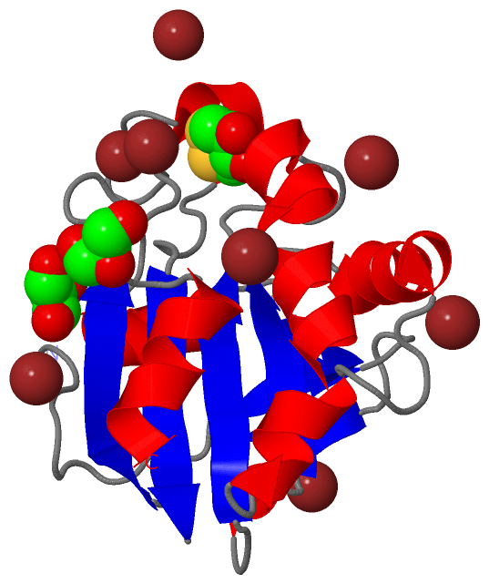
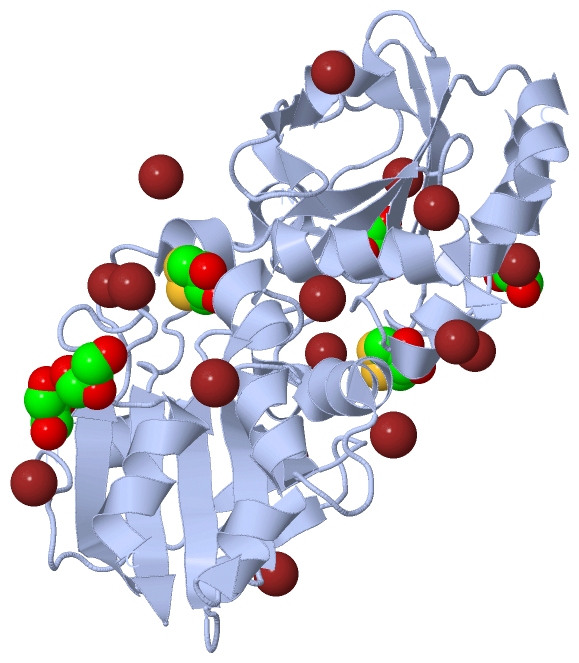
 Description
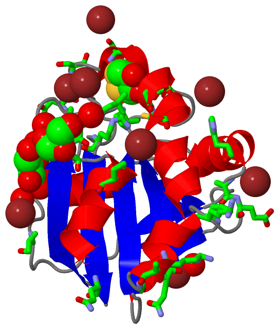
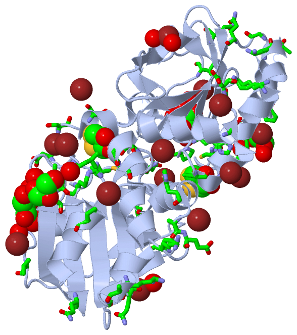