|
|
|
|
 Description
|
|
Compounds
|
||||||||||||||||||||||||||||||||||||||||||||||||
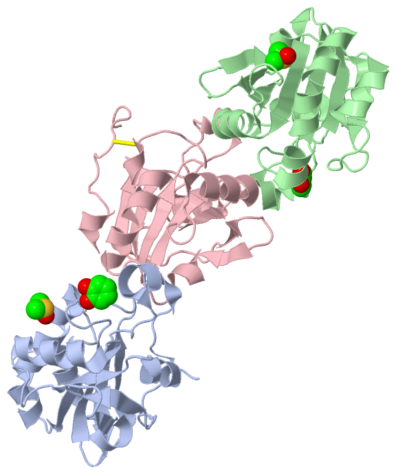
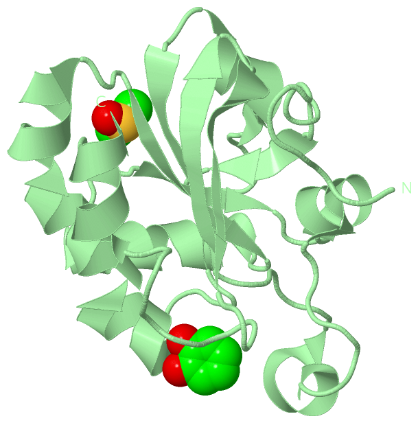
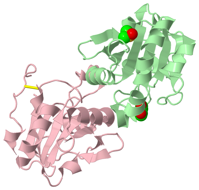
Chains, Units
Summary Information (see also Sequences/Alignments below) |
Ligands, Modified Residues, Ions (2, 4)| Asymmetric Unit (2, 4) Biological Unit 1 (2, 2) Biological Unit 2 (2, 2) Biological Unit 3 (0, 0) Biological Unit 4 (2, 4) Biological Unit 5 (2, 2) |
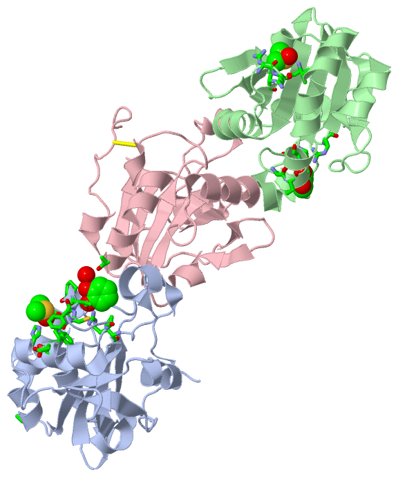
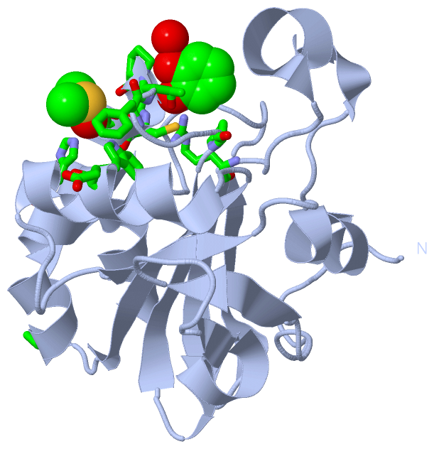
Sites (4, 4)
Asymmetric Unit (4, 4)
|
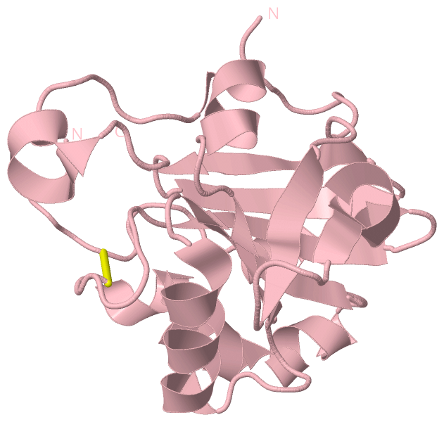
SS Bonds (1, 1)
Asymmetric Unit
|
||||||||
Cis Peptide Bonds (0, 0)| (no "Cis Peptide Bond" information available for 4K7I) |
SAPs(SNPs)/Variants (0, 0)| (no "SAP(SNP)/Variant" information available for 4K7I) |
PROSITE Motifs (0, 0)| (no "PROSITE Motif" information available for 4K7I) |
Exons (0, 0)| (no "Exon" information available for 4K7I) |
Sequences/Alignments
Asymmetric Unit
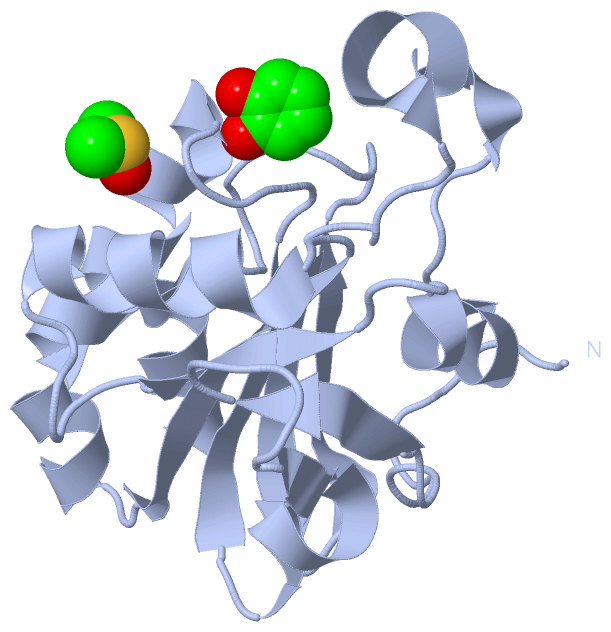
Chain A from PDB Type:PROTEIN Length:162
SCOP domains ------------------------------------------------------------------------------------------------------------------------------------------------------------------ SCOP domains
CATH domains ------------------------------------------------------------------------------------------------------------------------------------------------------------------ CATH domains
Pfam domains ------------------------------------------------------------------------------------------------------------------------------------------------------------------ Pfam domains
SAPs(SNPs) ------------------------------------------------------------------------------------------------------------------------------------------------------------------ SAPs(SNPs)
PROSITE ------------------------------------------------------------------------------------------------------------------------------------------------------------------ PROSITE
Transcript ------------------------------------------------------------------------------------------------------------------------------------------------------------------ Transcript
4k7i A 0 SAPIKVGDAIPAVEVFEGEPGNKVNLAELFKGKKGVLFGVPGAFTPGCSKTHLPGFVEQAEALKAKGVQVVACLSVNDAFVTGEWGRAHKAEGKVRLLADPTGAFGKETDLLLDDSLVSIFGNRRLKRFSMVVQDGIVKALNVEPDGTGLTCSLAPNIISQL 161
9 19 29 39 49 59 69 79 89 99 109 119 129 139 149 159
Chain B from PDB Type:PROTEIN Length:161
SCOP domains ----------------------------------------------------------------------------------------------------------------------------------------------------------------- SCOP domains
CATH domains ----------------------------------------------------------------------------------------------------------------------------------------------------------------- CATH domains
Pfam domains ----------------------------------------------------------------------------------------------------------------------------------------------------------------- Pfam domains
SAPs(SNPs) ----------------------------------------------------------------------------------------------------------------------------------------------------------------- SAPs(SNPs)
PROSITE ----------------------------------------------------------------------------------------------------------------------------------------------------------------- PROSITE
Transcript ----------------------------------------------------------------------------------------------------------------------------------------------------------------- Transcript
4k7i B 1 APIKVGDAIPAVEVFEGEPGNKVNLAELFKGKKGVLFGVPGAFTPGCSKTHLPGFVEQAEALKAKGVQVVACLSVNDAFVTGEWGRAHKAEGKVRLLADPTGAFGKETDLLLDDSLVSIFGNRRLKRFSMVVQDGIVKALNVEPDGTGLTCSLAPNIISQL 161
10 20 30 40 50 60 70 80 90 100 110 120 130 140 150 160
Chain C from PDB Type:PROTEIN Length:160
SCOP domains ---------------------------------------------------------------------------------------------------------------------------------------------------------------- SCOP domains
CATH domains ---------------------------------------------------------------------------------------------------------------------------------------------------------------- CATH domains
Pfam domains ---------------------------------------------------------------------------------------------------------------------------------------------------------------- Pfam domains
SAPs(SNPs) ---------------------------------------------------------------------------------------------------------------------------------------------------------------- SAPs(SNPs)
PROSITE ---------------------------------------------------------------------------------------------------------------------------------------------------------------- PROSITE
Transcript ---------------------------------------------------------------------------------------------------------------------------------------------------------------- Transcript
4k7i C 1 APIKVGDAIPAVEVFEGEPGNKVNLAELFKGKKGVLFGVPGAFTPGCSKTHLPGFVEQAEALKAKGVQVVACLSVNDAFVTGEWGRAHKAEGKVRLLADPTGAFGKETDLLLDDSLVSIFGNRRLKRFSMVVQDGIVKALNVEPGTGLTCSLAPNIISQL 161
10 20 30 40 50 60 70 80 90 100 110 120 130 140 || 151 161
144|
146
|
||||||||||||||||||||
SCOP Domains (0, 0)| (no "SCOP Domain" information available for 4K7I) |
CATH Domains (0, 0)| (no "CATH Domain" information available for 4K7I) |
Pfam Domains (0, 0)| (no "Pfam Domain" information available for 4K7I) |
Gene Ontology (38, 38)|
Asymmetric Unit(hide GO term definitions) |
Interactive Views
|
||||||||||||||||||||||||||||||||||||||||||||||||||||||||||||||||||||||||||||||||||||||||||||||||||||||||||||||||||||||||||||||||||||||||||||||||||||||||||||||||||||||||||||||||||||||||
Still Images
|
||||||||||||||||
Databases
|
||||||||||||||||||||||||||||||||||||||||||||||||||||||||||||||||||||||||||||||||||||||||||||||||||||||||||||||||||||||||||||||||||||||||||||||||||||||||||||||||
Analysis Tools
|
|||||||||||||||||||||||||||||||||||||||||||||||||||||||||||||
Entries Sharing at Least One Protein Chain (UniProt ID)
Related Entries Specified in the PDB File
|
|