Show PDB file:   
         Plain Text   HTML   (compressed file size)
QuickSearch:   
by PDB,NDB,UniProt,PROSITE Code or Search Term(s)  
(-)Asym./Biol. Unit
(-)Asym./Biol. Unit - sites
collapse expand < >
Image Asym./Biol. Unit
Asym./Biol. Unit  (Jmol Viewer)
Image Asym./Biol. Unit - sites
Asym./Biol. Unit - sites  (Jmol Viewer)

(-) Description

Title :  CRYSTAL STRUCTURE OF THE CATALYTIC DOMAIN OF ADAMTS-5 IN COMPLEX WITH AN AMINO-2-INDANOL COMPOUND
 
Authors :  H. -S. Shieh, J. M. Williams, N. Caspers
Date :  26 Jan 10  (Deposition) - 31 Mar 10  (Release) - 06 Apr 11  (Revision)
Method :  X-RAY DIFFRACTION
Resolution :  1.60
Chains :  Asym./Biol. Unit :  A
Keywords :  Alpha/Beta Structure, Central Five Stranded Beta-Sheet, Cleavage On Pair Of Basic Residues, Disulfide Bond, Extracellular Matrix, Glycoprotein, Metal-Binding, Metalloprotease, Protease, Secreted, Zymogen, Hydrolase (Keyword Search: [Gene Ontology, PubMed, Web (Google))
 
Reference :  H. S. Shieh, A. G. Tomasselli, K. J. Mathis, M. E. Schnute, S. S. Woodard N. Caspers, J. M. Williams, J. R. Kiefer, G. Munie, A. Wittwer, A. M. Malfait, M. D. Tortorella
Structure Analysis Reveals The Flexibility Of The Adamts-5 Active Site.
Protein Sci. V. 20 735 2011
PubMed-ID: 21370305  |  Reference-DOI: 10.1002/PRO.606

(-) Compounds

Molecule 1 - A DISINTEGRIN AND METALLOPROTEINASE WITH THROMBOSPONDIN MOTIFS 5
    ChainsA
    EC Number3.4.24.-
    EngineeredYES
    Expression SystemESCHERICHIA COLI
    Expression System PlasmidPPHA79257
    Expression System StrainMON208
    Expression System Taxid562
    Expression System Vector TypePLASMID
    FragmentCATALYTIC DOMAIN
    GeneADAMTS-5, ADAMTS11, ADAMTS5, ADMP2
    MutationYES
    Organism CommonHUMAN
    Organism ScientificHOMO SAPIENS
    Organism Taxid9606
    SynonymADAMTS-5, ADAM-TS 5, ADAM-TS5, AGGRECANASE-2, ADMP-2, A DISINTEGRIN AND METALLOPROTEINASE WITH THROMBOSPONDIN MOTIFS 11, ADAMTS-11, ADAM-TS 11

 Structural Features

(-) Chains, Units

  1
Asymmetric/Biological Unit A

Summary Information (see also Sequences/Alignments below)
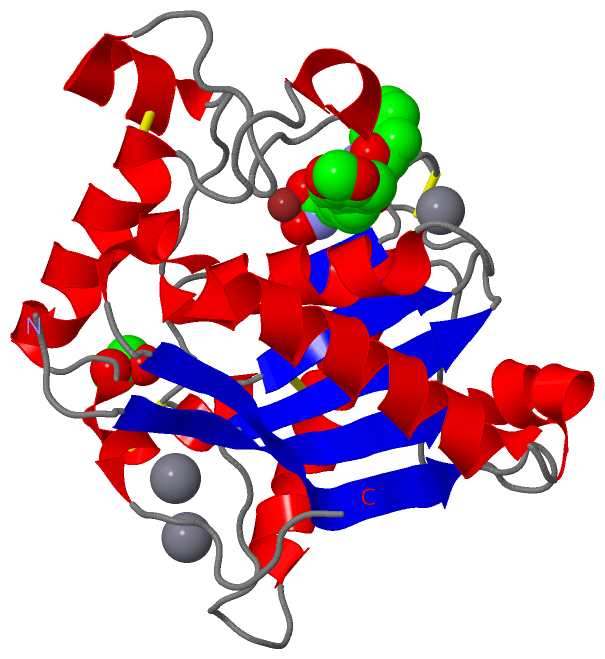
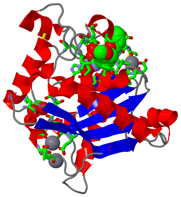
(-) Ligands, Modified Residues, Ions  (4, 6)

Asymmetric/Biological Unit (4, 6)
No.NameCountTypeFull Name
1CA3Ligand/IonCALCIUM ION
2EDO1Ligand/Ion1,2-ETHANEDIOL
3LA31Ligand/Ion(2R)-2-[4-(1,3-BENZODIOXOL-5-YL)BENZYL]-N~4~-HYDROXY-N~1~-[(1S,2R)-2-HYDROXY-2,3-DIHYDRO-1H-INDEN-1-YL]BUTANEDIAMIDE
4ZN1Ligand/IonZINC ION

(-) Sites  (6, 6)

Asymmetric Unit (6, 6)
No.NameEvidenceResiduesDescription
1AC1SOFTWAREASP A:377 , THR A:378 , LEU A:379 , GLY A:380 , PHE A:406 , THR A:407 , HIS A:410 , GLU A:411 , HIS A:414 , HIS A:420 , LEU A:438 , SER A:440 , SER A:441 , LEU A:443 , ILE A:446 , ZN A:901 , HOH A:5040 , HOH A:5296BINDING SITE FOR RESIDUE LA3 A 801
2AC2SOFTWAREGLY A:372 , GLY A:385 , THR A:386 , SER A:389 , HIS A:468 , HOH A:5006 , HOH A:5217BINDING SITE FOR RESIDUE EDO A 802
3AC3SOFTWAREHIS A:410 , HIS A:414 , HIS A:420 , LA3 A:801BINDING SITE FOR RESIDUE ZN A 901
4AC4SOFTWAREGLU A:270 , ASP A:360 , CYS A:471 , ASP A:474 , HOH A:5009 , HOH A:5031BINDING SITE FOR RESIDUE CA A 902
5AC5SOFTWAREGLU A:270 , ASP A:474 , LEU A:475 , HOH A:5068 , HOH A:5103 , HOH A:5157BINDING SITE FOR RESIDUE CA A 903
6AC6SOFTWAREASP A:369 , LEU A:370 , CYS A:376 , THR A:378 , GLU A:398 , HOH A:5014BINDING SITE FOR RESIDUE CA A 904

(-) SS Bonds  (4, 4)

Asymmetric/Biological Unit
No.Residues
1A:342 -A:394
2A:371 -A:376
3A:388 -A:471
4A:426 -A:455

(-) Cis Peptide Bonds  (0, 0)

(no "Cis Peptide Bond" information available for 3LJT)

 Sequence-Structure Mapping

(-) SAPs(SNPs)/Variants  (0, 0)

(no "SAP(SNP)/Variant" information available for 3LJT)

(-) PROSITE Motifs  (2, 2)

Asymmetric/Biological Unit (2, 2)
 PROSITEUniProtKBPDB
No.IDACDescriptionIDLocationCountLocation
1ADAM_MEPROPS50215 ADAM type metalloprotease domain profile.ATS5_HUMAN267-476  1A:267-476
2ZINC_PROTEASEPS00142 Neutral zinc metallopeptidases, zinc-binding region signature.ATS5_HUMAN407-416  1A:407-416

(-) Exons   (4, 4)

Asymmetric/Biological Unit (4, 4)
 ENSEMBLUniProtKBPDB
No.Transcript IDExonExon IDGenome LocationLengthIDLocationLengthCountLocationLength
1.1ENST000002849871ENSE00001017391chr21:28338832-283376071226ATS5_HUMAN1-3683681A:263-368106
1.2ENST000002849872ENSE00001017390chr21:28327190-28327058133ATS5_HUMAN369-413451A:369-41345
1.3ENST000002849873ENSE00001017386chr21:28315866-28315699168ATS5_HUMAN413-469571A:413-46957
1.4ENST000002849874ENSE00001017392chr21:28307068-28306785284ATS5_HUMAN469-563951A:469-48012
1.5ENST000002849875ENSE00001017389chr21:28305363-28305180184ATS5_HUMAN564-625620--
1.6ENST000002849876ENSE00001017388chr21:28304498-28304323176ATS5_HUMAN625-683590--
1.7ENST000002849877ENSE00001017393chr21:28302380-28302205176ATS5_HUMAN684-742590--
1.8ENST000002849878ENSE00001017387chr21:28296939-282902316709ATS5_HUMAN742-9301890--

(-) Sequences/Alignments

Asymmetric/Biological Unit
   Reformat: Number of residues per line =  ('0' or empty: single-line sequence representation)
  Number of residues per labelling interval =   
  UniProt sequence: complete  aligned part    
   Show mapping: SCOP domains CATH domains Pfam domains Secondary structure (by author)
SAPs(SNPs) PROSITE motifs Exons
(details for a mapped element are shown in a popup box when the mouse pointer rests over it)
Chain A from PDB  Type:PROTEIN  Length:218
 aligned with ATS5_HUMAN | Q9UNA0 from UniProtKB/Swiss-Prot  Length:930

    Alignment length:218
                                   272       282       292       302       312       322       332       342       352       362       372       382       392       402       412       422       432       442       452       462       472        
           ATS5_HUMAN   263 ISRARQVELLLVADASMARLYGRGLQHYLLTLASIANRLYSHASIENHIRLAVVKVVVLGDKDKSLEVSKNAATTLKNFCKWQHQHNQLGDDHEEHYDAAILFTREDLCGHHSCDTLGMADVGTICSPERSCAVIEDDGLHAAFTVAHEIGHLLGLSHDDSKFCEETFGSTEDKRLMSSILTSIDASKPWSKCTSATITEFLDDGHGNCLLDLPRKQI 480
               SCOP domains d3ljta_ A: automated matches                                                                                                                                                                                               SCOP domains
               CATH domains -------------------------------------------------------------------------------------------------------------------------------------------------------------------------------------------------------------------------- CATH domains
               Pfam domains ----Reprolysin-3ljtA01 A:267-476                                                                                                                                                                                      ---- Pfam domains
         Sec.struct. author ....eeeeeeeeehhhhhhhhh.hhhhhhhhhhhhhhhhhhhhhhh..eeeeeeeeee............hhhhhhhhhhhhhhhhhhh.........eeeeee.............eee......hhhh.eeeee....hhhhhhhhhhhhhh......hhhhhhhhh..........hhhhh......hhhhhhhhhhhhhh..hhhhh....... Sec.struct. author
                 SAPs(SNPs) -------------------------------------------------------------------------------------------------------------------------------------------------------------------------------------------------------------------------- SAPs(SNPs)
                PROSITE (1) ----ADAM_MEPRO  PDB: A:267-476 UniProt: 267-476                                                                                                                                                                       ---- PROSITE (1)
                PROSITE (2) ------------------------------------------------------------------------------------------------------------------------------------------------ZINC_PROTE---------------------------------------------------------------- PROSITE (2)
           Transcript 1 (1) Exon 1.1  PDB: A:263-368 UniProt: 1-368 [INCOMPLETE]                                                      Exon 1.2  PDB: A:369-413 UniProt: 369-413    -------------------------------------------------------Exon 1.4     Transcript 1 (1)
           Transcript 1 (2) ------------------------------------------------------------------------------------------------------------------------------------------------------Exon 1.3  PDB: A:413-469 UniProt: 413-469                ----------- Transcript 1 (2)
                 3ljt A 263 ASRARQVELLLVADASMARKYGRGLQHYLLTLASIANRLYSHASIENHIRLAVVKVVVLGDKDKSLEVSKNAATTLKNFCKWQHQHNQLGDDHEEHYDAAILFTREDLCGHHSCDTLGMADVGTICSPERSCAVIEDDGLHAAFTVAHEIGHLLGLSHDDSKFCEETFGSTEDKRLMSSILTSIDASKPWSKCTSATITEFLDDGHGNCLLDLPRKQI 480
                                   272       282       292       302       312       322       332       342       352       362       372       382       392       402       412       422       432       442       452       462       472        

   Legend:   → Mismatch (orange background)
  - → Gap (green background, '-', border residues have a numbering label)
    → Modified Residue (blue background, lower-case, 'x' indicates undefined single-letter code, labelled with number + name)
  x → Chemical Group (purple background, 'x', labelled with number + name, e.g. ACE or NH2)
  extra numbering lines below/above indicate numbering irregularities and modified residue names etc., number ends below/above '|'

 Classification and Annotation

(-) SCOP Domains  (1, 1)

Asymmetric/Biological Unit

(-) CATH Domains  (0, 0)

(no "CATH Domain" information available for 3LJT)

(-) Pfam Domains  (1, 1)

Asymmetric/Biological Unit

(-) Gene Ontology  (19, 19)

Asymmetric/Biological Unit(hide GO term definitions)
Chain A   (ATS5_HUMAN | Q9UNA0)
molecular function
    GO:0050840    extracellular matrix binding    Interacting selectively and non-covalently with a component of the extracellular matrix.
    GO:0008201    heparin binding    Interacting selectively and non-covalently with heparin, any member of a group of glycosaminoglycans found mainly as an intracellular component of mast cells and which consist predominantly of alternating alpha-(1->4)-linked D-galactose and N-acetyl-D-glucosamine-6-sulfate residues.
    GO:0016787    hydrolase activity    Catalysis of the hydrolysis of various bonds, e.g. C-O, C-N, C-C, phosphoric anhydride bonds, etc. Hydrolase is the systematic name for any enzyme of EC class 3.
    GO:0005178    integrin binding    Interacting selectively and non-covalently with an integrin.
    GO:0046872    metal ion binding    Interacting selectively and non-covalently with any metal ion.
    GO:0004222    metalloendopeptidase activity    Catalysis of the hydrolysis of internal, alpha-peptide bonds in a polypeptide chain by a mechanism in which water acts as a nucleophile, one or two metal ions hold the water molecule in place, and charged amino acid side chains are ligands for the metal ions.
    GO:0008237    metallopeptidase activity    Catalysis of the hydrolysis of peptide bonds by a mechanism in which water acts as a nucleophile, one or two metal ions hold the water molecule in place, and charged amino acid side chains are ligands for the metal ions.
    GO:0008233    peptidase activity    Catalysis of the hydrolysis of a peptide bond. A peptide bond is a covalent bond formed when the carbon atom from the carboxyl group of one amino acid shares electrons with the nitrogen atom from the amino group of a second amino acid.
    GO:0005515    protein binding    Interacting selectively and non-covalently with any protein or protein complex (a complex of two or more proteins that may include other nonprotein molecules).
    GO:0008270    zinc ion binding    Interacting selectively and non-covalently with zinc (Zn) ions.
biological process
    GO:0042742    defense response to bacterium    Reactions triggered in response to the presence of a bacterium that act to protect the cell or organism.
    GO:0022617    extracellular matrix disassembly    A process that results in the breakdown of the extracellular matrix.
    GO:0036066    protein O-linked fucosylation    The process of transferring a fucosyl group to a serine or threonine residues in a protein acceptor molecule, to form an O-linked protein-sugar linkage.
    GO:0006508    proteolysis    The hydrolysis of proteins into smaller polypeptides and/or amino acids by cleavage of their peptide bonds.
cellular component
    GO:0005788    endoplasmic reticulum lumen    The volume enclosed by the membranes of the endoplasmic reticulum.
    GO:0031012    extracellular matrix    A structure lying external to one or more cells, which provides structural support for cells or tissues.
    GO:0005576    extracellular region    The space external to the outermost structure of a cell. For cells without external protective or external encapsulating structures this refers to space outside of the plasma membrane. This term covers the host cell environment outside an intracellular parasite.
    GO:0005615    extracellular space    That part of a multicellular organism outside the cells proper, usually taken to be outside the plasma membranes, and occupied by fluid.
    GO:0005578    proteinaceous extracellular matrix    A layer consisting mainly of proteins (especially collagen) and glycosaminoglycans (mostly as proteoglycans) that forms a sheet underlying or overlying cells such as endothelial and epithelial cells. The proteins are secreted by cells in the vicinity. An example of this component is found in Mus musculus.

 Visualization

(-) Interactive Views

Asymmetric/Biological Unit
  Complete Structure
    Jena3D(integrated viewing of ligand, site, SAP, PROSITE, SCOP information)
    WebMol | AstexViewer[tm]@PDBe
(Java Applets, require no local installation except for Java; loading may be slow)
    STRAP
(Java WebStart application, automatic local installation, requires Java; full application with system access!)
    RasMol
(require local installation)
    Molscript (VRML)
(requires installation of a VRML viewer; select preferred view via VRML and generate a mono or stereo PDF format file)
 
  Ligands, Modified Residues, Ions
    CA  [ RasMol | Jena3D ]  +environment [ RasMol | Jena3D ]
    EDO  [ RasMol | Jena3D ]  +environment [ RasMol | Jena3D ]
    LA3  [ RasMol | Jena3D ]  +environment [ RasMol | Jena3D ]
    ZN  [ RasMol | Jena3D ]  +environment [ RasMol | Jena3D ]
 
  Sites
    AC1  [ RasMol ]  +environment [ RasMol ]
    AC2  [ RasMol ]  +environment [ RasMol ]
    AC3  [ RasMol ]  +environment [ RasMol ]
    AC4  [ RasMol ]  +environment [ RasMol ]
    AC5  [ RasMol ]  +environment [ RasMol ]
    AC6  [ RasMol ]  +environment [ RasMol ]
 
  Cis Peptide Bonds
(no "Cis Peptide Bonds" information available for 3ljt)
 

(-) Still Images

Jmol
  protein: cartoon or spacefill or dots and stick; nucleic acid: cartoon and stick; ligands: spacefill; active site: stick
Molscript
  protein, nucleic acid: cartoon; ligands: spacefill; active site: ball and stick

 Databases and Analysis Tools

(-) Databases

Access by PDB/NDB ID
  3ljt
    Family and Domain InformationProDom | SYSTERS
    General Structural InformationGlycoscienceDB | MMDB | NDB | OCA | PDB | PDBe | PDBj | PDBsum | PDBWiki | PQS | PROTEOPEDIA
    Orientation in MembranesOPM
    Protein SurfaceSURFACE
    Secondary StructureDSSP (structure derived) | HSSP (homology derived)
    Structural GenomicsGeneCensus
    Structural NeighboursCE | VAST
    Structure ClassificationCATH | Dali | SCOP
    Validation and Original DataBMRB Data View | BMRB Restraints Grid | EDS | PROCHECK | RECOORD | WHAT_CHECK
 
Access by UniProt ID/Accession number
  ATS5_HUMAN | Q9UNA0
    Comparative Protein Structure ModelsModBase
    Genomic InformationEnsembl
    Protein-protein InteractionDIP
    Sequence, Family and Domain InformationInterPro | Pfam | SMART | UniProtKB/SwissProt
 
Access by Enzyme Classificator   (EC Number)
  3.4.24.-
    General Enzyme InformationBRENDA | EC-PDB | Enzyme | IntEnz
    PathwayKEGG | MetaCyc
 
Access by Disease Identifier   (MIM ID)
  (no 'MIM ID' available)
    Disease InformationOMIM
 
Access by GenAge ID
  (no 'GenAge ID' available)
    Age Related InformationGenAge

(-) Analysis Tools

Access by PDB/NDB ID
    Domain InformationXDom
    Interatomic Contacts of Structural UnitsCSU
    Ligand-protein ContactsLPC
    Protein CavitiescastP
    Sequence and Secondary StructurePDBCartoon
    Structure AlignmentSTRAP(Java WebStart application, automatic local installation, requires Java; full application with system access!)
    Structure and Sequence BrowserSTING
 
Access by UniProt ID/Accession number
  ATS5_HUMAN | Q9UNA0
    Protein Disorder PredictionDisEMBL | FoldIndex | GLOBPLOT (for more information see DisProt)

 Related Entries

(-) Entries Sharing at Least One Protein Chain (UniProt ID)

UniProtKB/Swiss-Prot
        ATS5_HUMAN | Q9UNA02rjq 3b8z 3hy7 3hy9 3hyg

(-) Related Entries Specified in the PDB File

3hy9 THE SAME PROTEIN COMPLEXED WITH INHIBITOR OF SAME CHEMICAL CLASS
3hyg THE SAME PROTEIN COMPLEXED WITH INHIBITOR OF SAME CHEMICAL CLASS
3ljz