|
|
|
|
 Description
|
|
Compounds
|
||||||||||||||||||||||||||||||||||||||||||||||||||||||||
Chains, Units
Summary Information (see also Sequences/Alignments below) |
Ligands, Modified Residues, Ions (3, 10)
Asymmetric Unit (3, 10)
|
Sites (10, 10)
Asymmetric Unit (10, 10)
|
SS Bonds (8, 8)
Asymmetric Unit
|
||||||||||||||||||||||||||||||||||||
Cis Peptide Bonds (0, 0)| (no "Cis Peptide Bond" information available for 3HY9) |
SAPs(SNPs)/Variants (0, 0)| (no "SAP(SNP)/Variant" information available for 3HY9) |
PROSITE Motifs (2, 4)
Asymmetric Unit (2, 4)
|
||||||||||||||||||||||||||||||||||||||||||||||||||||||||||||||||||||||||||||||||||||||||||||||||
Exons (4, 8)
Asymmetric Unit (4, 8)
|
||||||||||||||||||||||||||||||||||||||||||||||||||||||||||||||||||||||||||||||||||||||||||||||||||||||||||||||||||||||||
Sequences/Alignments
Asymmetric UnitChain A from PDB Type:PROTEIN Length:217 aligned with ATS5_HUMAN | Q9UNA0 from UniProtKB/Swiss-Prot Length:930 Alignment length:217 273 283 293 303 313 323 333 343 353 363 373 383 393 403 413 423 433 443 453 463 473 ATS5_HUMAN 264 SRARQVELLLVADASMARLYGRGLQHYLLTLASIANRLYSHASIENHIRLAVVKVVVLGDKDKSLEVSKNAATTLKNFCKWQHQHNQLGDDHEEHYDAAILFTREDLCGHHSCDTLGMADVGTICSPERSCAVIEDDGLHAAFTVAHEIGHLLGLSHDDSKFCEETFGSTEDKRLMSSILTSIDASKPWSKCTSATITEFLDDGHGNCLLDLPRKQI 480 SCOP domains d3hy9a_ A: automated matches SCOP domains CATH domains ------------------------------------------------------------------------------------------------------------------------------------------------------------------------------------------------------------------------- CATH domains Pfam domains ------------------------------------------------------------------------------------------------------------------------------------------------------------------------------------------------------------------------- Pfam domains SAPs(SNPs) ------------------------------------------------------------------------------------------------------------------------------------------------------------------------------------------------------------------------- SAPs(SNPs) PROSITE (1) ---ADAM_MEPRO PDB: A:267-476 UniProt: 267-476 ---- PROSITE (1) PROSITE (2) -----------------------------------------------------------------------------------------------------------------------------------------------ZINC_PROTE---------------------------------------------------------------- PROSITE (2) Transcript 1 (1) Exon 1.1 PDB: A:264-368 UniProt: 1-368 [INCOMPLETE] Exon 1.2 PDB: A:369-413 UniProt: 369-413 -------------------------------------------------------Exon 1.4 Transcript 1 (1) Transcript 1 (2) -----------------------------------------------------------------------------------------------------------------------------------------------------Exon 1.3 PDB: A:413-469 UniProt: 413-469 ----------- Transcript 1 (2) 3hy9 A 264 SRARQVELLLVADASMARKYGRGLQHYLLTLASIANRLYSHASIENHIRLAVVKVVVLGDKDKSLEVSKNAATTLKNFCKWQHQHNQLGDDHEEHYDAAILFTREDLCGHHSCDTLGMADVGTICSPERSCAVIEDDGLHAAFTVAHEIGHLLGLSHDDSKFCEETFGSTEDKRLMSSILTSIDASKPWSKCTSATITEFLDDGHGNCLLDLPRKQI 480 273 283 293 303 313 323 333 343 353 363 373 383 393 403 413 423 433 443 453 463 473 Chain B from PDB Type:PROTEIN Length:220 aligned with ATS5_HUMAN | Q9UNA0 from UniProtKB/Swiss-Prot Length:930 Alignment length:220 270 280 290 300 310 320 330 340 350 360 370 380 390 400 410 420 430 440 450 460 470 480 ATS5_HUMAN 261 RSISRARQVELLLVADASMARLYGRGLQHYLLTLASIANRLYSHASIENHIRLAVVKVVVLGDKDKSLEVSKNAATTLKNFCKWQHQHNQLGDDHEEHYDAAILFTREDLCGHHSCDTLGMADVGTICSPERSCAVIEDDGLHAAFTVAHEIGHLLGLSHDDSKFCEETFGSTEDKRLMSSILTSIDASKPWSKCTSATITEFLDDGHGNCLLDLPRKQI 480 SCOP domains d3hy9b_ B: automated matches SCOP domains CATH domains ---------------------------------------------------------------------------------------------------------------------------------------------------------------------------------------------------------------------------- CATH domains Pfam domains ---------------------------------------------------------------------------------------------------------------------------------------------------------------------------------------------------------------------------- Pfam domains SAPs(SNPs) ---------------------------------------------------------------------------------------------------------------------------------------------------------------------------------------------------------------------------- SAPs(SNPs) PROSITE (1) ------ADAM_MEPRO PDB: B:267-476 UniProt: 267-476 ---- PROSITE (1) PROSITE (2) --------------------------------------------------------------------------------------------------------------------------------------------------ZINC_PROTE---------------------------------------------------------------- PROSITE (2) Transcript 1 (1) Exon 1.1 PDB: B:261-368 UniProt: 1-368 [INCOMPLETE] Exon 1.2 PDB: B:369-413 UniProt: 369-413 -------------------------------------------------------Exon 1.4 Transcript 1 (1) Transcript 1 (2) --------------------------------------------------------------------------------------------------------------------------------------------------------Exon 1.3 PDB: B:413-469 UniProt: 413-469 ----------- Transcript 1 (2) 3hy9 B 261 ASISRARQVELLLVADASMARKYGRGLQHYLLTLASIANRLYSHASIENHIRLAVVKVVVLGDKDKSLEVSKNAATTLKNFCKWQHQHNQLGDDHEEHYDAAILFTREDLCGHHSCDTLGMADVGTICSPERSCAVIEDDGLHAAFTVAHEIGHLLGLSHDDSKFCEETFGSTEDKRLMSSILTSIDASKPWSKCTSATITEFLDDGHGNCLLDLPRKQI 480 270 280 290 300 310 320 330 340 350 360 370 380 390 400 410 420 430 440 450 460 470 480
|
||||||||||||||||||||
SCOP Domains (1, 2)
Asymmetric Unit
 |
CATH Domains (0, 0)| (no "CATH Domain" information available for 3HY9) |
Pfam Domains (0, 0)| (no "Pfam Domain" information available for 3HY9) |
Gene Ontology (19, 19)|
Asymmetric Unit(hide GO term definitions) Chain A,B (ATS5_HUMAN | Q9UNA0)
|
||||||||||||||||||||||||||||||||||||||||||||||||||||||||||||||||||||||||||||||||||||||||||||||||||||||||||||||||||||||||||||||||||||
Interactive Views
|
||||||||||||||||||||||||||||||||||||||||||||||||||||||||||||||||||||||||||||||||||||||||||||||||||||||||||||||||||||||||||||||||||||||||||||||||||||||||||||||||||||||||||||||||||||||||||||||||||||||||||||||||||||||||||
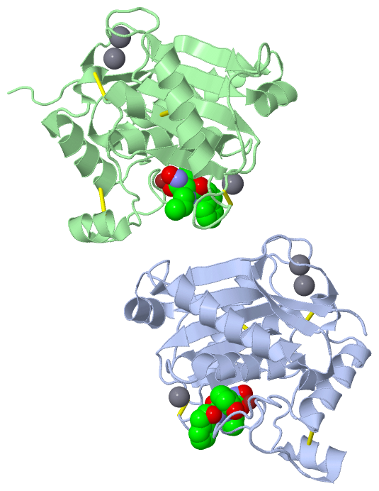
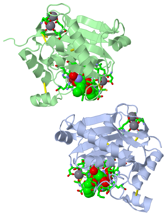
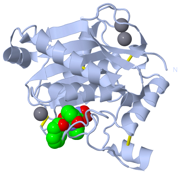
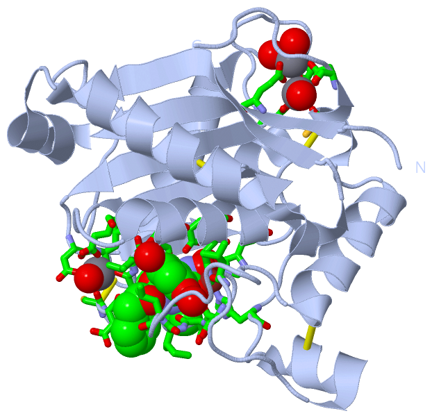
Still Images
|
||||||||||||||||
Databases
|
||||||||||||||||||||||||||||||||||||||||||||||||||||||||||||||||||||||||||||||||||||||||||||||||||||||||||||||||||||||||||||||||||||||||||||||||||||||||||||||||
Analysis Tools
|
|||||||||||||||||||||||||||||||||||||||||||||||||||||||||||||
Entries Sharing at Least One Protein Chain (UniProt ID)
Related Entries Specified in the PDB File
|
|