Asymmetric Unit(hide GO term definitions)
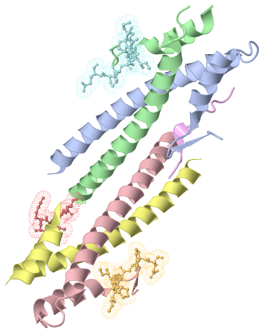
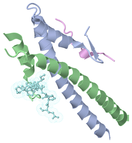
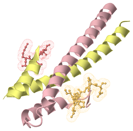
Chain A,B,C,D ( MARE1_HUMAN | Q15691)
| molecular function |
|---|
| | GO:0008017 | | microtubule binding | | Interacting selectively and non-covalently with microtubules, filaments composed of tubulin monomers. |
| | GO:0051010 | | microtubule plus-end binding | | Interacting selectively and non-covalently with the plus end of a microtubule. |
| | GO:0008022 | | protein C-terminus binding | | Interacting selectively and non-covalently with a protein C-terminus, the end of any peptide chain at which the 1-carboxy function of a constituent amino acid is not attached in peptide linkage to another amino-acid residue. |
| | GO:0005515 | | protein binding | | Interacting selectively and non-covalently with any protein or protein complex (a complex of two or more proteins that may include other nonprotein molecules). |
| biological process |
|---|
| | GO:0000086 | | G2/M transition of mitotic cell cycle | | The mitotic cell cycle transition by which a cell in G2 commits to M phase. The process begins when the kinase activity of M cyclin/CDK complex reaches a threshold high enough for the cell cycle to proceed. This is accomplished by activating a positive feedback loop that results in the accumulation of unphosphorylated and active M cyclin/CDK complex. |
| | GO:0007049 | | cell cycle | | The progression of biochemical and morphological phases and events that occur in a cell during successive cell replication or nuclear replication events. Canonically, the cell cycle comprises the replication and segregation of genetic material followed by the division of the cell, but in endocycles or syncytial cells nuclear replication or nuclear division may not be followed by cell division. |
| | GO:0051301 | | cell division | | The process resulting in division and partitioning of components of a cell to form more cells; may or may not be accompanied by the physical separation of a cell into distinct, individually membrane-bounded daughter cells. |
| | GO:0008283 | | cell proliferation | | The multiplication or reproduction of cells, resulting in the expansion of a cell population. |
| | GO:1904527 | | negative regulation of microtubule binding | | Any process that stops, prevents or reduces the frequency, rate or extent of microtubule binding. |
| | GO:0031115 | | negative regulation of microtubule polymerization | | Any process that stops, prevents, or reduces the frequency, rate or extent of microtubule polymerization. |
| | GO:0030335 | | positive regulation of cell migration | | Any process that activates or increases the frequency, rate or extent of cell migration. |
| | GO:1903033 | | positive regulation of microtubule plus-end binding | | Any process that activates or increases the frequency, rate or extent of microtubule plus-end binding. |
| | GO:0035372 | | protein localization to microtubule | | A process in which a protein is transported to, or maintained at, a microtubule. |
| | GO:0007062 | | sister chromatid cohesion | | The cell cycle process in which the sister chromatids of a replicated chromosome become tethered to each other. |
| cellular component |
|---|
| | GO:0005794 | | Golgi apparatus | | A compound membranous cytoplasmic organelle of eukaryotic cells, consisting of flattened, ribosome-free vesicles arranged in a more or less regular stack. The Golgi apparatus differs from the endoplasmic reticulum in often having slightly thicker membranes, appearing in sections as a characteristic shallow semicircle so that the convex side (cis or entry face) abuts the endoplasmic reticulum, secretory vesicles emerging from the concave side (trans or exit face). In vertebrate cells there is usually one such organelle, while in invertebrates and plants, where they are known usually as dictyosomes, there may be several scattered in the cytoplasm. The Golgi apparatus processes proteins produced on the ribosomes of the rough endoplasmic reticulum; such processing includes modification of the core oligosaccharides of glycoproteins, and the sorting and packaging of proteins for transport to a variety of cellular locations. Three different regions of the Golgi are now recognized both in terms of structure and function: cis, in the vicinity of the cis face, trans, in the vicinity of the trans face, and medial, lying between the cis and trans regions. |
| | GO:0042995 | | cell projection | | A prolongation or process extending from a cell, e.g. a flagellum or axon. |
| | GO:0031253 | | cell projection membrane | | The portion of the plasma membrane surrounding a plasma membrane bounded cell surface projection. |
| | GO:0005813 | | centrosome | | A structure comprised of a core structure (in most organisms, a pair of centrioles) and peripheral material from which a microtubule-based structure, such as a spindle apparatus, is organized. Centrosomes occur close to the nucleus during interphase in many eukaryotic cells, though in animal cells it changes continually during the cell-division cycle. |
| | GO:0030981 | | cortical microtubule cytoskeleton | | The portion of the microtubule cytoskeleton that lies just beneath the plasma membrane. |
| | GO:0005737 | | cytoplasm | | All of the contents of a cell excluding the plasma membrane and nucleus, but including other subcellular structures. |
| | GO:0005881 | | cytoplasmic microtubule | | Any microtubule in the cytoplasm of a cell. |
| | GO:0005856 | | cytoskeleton | | Any of the various filamentous elements that form the internal framework of cells, and typically remain after treatment of the cells with mild detergent to remove membrane constituents and soluble components of the cytoplasm. The term embraces intermediate filaments, microfilaments, microtubules, the microtrabecular lattice, and other structures characterized by a polymeric filamentous nature and long-range order within the cell. The various elements of the cytoskeleton not only serve in the maintenance of cellular shape but also have roles in other cellular functions, including cellular movement, cell division, endocytosis, and movement of organelles. |
| | GO:0005829 | | cytosol | | The part of the cytoplasm that does not contain organelles but which does contain other particulate matter, such as protein complexes. |
| | GO:0005874 | | microtubule | | Any of the long, generally straight, hollow tubes of internal diameter 12-15 nm and external diameter 24 nm found in a wide variety of eukaryotic cells; each consists (usually) of 13 protofilaments of polymeric tubulin, staggered in such a manner that the tubulin monomers are arranged in a helical pattern on the microtubular surface, and with the alpha/beta axes of the tubulin subunits parallel to the long axis of the tubule; exist in equilibrium with pool of tubulin monomers and can be rapidly assembled or disassembled in response to physiological stimuli; concerned with force generation, e.g. in the spindle. |
| | GO:0015630 | | microtubule cytoskeleton | | The part of the cytoskeleton (the internal framework of a cell) composed of microtubules and associated proteins. |
| | GO:0005815 | | microtubule organizing center | | An intracellular structure that can catalyze gamma-tubulin-dependent microtubule nucleation and that can anchor microtubules by interacting with their minus ends, plus ends or sides. |
| | GO:0035371 | | microtubule plus-end | | The growing (plus) end of a microtubule. In vitro, microtubules polymerize more quickly at the plus end than at the minus end. In vivo, microtubule growth occurs only at the plus end, and the plus end switches between periods of growth and shortening, a behavior known as dynamic instability. |
| | GO:0005819 | | spindle | | The array of microtubules and associated molecules that forms between opposite poles of a eukaryotic cell during mitosis or meiosis and serves to move the duplicated chromosomes apart. |
Chain E,F,G,H ( DYST_HUMAN | Q03001)
| molecular function |
|---|
| | GO:0003779 | | actin binding | | Interacting selectively and non-covalently with monomeric or multimeric forms of actin, including actin filaments. |
| | GO:0005509 | | calcium ion binding | | Interacting selectively and non-covalently with calcium ions (Ca2+). |
| | GO:0008092 | | cytoskeletal protein binding | | Interacting selectively and non-covalently with any protein component of any cytoskeleton (actin, microtubule, or intermediate filament cytoskeleton). |
| | GO:0005178 | | integrin binding | | Interacting selectively and non-covalently with an integrin. |
| | GO:0046872 | | metal ion binding | | Interacting selectively and non-covalently with any metal ion. |
| | GO:0051010 | | microtubule plus-end binding | | Interacting selectively and non-covalently with the plus end of a microtubule. |
| | GO:0008022 | | protein C-terminus binding | | Interacting selectively and non-covalently with a protein C-terminus, the end of any peptide chain at which the 1-carboxy function of a constituent amino acid is not attached in peptide linkage to another amino-acid residue. |
| | GO:0005515 | | protein binding | | Interacting selectively and non-covalently with any protein or protein complex (a complex of two or more proteins that may include other nonprotein molecules). |
| | GO:0042803 | | protein homodimerization activity | | Interacting selectively and non-covalently with an identical protein to form a homodimer. |
| biological process |
|---|
| | GO:0007155 | | cell adhesion | | The attachment of a cell, either to another cell or to an underlying substrate such as the extracellular matrix, via cell adhesion molecules. |
| | GO:0007050 | | cell cycle arrest | | A regulatory process that halts progression through the cell cycle during one of the normal phases (G1, S, G2, M). |
| | GO:0048870 | | cell motility | | Any process involved in the controlled self-propelled movement of a cell that results in translocation of the cell from one place to another. |
| | GO:0007010 | | cytoskeleton organization | | A process that is carried out at the cellular level which results in the assembly, arrangement of constituent parts, or disassembly of cytoskeletal structures. |
| | GO:0031581 | | hemidesmosome assembly | | Assembly of hemidesmosomes, integrin-containing protein complexes that bind to laminin in the basal lamina. Hemidesmosomes form the contact between the basal surface of epithelial cells and the underlying basal lamina. |
| | GO:0007229 | | integrin-mediated signaling pathway | | A series of molecular signals initiated by the binding of extracellular ligand to an integrin on the surface of a target cell, and ending with regulation of a downstream cellular process, e.g. transcription. |
| | GO:0045104 | | intermediate filament cytoskeleton organization | | A process that is carried out at the cellular level which results in the assembly, arrangement of constituent parts, or disassembly of cytoskeletal structures comprising intermediate filaments and their associated proteins. |
| | GO:0030011 | | maintenance of cell polarity | | The maintenance of established anisotropic intracellular organization or cell growth patterns. |
| | GO:0000226 | | microtubule cytoskeleton organization | | A process that is carried out at the cellular level which results in the assembly, arrangement of constituent parts, or disassembly of cytoskeletal structures comprising microtubules and their associated proteins. |
| | GO:0009611 | | response to wounding | | Any process that results in a change in state or activity of a cell or an organism (in terms of movement, secretion, enzyme production, gene expression, etc.) as a result of a stimulus indicating damage to the organism. |
| cellular component |
|---|
| | GO:0031673 | | H zone | | A relatively pale zone traversing the center of the A band of a sarcomere, visible in relaxed muscle fibers; consists of the central portion of thick (myosin) filaments that are not overlapped by thin (actin) filaments. |
| | GO:0030018 | | Z disc | | Platelike region of a muscle sarcomere to which the plus ends of actin filaments are attached. |
| | GO:0015629 | | actin cytoskeleton | | The part of the cytoskeleton (the internal framework of a cell) composed of actin and associated proteins. Includes actin cytoskeleton-associated complexes. |
| | GO:0030424 | | axon | | The long process of a neuron that conducts nerve impulses, usually away from the cell body to the terminals and varicosities, which are sites of storage and release of neurotransmitter. |
| | GO:0009925 | | basal plasma membrane | | The region of the plasma membrane located at the basal end of the cell. Often used in reference to animal polarized epithelial membranes, where the basal membrane is the part attached to the extracellular matrix, or in plant cells, where the basal membrane is defined with respect to the zygotic axis. |
| | GO:0005604 | | basement membrane | | A thin layer of dense material found in various animal tissues interposed between the cells and the adjacent connective tissue. It consists of the basal lamina plus an associated layer of reticulin fibers. |
| | GO:0005938 | | cell cortex | | The region of a cell that lies just beneath the plasma membrane and often, but not always, contains a network of actin filaments and associated proteins. |
| | GO:0030054 | | cell junction | | A cellular component that forms a specialized region of connection between two or more cells or between a cell and the extracellular matrix. At a cell junction, anchoring proteins extend through the plasma membrane to link cytoskeletal proteins in one cell to cytoskeletal proteins in neighboring cells or to proteins in the extracellular matrix. |
| | GO:0031252 | | cell leading edge | | The area of a motile cell closest to the direction of movement. |
| | GO:0042995 | | cell projection | | A prolongation or process extending from a cell, e.g. a flagellum or axon. |
| | GO:0005737 | | cytoplasm | | All of the contents of a cell excluding the plasma membrane and nucleus, but including other subcellular structures. |
| | GO:0005856 | | cytoskeleton | | Any of the various filamentous elements that form the internal framework of cells, and typically remain after treatment of the cells with mild detergent to remove membrane constituents and soluble components of the cytoplasm. The term embraces intermediate filaments, microfilaments, microtubules, the microtrabecular lattice, and other structures characterized by a polymeric filamentous nature and long-range order within the cell. The various elements of the cytoskeleton not only serve in the maintenance of cellular shape but also have roles in other cellular functions, including cellular movement, cell division, endocytosis, and movement of organelles. |
| | GO:0005829 | | cytosol | | The part of the cytoplasm that does not contain organelles but which does contain other particulate matter, such as protein complexes. |
| | GO:0005783 | | endoplasmic reticulum | | The irregular network of unit membranes, visible only by electron microscopy, that occurs in the cytoplasm of many eukaryotic cells. The membranes form a complex meshwork of tubular channels, which are often expanded into slitlike cavities called cisternae. The ER takes two forms, rough (or granular), with ribosomes adhering to the outer surface, and smooth (with no ribosomes attached). |
| | GO:0005789 | | endoplasmic reticulum membrane | | The lipid bilayer surrounding the endoplasmic reticulum. |
| | GO:0070062 | | extracellular exosome | | A vesicle that is released into the extracellular region by fusion of the limiting endosomal membrane of a multivesicular body with the plasma membrane. Extracellular exosomes, also simply called exosomes, have a diameter of about 40-100 nm. |
| | GO:0005925 | | focal adhesion | | Small region on the surface of a cell that anchors the cell to the extracellular matrix and that forms a point of termination of actin filaments. |
| | GO:0030056 | | hemidesmosome | | A cell-substrate junction (attachment structure) found in epithelial cells that links intermediate filaments to extracellular matrices via transmembrane complexes. In vertebrates, hemidesmosomes mediate contact between the basal side of epithelial cells and the basal lamina. In C. elegans, hemidesmosomes connect epithelial cells to distinct extracellular matrices on both the apical and basal cell surfaces. |
| | GO:0016021 | | integral component of membrane | | The component of a membrane consisting of the gene products and protein complexes having at least some part of their peptide sequence embedded in the hydrophobic region of the membrane. |
| | GO:0005882 | | intermediate filament | | A cytoskeletal structure that forms a distinct elongated structure, characteristically 10 nm in diameter, that occurs in the cytoplasm of eukaryotic cells. Intermediate filaments form a fibrous system, composed of chemically heterogeneous subunits and involved in mechanically integrating the various components of the cytoplasmic space. Intermediate filaments may be divided into five chemically distinct classes: Type I, acidic keratins; Type II, basic keratins; Type III, including desmin, vimentin and others; Type IV, neurofilaments and related filaments; and Type V, lamins. |
| | GO:0045111 | | intermediate filament cytoskeleton | | Cytoskeletal structure made from intermediate filaments, typically organized in the cytosol as an extended system that stretches from the nuclear envelope to the plasma membrane. Some intermediate filaments run parallel to the cell surface, while others traverse the cytosol; together they form an internal framework that helps support the shape and resilience of the cell. |
| | GO:0016020 | | membrane | | A lipid bilayer along with all the proteins and protein complexes embedded in it an attached to it. |
| | GO:0005874 | | microtubule | | Any of the long, generally straight, hollow tubes of internal diameter 12-15 nm and external diameter 24 nm found in a wide variety of eukaryotic cells; each consists (usually) of 13 protofilaments of polymeric tubulin, staggered in such a manner that the tubulin monomers are arranged in a helical pattern on the microtubular surface, and with the alpha/beta axes of the tubulin subunits parallel to the long axis of the tubule; exist in equilibrium with pool of tubulin monomers and can be rapidly assembled or disassembled in response to physiological stimuli; concerned with force generation, e.g. in the spindle. |
| | GO:0015630 | | microtubule cytoskeleton | | The part of the cytoskeleton (the internal framework of a cell) composed of microtubules and associated proteins. |
| | GO:0035371 | | microtubule plus-end | | The growing (plus) end of a microtubule. In vitro, microtubules polymerize more quickly at the plus end than at the minus end. In vivo, microtubule growth occurs only at the plus end, and the plus end switches between periods of growth and shortening, a behavior known as dynamic instability. |
| | GO:0005635 | | nuclear envelope | | The double lipid bilayer enclosing the nucleus and separating its contents from the rest of the cytoplasm; includes the intermembrane space, a gap of width 20-40 nm (also called the perinuclear space). |
| | GO:0005634 | | nucleus | | A membrane-bounded organelle of eukaryotic cells in which chromosomes are housed and replicated. In most cells, the nucleus contains all of the cell's chromosomes except the organellar chromosomes, and is the site of RNA synthesis and processing. In some species, or in specialized cell types, RNA metabolism or DNA replication may be absent. |
| | GO:0005886 | | plasma membrane | | The membrane surrounding a cell that separates the cell from its external environment. It consists of a phospholipid bilayer and associated proteins. |
|
 Description