Show PDB file:   
         Plain Text   HTML   (compressed file size)
QuickSearch:   
by PDB,NDB,UniProt,PROSITE Code or Search Term(s)  
(-)Biol.Unit 1 - manually
(-)Asymmetric Unit
(-)Asym. Unit - sites
(-)Biological Unit 1
(-)Biol. Unit 1 - sites
(-)Biological Unit 2
collapse expand < >
Image Biol.Unit 1 - manually
Biol.Unit 1 - manually  (Jmol Viewer)
Image Asymmetric Unit
Asymmetric Unit  (Jmol Viewer)
Image Asym. Unit - sites
Asym. Unit - sites  (Jmol Viewer)
Image Biological Unit 1
Biological Unit 1  (Jmol Viewer)
Image Biol. Unit 1 - sites
Biol. Unit 1 - sites  (Jmol Viewer)
Image Biological Unit 2
Biological Unit 2  (Jmol Viewer)

(-) Description

Title :  THE PAIRING GEOMETRY OF THE HYDROPHOBIC THYMINE ANALOG 2,4-DIFLUOROTOLUENE IN DUPLEX DNA AS ANALYZED BY X-RAY CRYSTALLOGRAPHY
 
Authors :  M. Egli, P. S. Pallan
Date :  09 Jul 09  (Deposition) - 01 Sep 09  (Release) - 13 Jul 11  (Revision)
Method :  X-RAY DIFFRACTION
Resolution :  1.61
Chains :  Asym. Unit :  A,B,C,D
Biol. Unit 1:  A,B  (2x)
Biol. Unit 2:  C,D  (2x)
Keywords :  Rnase-H, Rnase-H Dna-Complex, 2, 4-Difluorotoluene, Thymine Isostere, Hydrophobic Base, Endonuclease, Hydrolase, Magnesium, Manganese, Metal-Binding, Nuclease, Hydrolase-Dna Complex (Keyword Search: [Gene Ontology, PubMed, Web (Google))
 
Reference :  P. S. Pallan, M. Egli
Pairing Geometry Of The Hydrophobic Thymine Analogue 2, 4-Difluorotoluene In Duplex Dna As Analyzed By X-Ray Crystallography.
J. Am. Chem. Soc. V. 131 12548 2009
PubMed-ID: 19685868  |  Reference-DOI: 10.1021/JA905739J

(-) Compounds

Molecule 1 - RIBONUCLEASE H
    ChainsA, C
    EC Number3.1.26.4
    EngineeredYES
    Expression SystemESCHERICHIA COLI
    Expression System Taxid562
    FragmentRNASE-H
    GeneBACILLUS HALODURANS, BH0863, RNASE-H D132N MUTANT, RNHA
    MutationYES
    Organism ScientificBACILLUS HALODURANS
    Organism Taxid86665
    SynonymRNASE H
 
Molecule 2 - 5'-D(*CP*GP*CP*GP*AP*AP*TP*(DFT)P*CP*GP*CP*G)-3'
    ChainsB, D
    EngineeredYES
    SyntheticYES

 Structural Features

(-) Chains, Units

  1234
Asymmetric Unit ABCD
Biological Unit 1 (2x)AB  
Biological Unit 2 (2x)  CD

Summary Information (see also Sequences/Alignments below)

(-) Ligands, Modified Residues, Ions  (2, 6)

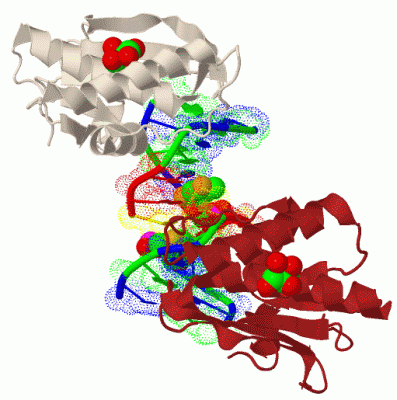
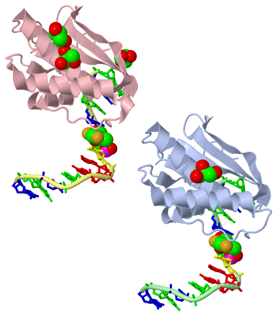
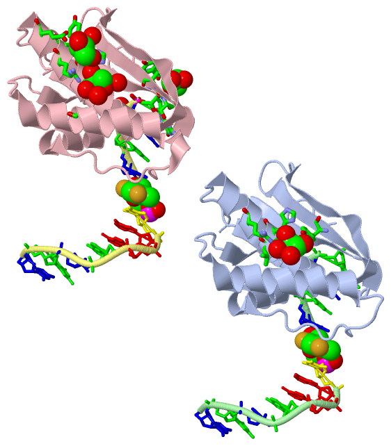
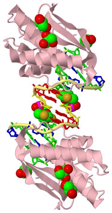
Asymmetric Unit (2, 6)
No.NameCountTypeFull Name
1DFT2Mod. Nucleotide1-[2-DEOXYRIBOFURANOSYL]-2,4-DIFLUORO-5-METHYL-BENZENE-5'MONOPHOSPHATE
2GOL4Ligand/IonGLYCEROL
Biological Unit 1 (2, 4)
No.NameCountTypeFull Name
1DFT2Mod. Nucleotide1-[2-DEOXYRIBOFURANOSYL]-2,4-DIFLUORO-5-METHYL-BENZENE-5'MONOPHOSPHATE
2GOL2Ligand/IonGLYCEROL
Biological Unit 2 (2, 8)
No.NameCountTypeFull Name
1DFT2Mod. Nucleotide1-[2-DEOXYRIBOFURANOSYL]-2,4-DIFLUORO-5-METHYL-BENZENE-5'MONOPHOSPHATE
2GOL6Ligand/IonGLYCEROL

(-) Sites  (4, 4)

Asymmetric Unit (4, 4)
No.NameEvidenceResiduesDescription
1AC1SOFTWAREHOH A:25 , TYR A:83 , VAL A:114 , HIS A:115 , ARG A:118BINDING SITE FOR RESIDUE GOL A 601
2AC2SOFTWAREHIS C:115 , ARG C:118 , ALA C:165 , HOH C:215 , HOH C:243 , GOL C:603BINDING SITE FOR RESIDUE GOL C 602
3AC3SOFTWAREHIS C:115 , TYR C:119 , GLU C:122 , HOH C:197 , HOH C:217 , GOL C:602BINDING SITE FOR RESIDUE GOL C 603
4AC4SOFTWAREGLY C:73 , SER C:74 , GLU C:82 , TYR C:193 , DG D:12BINDING SITE FOR RESIDUE GOL C 604

(-) SS Bonds  (0, 0)

(no "SS Bond" information available for 3I8D)

(-) Cis Peptide Bonds  (2, 2)

Asymmetric Unit
No.Residues
1Asn A:77 -Pro A:78
2Asn C:77 -Pro C:78

 Sequence-Structure Mapping

(-) SAPs(SNPs)/Variants  (0, 0)

(no "SAP(SNP)/Variant" information available for 3I8D)

(-) PROSITE Motifs  (0, 0)

(no "PROSITE Motif" information available for 3I8D)

(-) Exons   (0, 0)

(no "Exon" information available for 3I8D)

(-) Sequences/Alignments

Asymmetric Unit
   Reformat: Number of residues per line =  ('0' or empty: single-line sequence representation)
  Number of residues per labelling interval =   
  UniProt sequence: complete  aligned part    
   Show mapping: SCOP domains CATH domains Pfam domains Secondary structure (by author)
SAPs(SNPs) PROSITE motifs Exons
(details for a mapped element are shown in a popup box when the mouse pointer rests over it)
Chain A from PDB  Type:PROTEIN  Length:132
 aligned with RNH1_BACHD | Q9KEI9 from UniProtKB/Swiss-Prot  Length:196

    Alignment length:132
                                    71        81        91       101       111       121       131       141       151       161       171       181       191  
           RNH1_BACHD    62 EIIWESLSVDVGSQGNPGIVEYKGVDTKTGEVLFEREPIPIGTNNMGEFLAIVHGLRYLKERNSRKPIYSDSQTAIKWVKDKKAKSTLVRNEETALIWKLVDEAEEWLNTHTYETPILKWQTDKWGEIKADY 193
               SCOP domains d3i8da_ A: BH0863-like Ribonuclease H                                                                                                SCOP domains
               CATH domains ------------------------------------------------------------------------------------------------------------------------------------ CATH domains
               Pfam domains ------------------------------------------------------------------------------------------------------------------------------------ Pfam domains
         Sec.struct. author ......eeeeee.....eeeeeeeee.....eeeeeeeeeeehhhhhhhhhhhhhhhhhhhh.....eee.hhhhhhhhhhh...........hhhhhhhhhhhhhhhhhh.....eee.hhhhhh...... Sec.struct. author
                 SAPs(SNPs) ------------------------------------------------------------------------------------------------------------------------------------ SAPs(SNPs)
                    PROSITE ------------------------------------------------------------------------------------------------------------------------------------ PROSITE
                 Transcript ------------------------------------------------------------------------------------------------------------------------------------ Transcript
                 3i8d A  62 EIIWESLSVDVGSQGNPGIVEYKGVDTKTGEVLFEREPIPIGTNNMGEFLAIVHGLRYLKERNSRKPIYSNSQTAIKWVKDKKAKSTLVRNEETALIWKLVDEAEEWLNTHTYETPILKWQTDKWGEIKADY 193
                                    71        81        91       101       111       121       131       141       151       161       171       181       191  

Chain B from PDB  Type:DNA  Length:12
                                            
                 3i8d B   1 CGCGAATxCGCG  12
                                   |10  
                                   8-DFT

Chain C from PDB  Type:PROTEIN  Length:132
 aligned with RNH1_BACHD | Q9KEI9 from UniProtKB/Swiss-Prot  Length:196

    Alignment length:132
                                    71        81        91       101       111       121       131       141       151       161       171       181       191  
           RNH1_BACHD    62 EIIWESLSVDVGSQGNPGIVEYKGVDTKTGEVLFEREPIPIGTNNMGEFLAIVHGLRYLKERNSRKPIYSDSQTAIKWVKDKKAKSTLVRNEETALIWKLVDEAEEWLNTHTYETPILKWQTDKWGEIKADY 193
               SCOP domains d3i8dc_ C: BH0863-like Ribonuclease H                                                                                                SCOP domains
               CATH domains ------------------------------------------------------------------------------------------------------------------------------------ CATH domains
               Pfam domains ------------------------------------------------------------------------------------------------------------------------------------ Pfam domains
         Sec.struct. author ......eeeeeee....eeeeeeeee.....eeeeeeeeeeehhhhhhhhhhhhhhhhhhhh.....eee.hhhhhhhhhhh........hhhhhhhhhhhhhhhhhhhhh.....eee.hhhhhh...... Sec.struct. author
                 SAPs(SNPs) ------------------------------------------------------------------------------------------------------------------------------------ SAPs(SNPs)
                    PROSITE ------------------------------------------------------------------------------------------------------------------------------------ PROSITE
                 Transcript ------------------------------------------------------------------------------------------------------------------------------------ Transcript
                 3i8d C  62 EIIWESLSVDVGSQGNPGIVEYKGVDTKTGEVLFEREPIPIGTNNMGEFLAIVHGLRYLKERNSRKPIYSNSQTAIKWVKDKKAKSTLVRNEETALIWKLVDEAEEWLNTHTYETPILKWQTDKWGEIKADY 193
                                    71        81        91       101       111       121       131       141       151       161       171       181       191  

Chain D from PDB  Type:DNA  Length:12
                                            
                 3i8d D   1 CGCGAATxCGCG  12
                                   |10  
                                   8-DFT

   Legend:   → Mismatch (orange background)
  - → Gap (green background, '-', border residues have a numbering label)
    → Modified Residue (blue background, lower-case, 'x' indicates undefined single-letter code, labelled with number + name)
  x → Chemical Group (purple background, 'x', labelled with number + name, e.g. ACE or NH2)
  extra numbering lines below/above indicate numbering irregularities and modified residue names etc., number ends below/above '|'

 Classification and Annotation

(-) SCOP Domains  (1, 2)

Asymmetric Unit

(-) CATH Domains  (0, 0)

(no "CATH Domain" information available for 3I8D)

(-) Pfam Domains  (0, 0)

(no "Pfam Domain" information available for 3I8D)

(-) Gene Ontology  (9, 9)

Asymmetric Unit(hide GO term definitions)
Chain A,C   (RNH1_BACHD | Q9KEI9)
molecular function
    GO:0004523    RNA-DNA hybrid ribonuclease activity    Catalysis of the endonucleolytic cleavage of RNA in RNA-DNA hybrids to 5'-phosphomonoesters.
    GO:0004519    endonuclease activity    Catalysis of the hydrolysis of ester linkages within nucleic acids by creating internal breaks.
    GO:0016787    hydrolase activity    Catalysis of the hydrolysis of various bonds, e.g. C-O, C-N, C-C, phosphoric anhydride bonds, etc. Hydrolase is the systematic name for any enzyme of EC class 3.
    GO:0046872    metal ion binding    Interacting selectively and non-covalently with any metal ion.
    GO:0004518    nuclease activity    Catalysis of the hydrolysis of ester linkages within nucleic acids.
    GO:0003676    nucleic acid binding    Interacting selectively and non-covalently with any nucleic acid.
biological process
    GO:0090502    RNA phosphodiester bond hydrolysis, endonucleolytic    The chemical reactions and pathways involving the hydrolysis of internal 3',5'-phosphodiester bonds in one or two strands of ribonucleotides.
    GO:0090305    nucleic acid phosphodiester bond hydrolysis    The nucleic acid metabolic process in which the phosphodiester bonds between nucleotides are cleaved by hydrolysis.
cellular component
    GO:0005737    cytoplasm    All of the contents of a cell excluding the plasma membrane and nucleus, but including other subcellular structures.

 Visualization

(-) Interactive Views

Asymmetric Unit
  Complete Structure
    Jena3D(integrated viewing of ligand, site, SAP, PROSITE, SCOP information)
    WebMol | AstexViewer[tm]@PDBe
(Java Applets, require no local installation except for Java; loading may be slow)
    STRAP
(Java WebStart application, automatic local installation, requires Java; full application with system access!)
    RasMol
(require local installation)
    Molscript (VRML)
(requires installation of a VRML viewer; select preferred view via VRML and generate a mono or stereo PDF format file)
 
  Ligands, Modified Residues, Ions
    DFT  [ RasMol | Jena3D ]  +environment [ RasMol | Jena3D ]
    GOL  [ RasMol | Jena3D ]  +environment [ RasMol | Jena3D ]
 
  Sites
    AC1  [ RasMol ]  +environment [ RasMol ]
    AC2  [ RasMol ]  +environment [ RasMol ]
    AC3  [ RasMol ]  +environment [ RasMol ]
    AC4  [ RasMol ]  +environment [ RasMol ]
 
  Cis Peptide Bonds
    Asn A:77 - Pro A:78   [ RasMol ]  
    Asn C:77 - Pro C:78   [ RasMol ]  
 
Biological Units
  Complete Structure
    Biological Unit 1  [ Jena3D ]
    Biological Unit 2  [ Jena3D ]

(-) Still Images

Jmol
  protein: cartoon or spacefill or dots and stick; nucleic acid: cartoon and stick; ligands: spacefill; active site: stick
Molscript
  protein, nucleic acid: cartoon; ligands: spacefill; active site: ball and stick

 Databases and Analysis Tools

(-) Databases

Access by PDB/NDB ID
  3i8d
    Family and Domain InformationProDom | SYSTERS
    General Structural InformationGlycoscienceDB | MMDB | NDB | OCA | PDB | PDBe | PDBj | PDBsum | PDBWiki | PQS | PROTEOPEDIA
    Orientation in MembranesOPM
    Protein SurfaceSURFACE
    Secondary StructureDSSP (structure derived) | HSSP (homology derived)
    Structural GenomicsGeneCensus
    Structural NeighboursCE | VAST
    Structure ClassificationCATH | Dali | SCOP
    Validation and Original DataBMRB Data View | BMRB Restraints Grid | EDS | PROCHECK | RECOORD | WHAT_CHECK
 
Access by UniProt ID/Accession number
  RNH1_BACHD | Q9KEI9
    Comparative Protein Structure ModelsModBase
    Genomic InformationEnsembl
    Protein-protein InteractionDIP
    Sequence, Family and Domain InformationInterPro | Pfam | SMART | UniProtKB/SwissProt
 
Access by Enzyme Classificator   (EC Number)
  3.1.26.4
    General Enzyme InformationBRENDA | EC-PDB | Enzyme | IntEnz
    PathwayKEGG | MetaCyc
 
Access by Disease Identifier   (MIM ID)
  (no 'MIM ID' available)
    Disease InformationOMIM
 
Access by GenAge ID
  (no 'GenAge ID' available)
    Age Related InformationGenAge

(-) Analysis Tools

Access by PDB/NDB ID
    Domain InformationXDom
    Interatomic Contacts of Structural UnitsCSU
    Ligand-protein ContactsLPC
    Protein CavitiescastP
    Sequence and Secondary StructurePDBCartoon
    Structure AlignmentSTRAP(Java WebStart application, automatic local installation, requires Java; full application with system access!)
    Structure and Sequence BrowserSTING
 
Access by UniProt ID/Accession number
  RNH1_BACHD | Q9KEI9
    Protein Disorder PredictionDisEMBL | FoldIndex | GLOBPLOT (for more information see DisProt)

 Related Entries

(-) Entries Sharing at Least One Protein Chain (UniProt ID)

UniProtKB/Swiss-Prot
        RNH1_BACHD | Q9KEI91zbf 1zbi 1zbl 2g8f 2g8h 2g8i 2g8k 2g8u 2g8v 2g8w 2r7y 3d0p 3ey1 3twh 3uld 4htu 4hue 4huf 4hug 4opj 4opk 5swm 5us2 5usa 5use 5usg 5w7n 5w7o

(-) Related Entries Specified in the PDB File

(no "Related Entries Specified in the PDB File" available for 3I8D)