|
|
|
|
 Description
|
|
Compounds
|
||||||||||||||||||||||||||||||||||||||||||||||||||||||||||||||||||
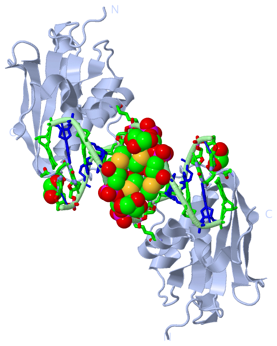
Chains, Units
Summary Information (see also Sequences/Alignments below) |
Ligands, Modified Residues, Ions (2, 5)| Asymmetric Unit (2, 5) Biological Unit 1 (2, 10) |
Sites (3, 3)
Asymmetric Unit (3, 3)
|
SS Bonds (0, 0)| (no "SS Bond" information available for 3EY1) |
Cis Peptide Bonds (2, 2)
Asymmetric Unit
|
||||||||||||
SAPs(SNPs)/Variants (0, 0)| (no "SAP(SNP)/Variant" information available for 3EY1) |
PROSITE Motifs (0, 0)| (no "PROSITE Motif" information available for 3EY1) |
Exons (0, 0)| (no "Exon" information available for 3EY1) |
Sequences/Alignments
Asymmetric UnitChain A from PDB Type:PROTEIN Length:137 aligned with RNH1_BACHD | Q9KEI9 from UniProtKB/Swiss-Prot Length:196 Alignment length:137 68 78 88 98 108 118 128 138 148 158 168 178 188 RNH1_BACHD 59 AKEEIIWESLSVDVGSQGNPGIVEYKGVDTKTGEVLFEREPIPIGTNNMGEFLAIVHGLRYLKERNSRKPIYSDSQTAIKWVKDKKAKSTLVRNEETALIWKLVDEAEEWLNTHTYETPILKWQTDKWGEIKADYGR 195 SCOP domains d3ey1a_ A: BH0863-like Ribonuclease H SCOP domains CATH domains ----------------------------------------------------------------------------------------------------------------------------------------- CATH domains Pfam domains ----------------------------------------------------------------------------------------------------------------------------------------- Pfam domains SAPs(SNPs) ----------------------------------------------------------------------------------------------------------------------------------------- SAPs(SNPs) PROSITE ----------------------------------------------------------------------------------------------------------------------------------------- PROSITE Transcript ----------------------------------------------------------------------------------------------------------------------------------------- Transcript 3ey1 A 59 AKEEIIWESLSVDVGSQGNPGIVEYKGVDTKTGEVLFEREPIPIGTNNMGEFLAIVHGLRYLKERNSRKPIYSNSQTAIKWVKDKKAKSTLVRNEETALIWKLVDEAEEWLNTHTYETPILKWQTDKWGEIKADYGR 195 68 78 88 98 108 118 128 138 148 158 168 178 188
Chain B from PDB  Type:DNA  Length:12

                 3ey1 B 301 CGCGAAxxCGCG 312
                                  |310
                                307-USM
                                 308-USM
|
||||||||||||||||||||
SCOP Domains (1, 1)
Asymmetric Unit
 |
CATH Domains (0, 0)| (no "CATH Domain" information available for 3EY1) |
Pfam Domains (0, 0)| (no "Pfam Domain" information available for 3EY1) |
Gene Ontology (9, 9)|
Asymmetric Unit(hide GO term definitions) Chain A (RNH1_BACHD | Q9KEI9)
|
||||||||||||||||||||||||||||||||||||||||||||||||||||||||||||||||||||||||
Interactive Views
|
|||||||||||||||||||||||||||||||||||||||||||||||||||||||||||||||||||||||||||||||||||||||||||||||||||||||||||||||||||||||||||||||||||||||||||||||||||||||||||||||||||||
Still Images
|
||||||||||||||||
Databases
|
||||||||||||||||||||||||||||||||||||||||||||||||||||||||||||||||||||||||||||||||||||||||||||||||||||||||||||||||||||||||||||||||||||||||||||||||||||||||||||||||
Analysis Tools
|
|||||||||||||||||||||||||||||||||||||||||||||||||||||||||||||
Entries Sharing at Least One Protein Chain (UniProt ID)
Related Entries Specified in the PDB File
|
|