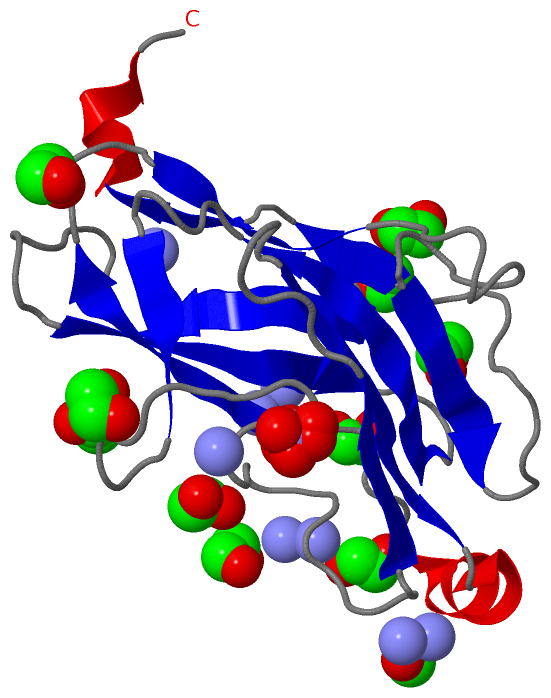
Show PDB file:   
         Plain Text   HTML   (compressed file size)
QuickSearch:   
by PDB,NDB,UniProt,PROSITE Code or Search Term(s)  
(-)Asym./Biol. Unit
(-)Asym./Biol. Unit - sites
collapse expand < >
Image Asym./Biol. Unit
Asym./Biol. Unit  (Jmol Viewer)
Image Asym./Biol. Unit - sites
Asym./Biol. Unit - sites  (Jmol Viewer)

(-) Description

Title :  CRYSTAL STRUCTURE ANALYSIS OF THE K171A MUTATION OF N-TERMINAL TYPE II COHESIN 1 FROM THE CELLULOSOMAL SCAB SUBUNIT OF ACETIVIBRIO CELLULOLYTICUS
 
Authors :  F. Frolow, A. Freeman, Y. Wine, A. Eppel, M. Shanzer, S. Stempler
Date :  30 Oct 08  (Deposition) - 02 Dec 08  (Release) - 02 Dec 08  (Revision)
Method :  X-RAY DIFFRACTION
Resolution :  1.85
Chains :  Asym./Biol. Unit :  A
Keywords :  Point Mutation, Structural Protein (Keyword Search: [Gene Ontology, PubMed, Web (Google))
 
Reference :  F. Frolow, A. Freeman, Y. Wine, A. Eppel, M. Shanzer, S. Stempler
Crystal Structure Analysis Of The K171A Mutation Of N-Terminal Type Ii Cohesin 1 From The Cellulosomal Scab Subunit Of Acetivibrio Cellulolyticus
To Be Published
PubMed: search
(for further references see the PDB file header)

(-) Compounds

Molecule 1 - CELLULOSOMAL SCAFFOLDIN ADAPTOR PROTEIN B
    ChainsA
    EngineeredYES
    Expression SystemESCHERICHIA COLI
    Expression System PlasmidPET28A+
    Expression System StrainBL21
    Expression System Taxid562
    Expression System Vector TypePLASMID
    FragmentN-TERMINAL TYPE II COHESIN 1 FROM THE CELLULOSOMAL SCAB SUBUNIT, UNP RESIDUES 28-199
    MutationYES
    Organism ScientificACETIVIBRIO CELLULOLYTICUS
    Organism Taxid35830

 Structural Features

(-) Chains, Units

  1
Asymmetric/Biological Unit A

Summary Information (see also Sequences/Alignments below)

(-) Ligands, Modified Residues, Ions  (3, 20)

Asymmetric/Biological Unit (3, 20)
No.NameCountTypeFull Name
1EDO11Ligand/Ion1,2-ETHANEDIOL
2NH48Ligand/IonAMMONIUM ION
3NO31Ligand/IonNITRATE ION

(-) Sites  (20, 20)

Asymmetric Unit (20, 20)
No.NameEvidenceResiduesDescription
01AC1SOFTWAREGLY A:55 , VAL A:56 , ALA A:57 , THR A:110 , HOH A:283BINDING SITE FOR RESIDUE EDO A 176
02AC2SOFTWAREASP A:73 , PHE A:74 , ASP A:100 , NH4 A:190 , HOH A:422BINDING SITE FOR RESIDUE EDO A 177
03AC3SOFTWAREPRO A:45 , ASN A:71 , LEU A:121 , HOH A:246BINDING SITE FOR RESIDUE EDO A 178
04AC4SOFTWAREASN A:75 , ILE A:140 , ASP A:141 , SER A:158 , VAL A:159 , HOH A:211 , HOH A:275BINDING SITE FOR RESIDUE EDO A 179
05AC5SOFTWARETYR A:101 , ALA A:106 , LYS A:119 , HOH A:250 , HOH A:282 , HOH A:370BINDING SITE FOR RESIDUE EDO A 180
06AC6SOFTWAREASN A:30 , GLN A:109 , THR A:110 , HOH A:241BINDING SITE FOR RESIDUE EDO A 181
07AC7SOFTWAREVAL A:51 , THR A:52 , SER A:53 , GLY A:55 , LYS A:117 , HOH A:224 , HOH A:330 , HOH A:353 , HOH A:364BINDING SITE FOR RESIDUE EDO A 182
08AC8SOFTWARESER A:5 , THR A:32 , ARG A:77 , VAL A:79 , GLY A:156 , HOH A:240 , HOH A:384BINDING SITE FOR RESIDUE EDO A 183
09AC9SOFTWAREASN A:71 , ASP A:73 , LYS A:122 , HOH A:279BINDING SITE FOR RESIDUE EDO A 184
10BC1SOFTWARETHR A:4 , PHE A:146 , GLY A:150 , HOH A:286 , HOH A:428BINDING SITE FOR RESIDUE EDO A 185
11BC2SOFTWARELYS A:13 , THR A:14 , VAL A:164 , HOH A:263BINDING SITE FOR RESIDUE NH4 A 186
12BC3SOFTWARETHR A:68 , LEU A:121 , NH4 A:188 , HOH A:246BINDING SITE FOR RESIDUE NH4 A 187
13BC4SOFTWARELEU A:70 , ASN A:71 , SER A:72 , TYR A:95 , GLU A:108 , NH4 A:187BINDING SITE FOR RESIDUE NH4 A 188
14BC5SOFTWAREASP A:73 , ASN A:75 , NH4 A:190 , HOH A:415BINDING SITE FOR RESIDUE NH4 A 189
15BC6SOFTWAREASN A:75 , SER A:97 , EDO A:177 , NH4 A:189BINDING SITE FOR RESIDUE NH4 A 190
16BC7SOFTWARETHR A:52 , TYR A:58 , MET A:63 , HOH A:215 , HOH A:260BINDING SITE FOR RESIDUE NH4 A 191
17BC8SOFTWAREASN A:27 , ILE A:28 , NH4 A:193BINDING SITE FOR RESIDUE NH4 A 192
18BC9SOFTWAREGLY A:111 , NH4 A:192 , HOH A:253BINDING SITE FOR RESIDUE NH4 A 193
19CC1SOFTWARESER A:53 , ALA A:66 , HOH A:215 , HOH A:362BINDING SITE FOR RESIDUE EDO A 194
20CC2SOFTWARETHR A:59 , LYS A:60 , SER A:61BINDING SITE FOR RESIDUE NO3 A 195

(-) SS Bonds  (0, 0)

(no "SS Bond" information available for 3F2L)

(-) Cis Peptide Bonds  (1, 1)

Asymmetric/Biological Unit
No.Residues
1Gln A:161 -Pro A:162

 Sequence-Structure Mapping

(-) SAPs(SNPs)/Variants  (0, 0)

(no "SAP(SNP)/Variant" information available for 3F2L)

(-) PROSITE Motifs  (0, 0)

(no "PROSITE Motif" information available for 3F2L)

(-) Exons   (0, 0)

(no "Exon" information available for 3F2L)

(-) Sequences/Alignments

Asymmetric/Biological Unit
   Reformat: Number of residues per line =  ('0' or empty: single-line sequence representation)
  Number of residues per labelling interval =   
  UniProt sequence: complete  aligned part    
   Show mapping: SCOP domains CATH domains Pfam domains Secondary structure (by author)
SAPs(SNPs) PROSITE motifs Exons
(details for a mapped element are shown in a popup box when the mouse pointer rests over it)
Chain A from PDB  Type:PROTEIN  Length:171
 aligned with Q7WYN3_9FIRM | Q7WYN3 from UniProtKB/TrEMBL  Length:942

    Alignment length:171
                                    37        47        57        67        77        87        97       107       117       127       137       147       157       167       177       187       197 
         Q7WYN3_9FIRM    28 APTSSIEIVLDKTTASVGEIVTASINIKNITNFSGCQLNMKYDPAVLQPVTSSGVAYTKSTMPGAGTILNSDFNLRQVADNDLEKGILNFSKAYVSLDDYRTAAAPEQTGTVAVVKFKVLKEETSSISFEDTTSVPNAIDGTVLFDWNGDRIQSGYSVIQPAVINLDMIKA 198
               SCOP domains d3f2la_ A: automated matches                                                                                                                                                SCOP domains
               CATH domains 3f2lA00 A:2-172  [code=2.60.40.680, no name defined]                                                                                                                        CATH domains
               Pfam domains --------------------------------------------------------------------------------------------------------------------------------------------------------------------------- Pfam domains
         Sec.struct. author ....eeeeee.........eeeeeeeee....eeeeeeeee.....eeee........................eeeee.......eeeeeeee.hhhhhhhhh.....eeeeeeeeee.....eeeee............eeeee..........eee..eeehhhhh.. Sec.struct. author
                 SAPs(SNPs) --------------------------------------------------------------------------------------------------------------------------------------------------------------------------- SAPs(SNPs)
                    PROSITE --------------------------------------------------------------------------------------------------------------------------------------------------------------------------- PROSITE
                 Transcript --------------------------------------------------------------------------------------------------------------------------------------------------------------------------- Transcript
                 3f2l A   2 APTSSIEIVLDKTTASVGEIVTASINIKNITNFSGCQLNMKYDPAVLQPVTSSGVAYTKSTMPGAGTILNSDFNLRQVADNDLEKGILNFSKAYVSLDDYRTAAAPEQTGTVAVVKFKVLKEETSSISFEDTTSVPNAIDGTVLFDWNGDRIQSGYSVIQPAVINLDMIAA 172
                                    11        21        31        41        51        61        71        81        91       101       111       121       131       141       151       161       171 

   Legend:   → Mismatch (orange background)
  - → Gap (green background, '-', border residues have a numbering label)
    → Modified Residue (blue background, lower-case, 'x' indicates undefined single-letter code, labelled with number + name)
  x → Chemical Group (purple background, 'x', labelled with number + name, e.g. ACE or NH2)
  extra numbering lines below/above indicate numbering irregularities and modified residue names etc., number ends below/above '|'

 Classification and Annotation

(-) SCOP Domains  (1, 1)

Asymmetric/Biological Unit

(-) CATH Domains  (1, 1)

Asymmetric/Biological Unit
(-)
Class: Mainly Beta (13760)

(-) Pfam Domains  (0, 0)

(no "Pfam Domain" information available for 3F2L)

(-) Gene Ontology  (3, 3)

Asymmetric/Biological Unit(hide GO term definitions)
Chain A   (Q7WYN3_9FIRM | Q7WYN3)
molecular function
    GO:0030246    carbohydrate binding    Interacting selectively and non-covalently with any carbohydrate, which includes monosaccharides, oligosaccharides and polysaccharides as well as substances derived from monosaccharides by reduction of the carbonyl group (alditols), by oxidation of one or more hydroxy groups to afford the corresponding aldehydes, ketones, or carboxylic acids, or by replacement of one or more hydroxy group(s) by a hydrogen atom. Cyclitols are generally not regarded as carbohydrates.
    GO:0046872    metal ion binding    Interacting selectively and non-covalently with any metal ion.
biological process
    GO:0000272    polysaccharide catabolic process    The chemical reactions and pathways resulting in the breakdown of a polysaccharide, a polymer of many (typically more than 10) monosaccharide residues linked glycosidically.

 Visualization

(-) Interactive Views

Asymmetric/Biological Unit
  Complete Structure
    Jena3D(integrated viewing of ligand, site, SAP, PROSITE, SCOP information)
    WebMol | AstexViewer[tm]@PDBe
(Java Applets, require no local installation except for Java; loading may be slow)
    STRAP
(Java WebStart application, automatic local installation, requires Java; full application with system access!)
    RasMol
(require local installation)
    Molscript (VRML)
(requires installation of a VRML viewer; select preferred view via VRML and generate a mono or stereo PDF format file)
 
  Ligands, Modified Residues, Ions
    EDO  [ RasMol | Jena3D ]  +environment [ RasMol | Jena3D ]
    NH4  [ RasMol | Jena3D ]  +environment [ RasMol | Jena3D ]
    NO3  [ RasMol | Jena3D ]  +environment [ RasMol | Jena3D ]
 
  Sites
    AC1  [ RasMol ]  +environment [ RasMol ]
    AC2  [ RasMol ]  +environment [ RasMol ]
    AC3  [ RasMol ]  +environment [ RasMol ]
    AC4  [ RasMol ]  +environment [ RasMol ]
    AC5  [ RasMol ]  +environment [ RasMol ]
    AC6  [ RasMol ]  +environment [ RasMol ]
    AC7  [ RasMol ]  +environment [ RasMol ]
    AC8  [ RasMol ]  +environment [ RasMol ]
    AC9  [ RasMol ]  +environment [ RasMol ]
    BC1  [ RasMol ]  +environment [ RasMol ]
    BC2  [ RasMol ]  +environment [ RasMol ]
    BC3  [ RasMol ]  +environment [ RasMol ]
    BC4  [ RasMol ]  +environment [ RasMol ]
    BC5  [ RasMol ]  +environment [ RasMol ]
    BC6  [ RasMol ]  +environment [ RasMol ]
    BC7  [ RasMol ]  +environment [ RasMol ]
    BC8  [ RasMol ]  +environment [ RasMol ]
    BC9  [ RasMol ]  +environment [ RasMol ]
    CC1  [ RasMol ]  +environment [ RasMol ]
    CC2  [ RasMol ]  +environment [ RasMol ]
 
  Cis Peptide Bonds
    Gln A:161 - Pro A:162   [ RasMol ]  
 

(-) Still Images

Jmol
  protein: cartoon or spacefill or dots and stick; nucleic acid: cartoon and stick; ligands: spacefill; active site: stick
Molscript
  protein, nucleic acid: cartoon; ligands: spacefill; active site: ball and stick

 Databases and Analysis Tools

(-) Databases

Access by PDB/NDB ID
  3f2l
    Family and Domain InformationProDom | SYSTERS
    General Structural InformationGlycoscienceDB | MMDB | NDB | OCA | PDB | PDBe | PDBj | PDBsum | PDBWiki | PQS | PROTEOPEDIA
    Orientation in MembranesOPM
    Protein SurfaceSURFACE
    Secondary StructureDSSP (structure derived) | HSSP (homology derived)
    Structural GenomicsGeneCensus
    Structural NeighboursCE | VAST
    Structure ClassificationCATH | Dali | SCOP
    Validation and Original DataBMRB Data View | BMRB Restraints Grid | EDS | PROCHECK | RECOORD | WHAT_CHECK
 
Access by UniProt ID/Accession number
  Q7WYN3_9FIRM | Q7WYN3
    Comparative Protein Structure ModelsModBase
    Genomic InformationEnsembl
    Protein-protein InteractionDIP
    Sequence, Family and Domain InformationInterPro | Pfam | SMART | UniProtKB/TrEMBL
 
Access by Enzyme Classificator   (EC Number)
  (no 'Enzyme Classificator' available)
    General Enzyme InformationBRENDA | EC-PDB | Enzyme | IntEnz
    PathwayKEGG | MetaCyc
 
Access by Disease Identifier   (MIM ID)
  (no 'MIM ID' available)
    Disease InformationOMIM
 
Access by GenAge ID
  (no 'GenAge ID' available)
    Age Related InformationGenAge

(-) Analysis Tools

Access by PDB/NDB ID
    Domain InformationXDom
    Interatomic Contacts of Structural UnitsCSU
    Ligand-protein ContactsLPC
    Protein CavitiescastP
    Sequence and Secondary StructurePDBCartoon
    Structure AlignmentSTRAP(Java WebStart application, automatic local installation, requires Java; full application with system access!)
    Structure and Sequence BrowserSTING
 
Access by UniProt ID/Accession number
  Q7WYN3_9FIRM | Q7WYN3
    Protein Disorder PredictionDisEMBL | FoldIndex | GLOBPLOT (for more information see DisProt)

 Related Entries

(-) Entries Sharing at Least One Protein Chain (UniProt ID)

UniProtKB/TrEMBL
        Q7WYN3_9FIRM | Q7WYN31qzn 1zv9 3bwz 3fnk 3ghp 3l8q 4u3s 4uyp 4uyq 4wi0

(-) Related Entries Specified in the PDB File

1qzn WILD TYPE STRUCTURE