Show PDB file:   
         Plain Text   HTML   (compressed file size)
QuickSearch:   
by PDB,NDB,UniProt,PROSITE Code or Search Term(s)  
(-)Asymmetric Unit
(-)Asym. Unit - sites
(-)Biological Unit 1
(-)Biol. Unit 1 - sites
(-)Biological Unit 2
collapse expand < >
Image Asymmetric Unit
Asymmetric Unit  (Jmol Viewer)
Image Asym. Unit - sites
Asym. Unit - sites  (Jmol Viewer)
Image Biological Unit 1
Biological Unit 1  (Jmol Viewer)
Image Biol. Unit 1 - sites
Biol. Unit 1 - sites  (Jmol Viewer)
Image Biological Unit 2
Biological Unit 2  (Jmol Viewer)

(-) Description

Title :  STRUCTURE OF THE SECOND TYPE II COHESIN MODULE FROM THE ADAPTOR SCAA SCAFFOLDIN OF ACETIVIBRIO CELLULOLYTICUS (INCLUDING LONG C-TERMINAL LINKER)
 
Authors :  I. Noach, F. Frolow, E. A. Bayer
Date :  04 Mar 09  (Deposition) - 23 Jun 09  (Release) - 12 Feb 14  (Revision)
Method :  X-RAY DIFFRACTION
Resolution :  2.49
Chains :  Asym. Unit :  A,B
Biol. Unit 1:  A  (1x)
Biol. Unit 2:  B  (1x)
Keywords :  Linker Segments, Beta Barrel, Alpha Helix, Beta Flaps, Structural Protein (Keyword Search: [Gene Ontology, PubMed, Web (Google))
 
Reference :  I. Noach, F. Frolow, O. Alber, R. Lamed, L. J. W. Shimon, E. A. Bayer
Intermodular Linker Flexibility Revealed From Crystal Structures Of Adjacent Cellulosomal Cohesins Of Acetivibrio Cellulolyticus
J. Mol. Biol. V. 391 86 2009
PubMed-ID: 19501595  |  Reference-DOI: 10.1016/J.JMB.2009.06.006
(for further references see the PDB file header)

(-) Compounds

Molecule 1 - CELLULOSOMAL SCAFFOLDIN ADAPTOR PROTEIN B
    ChainsA, B
    EngineeredYES
    Expression SystemESCHERICHIA COLI BL21(DE3)
    Expression System PlasmidPET28A
    Expression System StrainBL21(DE3)PLYSS
    Expression System Taxid469008
    Expression System Vector TypePLASMID
    FragmentCOHESIN MODULE RESIDUES 190-408
    GeneSCAB
    Organism ScientificACETIVIBRIO CELLULOLYTICUS
    Organism Taxid35830

 Structural Features

(-) Chains, Units

  12
Asymmetric Unit AB
Biological Unit 1 (1x)A 
Biological Unit 2 (1x) B

Summary Information (see also Sequences/Alignments below)

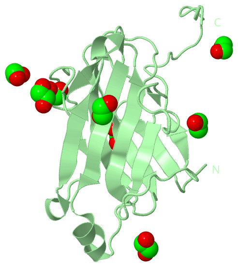
(-) Ligands, Modified Residues, Ions  (1, 14)

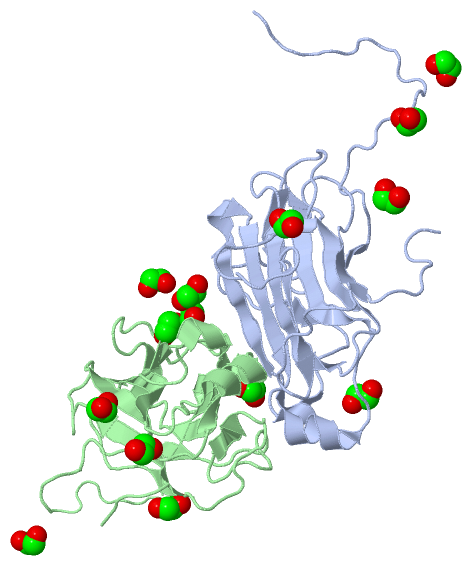
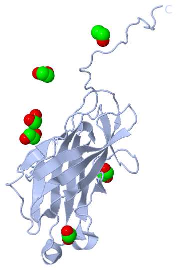
Asymmetric Unit (1, 14)
No.NameCountTypeFull Name
1EDO14Ligand/Ion1,2-ETHANEDIOL
Biological Unit 1 (1, 6)
No.NameCountTypeFull Name
1EDO6Ligand/Ion1,2-ETHANEDIOL
Biological Unit 2 (1, 8)
No.NameCountTypeFull Name
1EDO8Ligand/Ion1,2-ETHANEDIOL

(-) Sites  (10, 10)

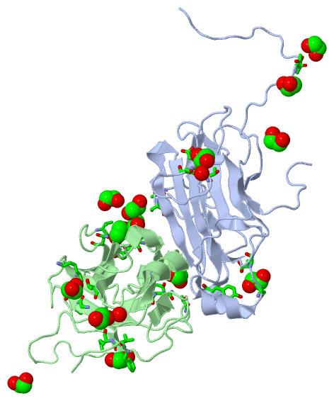
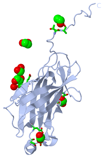
Asymmetric Unit (10, 10)
No.NameEvidenceResiduesDescription
01AC1SOFTWARETYR A:81 , SER A:113 , GLU A:115 , EDO B:230BINDING SITE FOR RESIDUE EDO A 228
02AC2SOFTWARETHR A:179 , ALA A:180BINDING SITE FOR RESIDUE EDO A 229
03AC3SOFTWARETHR A:65 , SER A:67 , EDO B:229BINDING SITE FOR RESIDUE EDO A 232
04AC4SOFTWAREGLU A:138 , ASP A:139 , THR A:148 , HOH A:241BINDING SITE FOR RESIDUE EDO A 233
05AC5SOFTWAREASN B:57 , VAL B:62 , ALA B:63 , TYR B:64 , SER B:68 , EDO B:229BINDING SITE FOR RESIDUE EDO B 228
06AC6SOFTWAREEDO A:232 , SER B:68 , LEU B:69 , EDO B:228BINDING SITE FOR RESIDUE EDO B 229
07AC7SOFTWAREEDO A:228 , GLY B:15 , TYR B:16 , LYS B:28 , THR B:30BINDING SITE FOR RESIDUE EDO B 230
08AC8SOFTWAREGLN B:43 , ASN B:97 , SER B:99 , THR B:142BINDING SITE FOR RESIDUE EDO B 231
09AC9SOFTWAREPHE B:165 , VAL B:166 , ILE B:167BINDING SITE FOR RESIDUE EDO B 232
10BC1SOFTWAREHOH B:262BINDING SITE FOR RESIDUE EDO B 235

(-) SS Bonds  (0, 0)

(no "SS Bond" information available for 3GHP)

(-) Cis Peptide Bonds  (5, 5)

Asymmetric Unit
No.Residues
1Ala A:188 -Phe A:189
2Ile B:7 -Lys B:8
3Gln B:168 -Pro B:169
4Pro B:169 -Gly B:170
5Ile B:172 -Asn B:173

 Sequence-Structure Mapping

(-) SAPs(SNPs)/Variants  (0, 0)

(no "SAP(SNP)/Variant" information available for 3GHP)

(-) PROSITE Motifs  (0, 0)

(no "PROSITE Motif" information available for 3GHP)

(-) Exons   (0, 0)

(no "Exon" information available for 3GHP)

(-) Sequences/Alignments

Asymmetric Unit
   Reformat: Number of residues per line =  ('0' or empty: single-line sequence representation)
  Number of residues per labelling interval =   
  UniProt sequence: complete  aligned part    
   Show mapping: SCOP domains CATH domains Pfam domains Secondary structure (by author)
SAPs(SNPs) PROSITE motifs Exons
(details for a mapped element are shown in a popup box when the mouse pointer rests over it)
Chain A from PDB  Type:PROTEIN  Length:190
 aligned with Q7WYN3_9FIRM | Q7WYN3 from UniProtKB/TrEMBL  Length:942

    Alignment length:190
                                   201       211       221       231       241       251       261       271       281       291       301       311       321       331       341       351       361       371       381
         Q7WYN3_9FIRM   192 NLDMIKASYITMGYDKNAAEVGEIIKATVKINKITNFSGYQVNIKYDPTVLQAVNPKTGVAYTNSSLPTSGELLVSEDYGPIVQGVHKISEGILNLSRSYTALEVYRASESPEETGTLAVVGFKVLQKKATTVVFEDSETMPNGITGTTLFNWYGNRIQSGYFVIQPGEINSAPIATATPTTKPTAFAST 381
               SCOP domains ---------------------------------------------------------------------------------------------------------------------------------------------------------------------------------------------- SCOP domains
               CATH domains 3ghpA00 A:3-192  [code=2.60.40.680, no name defined]                                                                                                                                           CATH domains
               Pfam domains ---------------------------------------------------------------------------------------------------------------------------------------------------------------------------------------------- Pfam domains
         Sec.struct. author ........eeeeee.........eeeeeeeee....eeeeeeeee.....eeee..............ee.....hhhhh.eeeeeeehhh.eeeeeee..hhhhhhhh......eeeeeeeeee........ee............eeeee..........eee......................... Sec.struct. author
                 SAPs(SNPs) ---------------------------------------------------------------------------------------------------------------------------------------------------------------------------------------------- SAPs(SNPs)
                    PROSITE ---------------------------------------------------------------------------------------------------------------------------------------------------------------------------------------------- PROSITE
                 Transcript ---------------------------------------------------------------------------------------------------------------------------------------------------------------------------------------------- Transcript
                 3ghp A   3 NLDMIKASYITMGYDKNAAEVGEIIKATVKINKITNFSGYQVNIKYDPTVLQAVNPKTGVAYTNSSLPTSGELLVSEDYGPIVQGVHKISEGILNLSRSYTALEVYRASESPEETGTLAVVGFKVLQKKATTVVFEDSETMPNGITGTTLFNWYGNRIQSGYFVIQPGEINSAPIATATPTTKPTAFAST 192
                                    12        22        32        42        52        62        72        82        92       102       112       122       132       142       152       162       172       182       192

Chain B from PDB  Type:PROTEIN  Length:183
 aligned with Q7WYN3_9FIRM | Q7WYN3 from UniProtKB/TrEMBL  Length:942

    Alignment length:183
                                   204       214       224       234       244       254       264       274       284       294       304       314       324       334       344       354       364       374   
         Q7WYN3_9FIRM   195 MIKASYITMGYDKNAAEVGEIIKATVKINKITNFSGYQVNIKYDPTVLQAVNPKTGVAYTNSSLPTSGELLVSEDYGPIVQGVHKISEGILNLSRSYTALEVYRASESPEETGTLAVVGFKVLQKKATTVVFEDSETMPNGITGTTLFNWYGNRIQSGYFVIQPGEINSAPIATATPTTKPTA 377
               SCOP domains d3ghpb_ B: Cellulosomal scaffoldin adaptor protein B, ScaB                                                                                                                              SCOP domains
               CATH domains 3ghpB00 B:6-188  [code=2.60.40.680, no name defined]                                                                                                                                    CATH domains
               Pfam domains --------------------------------------------------------------------------------------------------------------------------------------------------------------------------------------- Pfam domains
         Sec.struct. author .....eeeeee.........eeeeeeeee....eeeeeeeee.....eeee..............ee.....hhhh.eeeee..hhhh.eeeeeeee.hhhhhhhh......eeeeeeeeee........ee............eeeee..........eee..................... Sec.struct. author
                 SAPs(SNPs) --------------------------------------------------------------------------------------------------------------------------------------------------------------------------------------- SAPs(SNPs)
                    PROSITE --------------------------------------------------------------------------------------------------------------------------------------------------------------------------------------- PROSITE
                 Transcript --------------------------------------------------------------------------------------------------------------------------------------------------------------------------------------- Transcript
                 3ghp B   6 MIKASYITMGYDKNAAEVGEIIKATVKINKITNFSGYQVNIKYDPTVLQAVNPKTGVAYTNSSLPTSGELLVSEDYGPIVQGVHKISEGILNLSRSYTALEVYRASESPEETGTLAVVGFKVLQKKATTVVFEDSETMPNGITGTTLFNWYGNRIQSGYFVIQPGEINSAPIATATPTTKPTA 188
                                    15        25        35        45        55        65        75        85        95       105       115       125       135       145       155       165       175       185   

   Legend:   → Mismatch (orange background)
  - → Gap (green background, '-', border residues have a numbering label)
    → Modified Residue (blue background, lower-case, 'x' indicates undefined single-letter code, labelled with number + name)
  x → Chemical Group (purple background, 'x', labelled with number + name, e.g. ACE or NH2)
  extra numbering lines below/above indicate numbering irregularities and modified residue names etc., number ends below/above '|'

 Classification and Annotation

(-) SCOP Domains  (1, 1)

Asymmetric Unit

(-) CATH Domains  (1, 2)

Asymmetric Unit
(-)
Class: Mainly Beta (13760)

(-) Pfam Domains  (0, 0)

(no "Pfam Domain" information available for 3GHP)

(-) Gene Ontology  (3, 3)

Asymmetric Unit(hide GO term definitions)
Chain A,B   (Q7WYN3_9FIRM | Q7WYN3)
molecular function
    GO:0030246    carbohydrate binding    Interacting selectively and non-covalently with any carbohydrate, which includes monosaccharides, oligosaccharides and polysaccharides as well as substances derived from monosaccharides by reduction of the carbonyl group (alditols), by oxidation of one or more hydroxy groups to afford the corresponding aldehydes, ketones, or carboxylic acids, or by replacement of one or more hydroxy group(s) by a hydrogen atom. Cyclitols are generally not regarded as carbohydrates.
    GO:0046872    metal ion binding    Interacting selectively and non-covalently with any metal ion.
biological process
    GO:0000272    polysaccharide catabolic process    The chemical reactions and pathways resulting in the breakdown of a polysaccharide, a polymer of many (typically more than 10) monosaccharide residues linked glycosidically.

 Visualization

(-) Interactive Views

Asymmetric Unit
  Complete Structure
    Jena3D(integrated viewing of ligand, site, SAP, PROSITE, SCOP information)
    WebMol | AstexViewer[tm]@PDBe
(Java Applets, require no local installation except for Java; loading may be slow)
    STRAP
(Java WebStart application, automatic local installation, requires Java; full application with system access!)
    RasMol
(require local installation)
    Molscript (VRML)
(requires installation of a VRML viewer; select preferred view via VRML and generate a mono or stereo PDF format file)
 
  Ligands, Modified Residues, Ions
    EDO  [ RasMol | Jena3D ]  +environment [ RasMol | Jena3D ]
 
  Sites
    AC1  [ RasMol ]  +environment [ RasMol ]
    AC2  [ RasMol ]  +environment [ RasMol ]
    AC3  [ RasMol ]  +environment [ RasMol ]
    AC4  [ RasMol ]  +environment [ RasMol ]
    AC5  [ RasMol ]  +environment [ RasMol ]
    AC6  [ RasMol ]  +environment [ RasMol ]
    AC7  [ RasMol ]  +environment [ RasMol ]
    AC8  [ RasMol ]  +environment [ RasMol ]
    AC9  [ RasMol ]  +environment [ RasMol ]
    BC1  [ RasMol ]  +environment [ RasMol ]
 
  Cis Peptide Bonds
    Ala A:188 - Phe A:189   [ RasMol ]  
    Gln B:168 - Pro B:169   [ RasMol ]  
    Ile B:172 - Asn B:173   [ RasMol ]  
    Ile B:7 - Lys B:8   [ RasMol ]  
    Pro B:169 - Gly B:170   [ RasMol ]  
 
Biological Units
  Complete Structure
    Biological Unit 1  [ Jena3D ]
    Biological Unit 2  [ Jena3D ]

(-) Still Images

Jmol
  protein: cartoon or spacefill or dots and stick; nucleic acid: cartoon and stick; ligands: spacefill; active site: stick
Molscript
  protein, nucleic acid: cartoon; ligands: spacefill; active site: ball and stick

 Databases and Analysis Tools

(-) Databases

Access by PDB/NDB ID
  3ghp
    Family and Domain InformationProDom | SYSTERS
    General Structural InformationGlycoscienceDB | MMDB | NDB | OCA | PDB | PDBe | PDBj | PDBsum | PDBWiki | PQS | PROTEOPEDIA
    Orientation in MembranesOPM
    Protein SurfaceSURFACE
    Secondary StructureDSSP (structure derived) | HSSP (homology derived)
    Structural GenomicsGeneCensus
    Structural NeighboursCE | VAST
    Structure ClassificationCATH | Dali | SCOP
    Validation and Original DataBMRB Data View | BMRB Restraints Grid | EDS | PROCHECK | RECOORD | WHAT_CHECK
 
Access by UniProt ID/Accession number
  Q7WYN3_9FIRM | Q7WYN3
    Comparative Protein Structure ModelsModBase
    Genomic InformationEnsembl
    Protein-protein InteractionDIP
    Sequence, Family and Domain InformationInterPro | Pfam | SMART | UniProtKB/TrEMBL
 
Access by Enzyme Classificator   (EC Number)
  (no 'Enzyme Classificator' available)
    General Enzyme InformationBRENDA | EC-PDB | Enzyme | IntEnz
    PathwayKEGG | MetaCyc
 
Access by Disease Identifier   (MIM ID)
  (no 'MIM ID' available)
    Disease InformationOMIM
 
Access by GenAge ID
  (no 'GenAge ID' available)
    Age Related InformationGenAge

(-) Analysis Tools

Access by PDB/NDB ID
    Domain InformationXDom
    Interatomic Contacts of Structural UnitsCSU
    Ligand-protein ContactsLPC
    Protein CavitiescastP
    Sequence and Secondary StructurePDBCartoon
    Structure AlignmentSTRAP(Java WebStart application, automatic local installation, requires Java; full application with system access!)
    Structure and Sequence BrowserSTING
 
Access by UniProt ID/Accession number
  Q7WYN3_9FIRM | Q7WYN3
    Protein Disorder PredictionDisEMBL | FoldIndex | GLOBPLOT (for more information see DisProt)

 Related Entries

(-) Entries Sharing at Least One Protein Chain (UniProt ID)

UniProtKB/TrEMBL
        Q7WYN3_9FIRM | Q7WYN31qzn 1zv9 3bwz 3f2l 3fnk 3l8q 4u3s 4uyp 4uyq 4wi0

(-) Related Entries Specified in the PDB File

1qzn CRYSTAL STRUCTURE ANALYSIS OF A TYPE II COHESIN DOMAIN FROM THE CELLULOSOME OF ACETIVIBRIO CELLULOLYTICUS
1tyj CRYSTAL STRUCTURE ANALYSIS OF TYPE II COHESIN A11 FROM BACTEROIDES CELLULOSOLVENS
1zv9 CRYSTAL STRUCTURE ANALYSIS OF A TYPE II CRYSTAL STRUCTURE OF A TYPE II COHESIN MODULE FROM THE CELLULOSOME OF ACETIVIBRIO CELLULOLYTICUS - SEMET DERIVATIVE
2bm3 STRUCTURE OF THE TYPE II COHESIN FROM CLOSTRIDIUM THERMOCELLUM SDBA
3bwz CRYSTAL STRUCTURE OF A TYPE II COHESIN MODULE FROM THE CELLULOSOME OF ACETIVIBRIO CELLULOLYTICUS
3fnk CRYSTAL STRUCTURE OF THE SECOND TYPE II COHESIN MODULE FROM THE CELLULOSOMAL ADAPTOR SCAA SCAFFOLDIN OF ACETIVIBRIO CELLULOLYTICUS