|
|
|
|
 Description
|
|
Compounds
|
||||||||||||||||||||||||||||||||||||||||||||||||
Chains, Units
Summary Information (see also Sequences/Alignments below) |
Ligands, Modified Residues, Ions (0, 0)| (no "Ligand,Modified Residues,Ions" information available for 1QZN) |
Sites (0, 0)| (no "Site" information available for 1QZN) |
SS Bonds (0, 0)| (no "SS Bond" information available for 1QZN) |
Cis Peptide Bonds (1, 1)
Asymmetric/Biological Unit
|
||||||||
SAPs(SNPs)/Variants (0, 0)| (no "SAP(SNP)/Variant" information available for 1QZN) |
PROSITE Motifs (0, 0)| (no "PROSITE Motif" information available for 1QZN) |
Exons (0, 0)| (no "Exon" information available for 1QZN) |
Sequences/Alignments
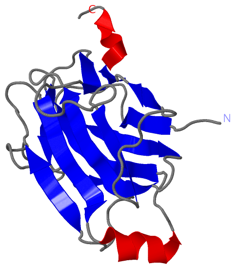
Asymmetric/Biological UnitChain A from PDB Type:PROTEIN Length:171 aligned with Q7WYN3_9FIRM | Q7WYN3 from UniProtKB/TrEMBL Length:942 Alignment length:171 38 48 58 68 78 88 98 108 118 128 138 148 158 168 178 188 198 Q7WYN3_9FIRM 29 PTSSIEIVLDKTTASVGEIVTASINIKNITNFSGCQLNMKYDPAVLQPVTSSGVAYTKSTMPGAGTILNSDFNLRQVADNDLEKGILNFSKAYVSLDDYRTAAAPEQTGTVAVVKFKVLKEETSSISFEDTTSVPNAIDGTVLFDWNGDRIQSGYSVIQPAVINLDMIKAS 199 SCOP domains d1qzna_ A: Cellulosomal scaffoldin adaptor protein B, ScaB SCOP domains CATH domains 1qznA00 A:3-173 [code=2.60.40.680, no name defined] CATH domains Pfam domains --------------------------------------------------------------------------------------------------------------------------------------------------------------------------- Pfam domains SAPs(SNPs) --------------------------------------------------------------------------------------------------------------------------------------------------------------------------- SAPs(SNPs) PROSITE --------------------------------------------------------------------------------------------------------------------------------------------------------------------------- PROSITE Transcript --------------------------------------------------------------------------------------------------------------------------------------------------------------------------- Transcript 1qzn A 3 PTSSIEIVLDKTTASVGEIVTASINIKNITNFSGCQLNMKYDPAVLQPVTSSGVAYTKSTMPGAGTILNSDFNLRQVADNDLEKGILNFSKAYVSLDDYRTAAAPEQTGTVAVVKFKVLKEETSSISFEDTTSVPNAIDGTVLFDWNGDRIQSGYSVIQPAVINLDMIKAS 173 12 22 32 42 52 62 72 82 92 102 112 122 132 142 152 162 172
|
||||||||||||||||||||
SCOP Domains (1, 1)
Asymmetric/Biological Unit
 |
CATH Domains (1, 1)
Asymmetric/Biological Unit
|
Pfam Domains (0, 0)| (no "Pfam Domain" information available for 1QZN) |
Gene Ontology (3, 3)|
Asymmetric/Biological Unit(hide GO term definitions) Chain A (Q7WYN3_9FIRM | Q7WYN3)
|
||||||||||||||||||||||||||||||
Interactive Views
|
|||||||||||||||||||||||||||||||||||||||||||||||||||||||||||||||||||||||||||||||||||||||||||||||||||||||||||||||||||||
Still Images
|
||||||||||||||||
Databases
|
||||||||||||||||||||||||||||||||||||||||||||||||||||||||||||||||||||||||||||||||||||||||||||||||||||||||||||||||||||||||||||||||||||||||||||||||||||||||||||||||
Analysis Tools
|
|||||||||||||||||||||||||||||||||||||||||||||||||||||||||||||
Entries Sharing at Least One Protein Chain (UniProt ID)
Related Entries Specified in the PDB File
|
|