|
|
|
|
 Description
|
|
Compounds
|
||||||||||||||||||||||||||||||||||||||||||||||||
Chains, Units
Summary Information (see also Sequences/Alignments below) |
Ligands, Modified Residues, Ions (0, 0)| (no "Ligand,Modified Residues,Ions" information available for 3ETZ) |
Sites (0, 0)| (no "Site" information available for 3ETZ) |
SS Bonds (0, 0)| (no "SS Bond" information available for 3ETZ) |
Cis Peptide Bonds (0, 0)| (no "Cis Peptide Bond" information available for 3ETZ) |
SAPs(SNPs)/Variants (0, 0)| (no "SAP(SNP)/Variant" information available for 3ETZ) |
PROSITE Motifs (0, 0)| (no "PROSITE Motif" information available for 3ETZ) |
Exons (0, 0)| (no "Exon" information available for 3ETZ) |
Sequences/Alignments
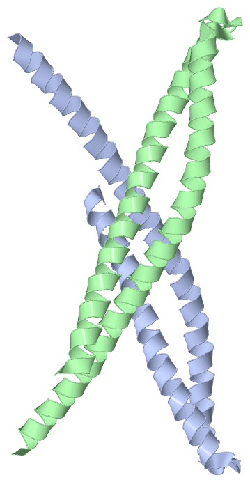
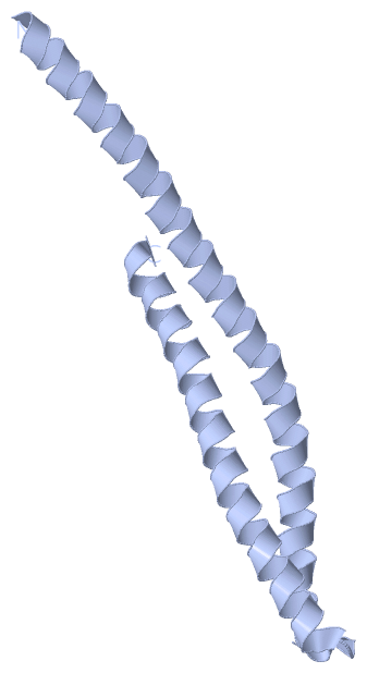
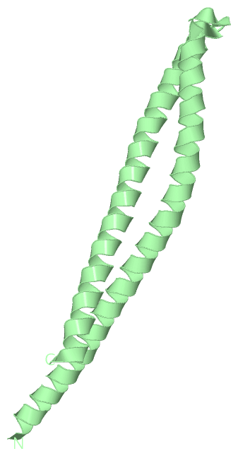
Asymmetric UnitChain A from PDB Type:PROTEIN Length:106 aligned with Q5I6B0_FUSNU | Q5I6B0 from UniProtKB/TrEMBL Length:129 Alignment length:106 31 41 51 61 71 81 91 101 111 121 Q5I6B0_FUSNU 22 AASLVGELQALDAEYQNLANQEEARFNEERAQADAARQALAQNEQVYNELSQRAQRLQAEANTRFYKSQYQELASKYEDALKKLEAEMEQQKAVISDFEKIQALRA 127 SCOP domains ---------------------------------------------------------------------------------------------------------- SCOP domains CATH domains ---------------------------------------------------------------------------------------------------------- CATH domains Pfam domains ---------------------------------------------------------------------------------------------------------- Pfam domains SAPs(SNPs) ---------------------------------------------------------------------------------------------------------- SAPs(SNPs) PROSITE ---------------------------------------------------------------------------------------------------------- PROSITE Transcript ---------------------------------------------------------------------------------------------------------- Transcript 3etz A 4 AASLVGELQALDAEYQNLANQEEARFNEERAQADAARQALAQNEQVYNELSQRAQRLQAEANTRFYKSQYQEAASKYEDALKKLEAEMEQQKAVISDFEKIQALRA 109 13 23 33 43 53 63 73 83 93 103 Chain B from PDB Type:PROTEIN Length:115 aligned with Q5I6B0_FUSNU | Q5I6B0 from UniProtKB/TrEMBL Length:129 Alignment length:115 129 31 41 51 61 71 81 91 101 111 121 | - Q5I6B0_FUSNU 22 AASLVGELQALDAEYQNLANQEEARFNEERAQADAARQALAQNEQVYNELSQRAQRLQAEANTRFYKSQYQELASKYEDALKKLEAEMEQQKAVISDFEKIQALRAGN------- - SCOP domains ------------------------------------------------------------------------------------------------------------------- SCOP domains CATH domains ------------------------------------------------------------------------------------------------------------------- CATH domains Pfam domains ------------------------------------------------------------------------------------------------------------------- Pfam domains SAPs(SNPs) ------------------------------------------------------------------------------------------------------------------- SAPs(SNPs) PROSITE ------------------------------------------------------------------------------------------------------------------- PROSITE Transcript ------------------------------------------------------------------------------------------------------------------- Transcript 3etz B 4 AASLVGELQALDAEYQNLANQEEARFNEERAQADAARQALAQNEQVYNELSQRAQRLQAEANTRFYKSQYQEAASKYEDALKKLEAEMEQQKAVISDFEKIQALRAGNLEHHHHH 118 13 23 33 43 53 63 73 83 93 103 113
|
||||||||||||||||||||
SCOP Domains (0, 0)| (no "SCOP Domain" information available for 3ETZ) |
CATH Domains (0, 0)| (no "CATH Domain" information available for 3ETZ) |
Pfam Domains (0, 0)| (no "Pfam Domain" information available for 3ETZ) |
Gene Ontology (0, 0)|
Asymmetric Unit(hide GO term definitions)
(no "Gene Ontology" information available for 3ETZ)
|
Interactive Views
|
|||||||||||||||||||||||||||||||||||||||||||||||||||||||||||||||||||||||||||||||||||||||||||||||||||||||||||||||||||||||||||||||||||||||||||
Still Images
|
||||||||||||||||
Databases
|
||||||||||||||||||||||||||||||||||||||||||||||||||||||||||||||||||||||||||||||||||||||||||||||||||||||||||||||||||||||||||||||||||||||||||||||||||||||||||||||||
Analysis Tools
|
|||||||||||||||||||||||||||||||||||||||||||||||||||||||||||||
Entries Sharing at Least One Protein Chain (UniProt ID)
Related Entries Specified in the PDB File
|
|