|
|
|
|
 Description
|
|
Compounds
|
||||||||||||||||||||||||||||||||||||||||||||||||
Chains, Units
Summary Information (see also Sequences/Alignments below) |
Ligands, Modified Residues, Ions (0, 0)| (no "Ligand,Modified Residues,Ions" information available for 3ETX) |
Sites (0, 0)| (no "Site" information available for 3ETX) |
SS Bonds (0, 0)| (no "SS Bond" information available for 3ETX) |
Cis Peptide Bonds (0, 0)| (no "Cis Peptide Bond" information available for 3ETX) |
SAPs(SNPs)/Variants (0, 0)| (no "SAP(SNP)/Variant" information available for 3ETX) |
PROSITE Motifs (0, 0)| (no "PROSITE Motif" information available for 3ETX) |
Exons (0, 0)| (no "Exon" information available for 3ETX) |
Sequences/Alignments
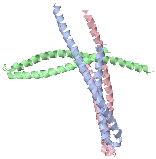
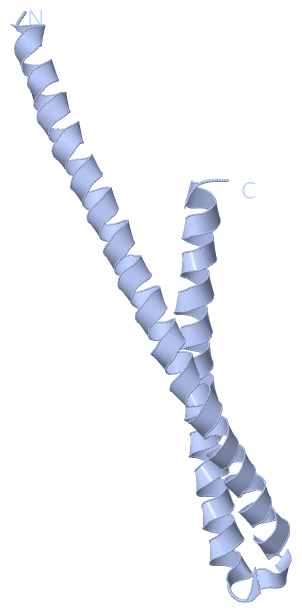
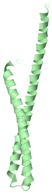
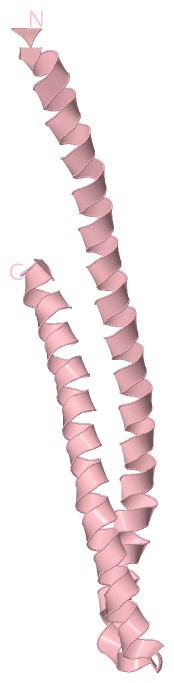
Asymmetric UnitChain A from PDB Type:PROTEIN Length:106 aligned with Q5I6B0_FUSNU | Q5I6B0 from UniProtKB/TrEMBL Length:129 Alignment length:106 32 42 52 62 72 82 92 102 112 122 Q5I6B0_FUSNU 23 ASLVGELQALDAEYQNLANQEEARFNEERAQADAARQALAQNEQVYNELSQRAQRLQAEANTRFYKSQYQELASKYEDALKKLEAEMEQQKAVISDFEKIQALRAG 128 SCOP domains ---------------------------------------------------------------------------------------------------------- SCOP domains CATH domains ---------------------------------------------------------------------------------------------------------- CATH domains Pfam domains ---------------------------------------------------------------------------------------------------------- Pfam domains SAPs(SNPs) ---------------------------------------------------------------------------------------------------------- SAPs(SNPs) PROSITE ---------------------------------------------------------------------------------------------------------- PROSITE Transcript ---------------------------------------------------------------------------------------------------------- Transcript 3etx A 5 ASLVGELQAADAEYQNLANQEEARFNEERAQADAARQALAQNEQVYNELSQRAQRLQAEANTRFYKSQYQELASKYEDALKKLEAEMEQQKAVISDFEKIQALRAG 110 14 24 34 44 54 64 74 84 94 104 Chain B from PDB Type:PROTEIN Length:107 aligned with Q5I6B0_FUSNU | Q5I6B0 from UniProtKB/TrEMBL Length:129 Alignment length:107 32 42 52 62 72 82 92 102 112 122 Q5I6B0_FUSNU 23 ASLVGELQALDAEYQNLANQEEARFNEERAQADAARQALAQNEQVYNELSQRAQRLQAEANTRFYKSQYQELASKYEDALKKLEAEMEQQKAVISDFEKIQALRAGN 129 SCOP domains ----------------------------------------------------------------------------------------------------------- SCOP domains CATH domains ----------------------------------------------------------------------------------------------------------- CATH domains Pfam domains ----------------------------------------------------------------------------------------------------------- Pfam domains SAPs(SNPs) ----------------------------------------------------------------------------------------------------------- SAPs(SNPs) PROSITE ----------------------------------------------------------------------------------------------------------- PROSITE Transcript ----------------------------------------------------------------------------------------------------------- Transcript 3etx B 5 ASLVGELQAADAEYQNLANQEEARFNEERAQADAARQALAQNEQVYNELSQRAQRLQAEANTRFYKSQYQELASKYEDALKKLEAEMEQQKAVISDFEKIQALRAGN 111 14 24 34 44 54 64 74 84 94 104 Chain C from PDB Type:PROTEIN Length:106 aligned with Q5I6B0_FUSNU | Q5I6B0 from UniProtKB/TrEMBL Length:129 Alignment length:106 32 42 52 62 72 82 92 102 112 122 Q5I6B0_FUSNU 23 ASLVGELQALDAEYQNLANQEEARFNEERAQADAARQALAQNEQVYNELSQRAQRLQAEANTRFYKSQYQELASKYEDALKKLEAEMEQQKAVISDFEKIQALRAG 128 SCOP domains ---------------------------------------------------------------------------------------------------------- SCOP domains CATH domains ---------------------------------------------------------------------------------------------------------- CATH domains Pfam domains ---------------------------------------------------------------------------------------------------------- Pfam domains SAPs(SNPs) ---------------------------------------------------------------------------------------------------------- SAPs(SNPs) PROSITE ---------------------------------------------------------------------------------------------------------- PROSITE Transcript ---------------------------------------------------------------------------------------------------------- Transcript 3etx C 5 ASLVGELQAADAEYQNLANQEEARFNEERAQADAARQALAQNEQVYNELSQRAQRLQAEANTRFYKSQYQELASKYEDALKKLEAEMEQQKAVISDFEKIQALRAG 110 14 24 34 44 54 64 74 84 94 104
|
||||||||||||||||||||
SCOP Domains (0, 0)| (no "SCOP Domain" information available for 3ETX) |
CATH Domains (0, 0)| (no "CATH Domain" information available for 3ETX) |
Pfam Domains (0, 0)| (no "Pfam Domain" information available for 3ETX) |
Gene Ontology (0, 0)|
Asymmetric Unit(hide GO term definitions)
(no "Gene Ontology" information available for 3ETX)
|
Interactive Views
|
||||||||||||||||||||||||||||||||||||||||||||||||||||||||||||||||||||||||||||||||||||||||||||||||||||||||||||||||||||||||||||||||||||||||||||||||
Still Images
|
||||||||||||||||
Databases
|
||||||||||||||||||||||||||||||||||||||||||||||||||||||||||||||||||||||||||||||||||||||||||||||||||||||||||||||||||||||||||||||||||||||||||||||||||||||||||||||||
Analysis Tools
|
|||||||||||||||||||||||||||||||||||||||||||||||||||||||||||||
Entries Sharing at Least One Protein Chain (UniProt ID)
Related Entries Specified in the PDB File
|
|