| molecular function |
|---|
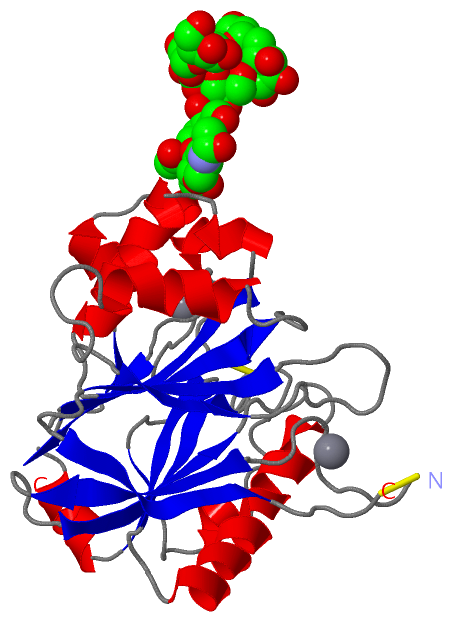
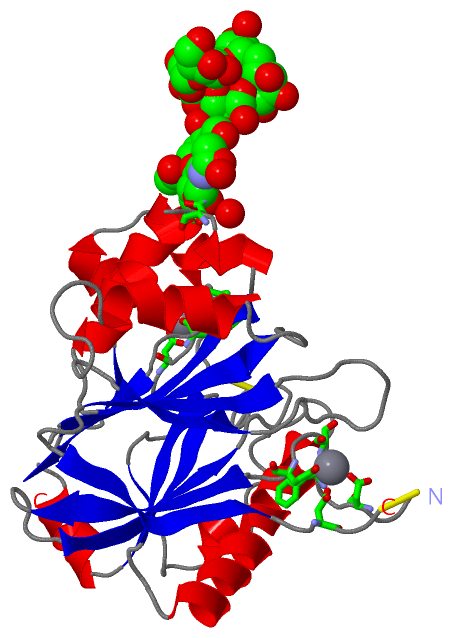
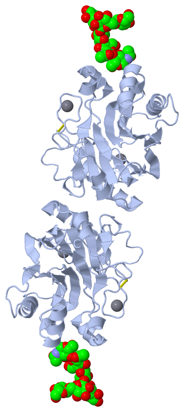
| | GO:0003779 | | actin binding | | Interacting selectively and non-covalently with monomeric or multimeric forms of actin, including actin filaments. |
| | GO:0004530 | | deoxyribonuclease I activity | | Catalysis of the endonucleolytic cleavage of DNA to 5'-phosphodinucleotide and 5'-phosphooligonucleotide end products. |
| | GO:0004536 | | deoxyribonuclease activity | | Catalysis of the hydrolysis of ester linkages within deoxyribonucleic acid. |
| | GO:0004519 | | endonuclease activity | | Catalysis of the hydrolysis of ester linkages within nucleic acids by creating internal breaks. |
| | GO:0016787 | | hydrolase activity | | Catalysis of the hydrolysis of various bonds, e.g. C-O, C-N, C-C, phosphoric anhydride bonds, etc. Hydrolase is the systematic name for any enzyme of EC class 3. |
| | GO:0004518 | | nuclease activity | | Catalysis of the hydrolysis of ester linkages within nucleic acids. |
| biological process |
|---|
| | GO:0006308 | | DNA catabolic process | | The cellular DNA metabolic process resulting in the breakdown of DNA, deoxyribonucleic acid, one of the two main types of nucleic acid, consisting of a long unbranched macromolecule formed from one or two strands of linked deoxyribonucleotides, the 3'-phosphate group of each constituent deoxyribonucleotide being joined in 3',5'-phosphodiester linkage to the 5'-hydroxyl group of the deoxyribose moiety of the next one. |
| | GO:0006915 | | apoptotic process | | A programmed cell death process which begins when a cell receives an internal (e.g. DNA damage) or external signal (e.g. an extracellular death ligand), and proceeds through a series of biochemical events (signaling pathway phase) which trigger an execution phase. The execution phase is the last step of an apoptotic process, and is typically characterized by rounding-up of the cell, retraction of pseudopodes, reduction of cellular volume (pyknosis), chromatin condensation, nuclear fragmentation (karyorrhexis), plasma membrane blebbing and fragmentation of the cell into apoptotic bodies. When the execution phase is completed, the cell has died. |
| | GO:0090305 | | nucleic acid phosphodiester bond hydrolysis | | The nucleic acid metabolic process in which the phosphodiester bonds between nucleotides are cleaved by hydrolysis. |
| cellular component |
|---|
| | GO:0005576 | | extracellular region | | The space external to the outermost structure of a cell. For cells without external protective or external encapsulating structures this refers to space outside of the plasma membrane. This term covers the host cell environment outside an intracellular parasite. |
| | GO:0005635 | | nuclear envelope | | The double lipid bilayer enclosing the nucleus and separating its contents from the rest of the cytoplasm; includes the intermembrane space, a gap of width 20-40 nm (also called the perinuclear space). |
| | GO:0005634 | | nucleus | | A membrane-bounded organelle of eukaryotic cells in which chromosomes are housed and replicated. In most cells, the nucleus contains all of the cell's chromosomes except the organellar chromosomes, and is the site of RNA synthesis and processing. In some species, or in specialized cell types, RNA metabolism or DNA replication may be absent. |
 Description