|
|
|
|
 Description
|
|
Compounds
|
||||||||||||||||||||||||||||||||||||||||||||||||
Chains, Units
Summary Information (see also Sequences/Alignments below) |
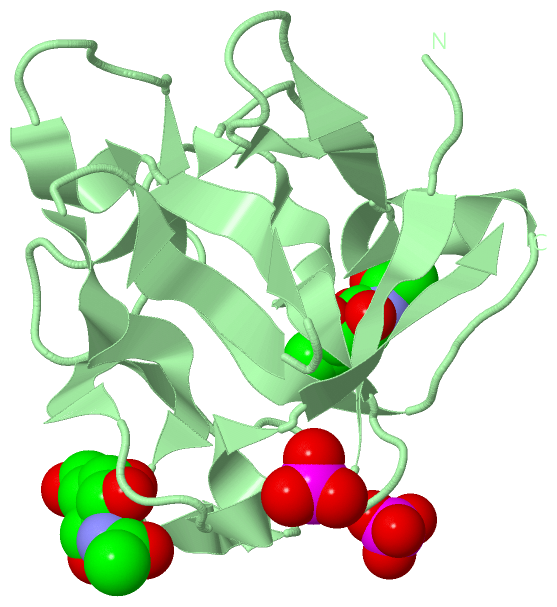
Ligands, Modified Residues, Ions (4, 9)
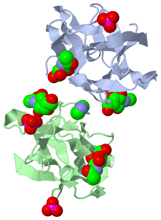
Asymmetric Unit (4, 9)
|
Sites (9, 9)
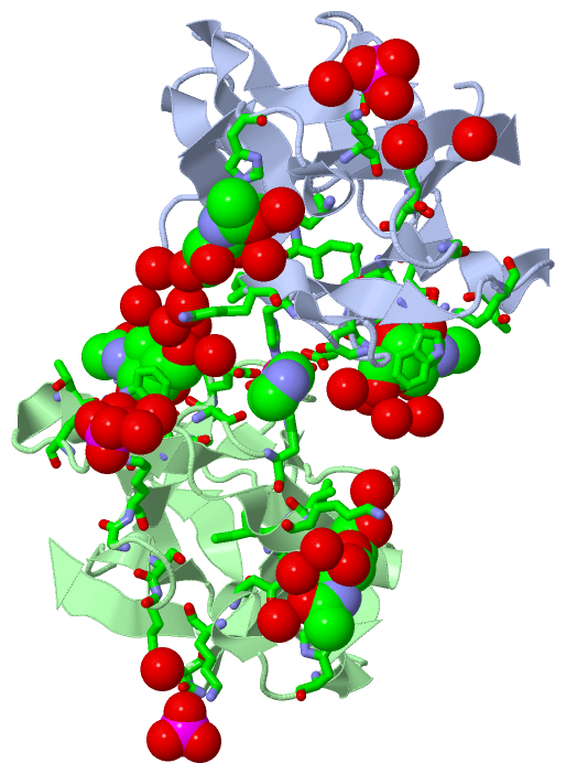
Asymmetric Unit (9, 9)
|
SS Bonds (0, 0)| (no "SS Bond" information available for 2ZQO) |
Cis Peptide Bonds (0, 0)| (no "Cis Peptide Bond" information available for 2ZQO) |
SAPs(SNPs)/Variants (0, 0)| (no "SAP(SNP)/Variant" information available for 2ZQO) |
PROSITE Motifs (0, 0)| (no "PROSITE Motif" information available for 2ZQO) |
Exons (0, 0)| (no "Exon" information available for 2ZQO) |
Sequences/Alignments
Asymmetric UnitChain A from PDB Type:PROTEIN Length:130 aligned with O96048_LUMTE | O96048 from UniProtKB/TrEMBL Length:260 Alignment length:130 140 150 160 170 180 190 200 210 220 230 240 250 260 O96048_LUMTE 131 PKFFYIKSELNGKVLDIEGQNPAPGSKIITWDQKKGPTAVNQLWYTDQQGVIRSKLNDFAIDASHEQIETQPFDPNNPKRAWIVSGNTIAQLSDRDIVLDIIKSDKEAGAHICAWKQHGGPNQKFIIESE 260 SCOP domains d2zqoa1 A:131-260 29-kDa galactose-binding lectin SCOP domains CATH domains ---------------------------------------------------------------------------------------------------------------------------------- CATH domains Pfam domains ---------------------------------------------------------------------------------------------------------------------------------- Pfam domains SAPs(SNPs) ---------------------------------------------------------------------------------------------------------------------------------- SAPs(SNPs) PROSITE ---------------------------------------------------------------------------------------------------------------------------------- PROSITE Transcript ---------------------------------------------------------------------------------------------------------------------------------- Transcript 2zqo A 131 PKFFYIKSELNGKVLDIEGQNPAPGSKIITWDQKKGPTAVNQLWYTDQQGVIRSKLNDFAIDASHEQIETQPFDPNNPKRAWIVSGNTIAQLSDRDIVLDIIKSDKEAGAHICAWKQHGGPNQKFIIESE 260 140 150 160 170 180 190 200 210 220 230 240 250 260 Chain B from PDB Type:PROTEIN Length:130 aligned with O96048_LUMTE | O96048 from UniProtKB/TrEMBL Length:260 Alignment length:130 140 150 160 170 180 190 200 210 220 230 240 250 260 O96048_LUMTE 131 PKFFYIKSELNGKVLDIEGQNPAPGSKIITWDQKKGPTAVNQLWYTDQQGVIRSKLNDFAIDASHEQIETQPFDPNNPKRAWIVSGNTIAQLSDRDIVLDIIKSDKEAGAHICAWKQHGGPNQKFIIESE 260 SCOP domains d2zqob_ B: 29-kDa galactose-binding lectin SCOP domains CATH domains ---------------------------------------------------------------------------------------------------------------------------------- CATH domains Pfam domains (1) Ricin_B_lectin-2zqoB01 B:131-255 ----- Pfam domains (1) Pfam domains (2) Ricin_B_lectin-2zqoB02 B:131-255 ----- Pfam domains (2) SAPs(SNPs) ---------------------------------------------------------------------------------------------------------------------------------- SAPs(SNPs) PROSITE ---------------------------------------------------------------------------------------------------------------------------------- PROSITE Transcript ---------------------------------------------------------------------------------------------------------------------------------- Transcript 2zqo B 131 PKFFYIKSELNGKVLDIEGQNPAPGSKIITWDQKKGPTAVNQLWYTDQQGVIRSKLNDFAIDASHEQIETQPFDPNNPKRAWIVSGNTIAQLSDRDIVLDIIKSDKEAGAHICAWKQHGGPNQKFIIESE 260 140 150 160 170 180 190 200 210 220 230 240 250 260
|
||||||||||||||||||||
SCOP Domains (1, 2)
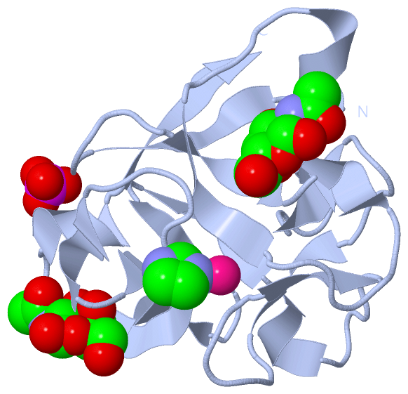
Asymmetric Unit
 |
CATH Domains (0, 0)| (no "CATH Domain" information available for 2ZQO) |
Pfam Domains (1, 2)
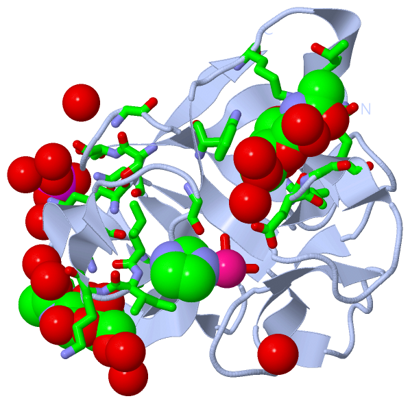
Asymmetric Unit
|
Gene Ontology (1, 1)|
Asymmetric Unit(hide GO term definitions) Chain A,B (O96048_LUMTE | O96048)
|
||||||||||||
Interactive Views
|
||||||||||||||||||||||||||||||||||||||||||||||||||||||||||||||||||||||||||||||||||||||||||||||||||||||||||||||||||||||||||||||||||||||||||||||||||||||||||||||||||||||||||||||||||||||||||||||||||||||||||||||||||||||||||
Still Images
|
||||||||||||||||
Databases
|
||||||||||||||||||||||||||||||||||||||||||||||||||||||||||||||||||||||||||||||||||||||||||||||||||||||||||||||||||||||||||||||||||||||||||||||||||||||||||||||||
Analysis Tools
|
|||||||||||||||||||||||||||||||||||||||||||||||||||||||||||||
Entries Sharing at Least One Protein Chain (UniProt ID)
Related Entries Specified in the PDB File
|
|