Show PDB file:   
         Plain Text   HTML   (compressed file size)
QuickSearch:   
by PDB,NDB,UniProt,PROSITE Code or Search Term(s)  
(-)Asymmetric Unit
(-)Asym. Unit - sites
(-)Biological Unit 1
(-)Biol. Unit 1 - sites
(-)Biological Unit 2
collapse expand < >
Image Asymmetric Unit
Asymmetric Unit  (Jmol Viewer)
Image Asym. Unit - sites
Asym. Unit - sites  (Jmol Viewer)
Image Biological Unit 1
Biological Unit 1  (Jmol Viewer)
Image Biol. Unit 1 - sites
Biol. Unit 1 - sites  (Jmol Viewer)
Image Biological Unit 2
Biological Unit 2  (Jmol Viewer)

(-) Description

Title :  CRYSTAL STRUCTURE OF THE EARTHWORM LECTIN C-TERMINAL DOMAIN MUTANT IN COMPLEX WITH 6'-SIALYLLACTOSE
 
Authors :  R. Suzuki, Z. Fujimoto
Date :  16 Jun 06  (Deposition) - 06 Feb 07  (Release) - 04 Aug 09  (Revision)
Method :  X-RAY DIFFRACTION
Resolution :  1.80
Chains :  Asym. Unit :  A,B
Biol. Unit 1:  A  (1x)
Biol. Unit 2:  B  (1x)
Keywords :  Earthworm Lumbricus Terrestris, Sialic Acid, Galactose, In Vitro Evolution, Beta-Trefoil Fold, Sugar Complex, Sugar Binding Protein (Keyword Search: [Gene Ontology, PubMed, Web (Google))
 
Reference :  R. Yabe, R. Suzuki, A. Kuno, Z. Fujimoto, Y. Jigami, J. Hirabayashi
Tailoring A Novel Sialic Acid-Binding Lectin From A Ricin-B Chain-Like Galactose-Binding Protein By Natural Evolution-Mimicry
J. Biochem. V. 141 389 2007
PubMed-ID: 17234683  |  Reference-DOI: 10.1093/JB/MVM043
(for further references see the PDB file header)

(-) Compounds

Molecule 1 - 29-KDA GALACTOSE-BINDING LECTIN
    ChainsA, B
    EngineeredYES
    Expression SystemESCHERICHIA COLI BL21(DE3)
    Expression System PlasmidPET27
    Expression System StrainBL21 DE3
    Expression System Taxid469008
    Expression System Vector TypePLASMID
    FragmentC-TERMINAL DOMAIN
    MutationYES
    Organism CommonCOMMON EARTHWORM
    Organism ScientificLUMBRICUS TERRESTRIS
    Organism Taxid6398

 Structural Features

(-) Chains, Units

  12
Asymmetric Unit AB
Biological Unit 1 (1x)A 
Biological Unit 2 (1x) B

Summary Information (see also Sequences/Alignments below)

(-) Ligands, Modified Residues, Ions  (4, 11)

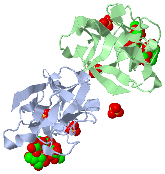
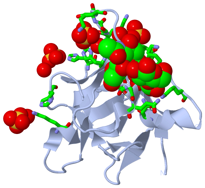
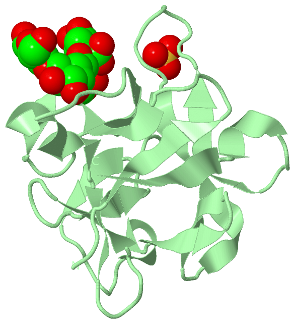
Asymmetric Unit (4, 11)
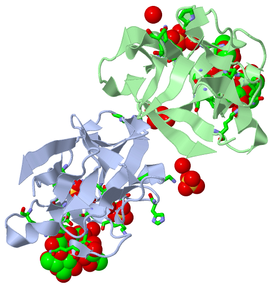
No.NameCountTypeFull Name
1BGC2Ligand/IonBETA-D-GLUCOSE
2GAL2Ligand/IonBETA-D-GALACTOSE
3SIA2Ligand/IonO-SIALIC ACID
4SO45Ligand/IonSULFATE ION
Biological Unit 1 (4, 7)
No.NameCountTypeFull Name
1BGC1Ligand/IonBETA-D-GLUCOSE
2GAL1Ligand/IonBETA-D-GALACTOSE
3SIA1Ligand/IonO-SIALIC ACID
4SO44Ligand/IonSULFATE ION
Biological Unit 2 (4, 4)
No.NameCountTypeFull Name
1BGC1Ligand/IonBETA-D-GLUCOSE
2GAL1Ligand/IonBETA-D-GALACTOSE
3SIA1Ligand/IonO-SIALIC ACID
4SO41Ligand/IonSULFATE ION

(-) Sites  (11, 11)

Asymmetric Unit (11, 11)
No.NameEvidenceResiduesDescription
01AC1SOFTWAREGLY A:148 , GLY A:149 , ILE A:159 , SER A:239 , GAL A:281 , HOH A:1061BINDING SITE FOR RESIDUE SIA A 280
02AC2SOFTWAREASP A:146 , GLY A:148 , GLY A:149 , GLN A:150 , TRP A:161 , LYS A:164 , ASN A:171 , SIA A:280 , BGC A:282BINDING SITE FOR RESIDUE GAL A 281
03AC3SOFTWAREGAL A:281BINDING SITE FOR RESIDUE BGC A 282
04AC4SOFTWAREHIS A:248 , HOH A:1082 , PRO B:208 , GLN B:221 , SER B:223 , HOH B:1023BINDING SITE FOR RESIDUE SO4 A 1001
05AC5SOFTWAREASN A:141 , LYS A:143 , LYS A:233 , HOH A:1007 , HOH A:1016BINDING SITE FOR RESIDUE SO4 A 1002
06AC6SOFTWARESER A:234 , ASP A:235 , LYS A:236 , HIS A:241 , HOH A:1012BINDING SITE FOR RESIDUE SO4 A 1003
07AC7SOFTWARELYS A:209 , HOH A:1075BINDING SITE FOR RESIDUE SO4 A 1004
08AC8SOFTWAREGLY B:148 , LYS B:157 , ILE B:159 , TRP B:161 , SER B:239 , GAL B:271 , HOH B:1079BINDING SITE FOR RESIDUE SIA B 270
09AC9SOFTWAREASP B:146 , ILE B:147 , GLY B:148 , GLY B:149 , GLN B:150 , TRP B:161 , LYS B:164 , ASN B:171 , SIA B:270 , BGC B:272 , HOH B:1089 , HOH B:1095BINDING SITE FOR RESIDUE GAL B 271
10BC1SOFTWAREGAL B:271BINDING SITE FOR RESIDUE BGC B 272
11BC2SOFTWAREHIS A:195 , SER B:234 , ASP B:235 , HIS B:241 , HOH B:1105BINDING SITE FOR RESIDUE SO4 B 1005

(-) SS Bonds  (0, 0)

(no "SS Bond" information available for 2DS0)

(-) Cis Peptide Bonds  (0, 0)

(no "Cis Peptide Bond" information available for 2DS0)

 Sequence-Structure Mapping

(-) SAPs(SNPs)/Variants  (0, 0)

(no "SAP(SNP)/Variant" information available for 2DS0)

(-) PROSITE Motifs  (0, 0)

(no "PROSITE Motif" information available for 2DS0)

(-) Exons   (0, 0)

(no "Exon" information available for 2DS0)

(-) Sequences/Alignments

Asymmetric Unit
   Reformat: Number of residues per line =  ('0' or empty: single-line sequence representation)
  Number of residues per labelling interval =   
  UniProt sequence: complete  aligned part    
   Show mapping: SCOP domains CATH domains Pfam domains Secondary structure (by author)
SAPs(SNPs) PROSITE motifs Exons
(details for a mapped element are shown in a popup box when the mouse pointer rests over it)
Chain A from PDB  Type:PROTEIN  Length:130
 aligned with O96048_LUMTE | O96048 from UniProtKB/TrEMBL  Length:260

    Alignment length:130
                                   140       150       160       170       180       190       200       210       220       230       240       250       260
         O96048_LUMTE   131 PKFFYIKSELNGKVLDIEGQNPAPGSKIITWDQKKGPTAVNQLWYTDQQGVIRSKLNDFAIDASHEQIETQPFDPNNPKRAWIVSGNTIAQLSDRDIVLDIIKSDKEAGAHICAWKQHGGPNQKFIIESE 260
               SCOP domains d2ds0a_ A: automated matches                                                                                                       SCOP domains
               CATH domains ---------------------------------------------------------------------------------------------------------------------------------- CATH domains
               Pfam domains ---------------------------------------------------------------------------------------------------------------------------------- Pfam domains
         Sec.struct. author ...eeeee.....eeee.hhh......eeee....hhhhhhh.eee.....eee.....eee......eee.....hhhh.eeee..eeee..eeeeeeeee........eeeeee...hhhh.eeeee. Sec.struct. author
                 SAPs(SNPs) ---------------------------------------------------------------------------------------------------------------------------------- SAPs(SNPs)
                    PROSITE ---------------------------------------------------------------------------------------------------------------------------------- PROSITE
                 Transcript ---------------------------------------------------------------------------------------------------------------------------------- Transcript
                 2ds0 A 131 PKFFYIKSELNGKVLDIGGQNPAPGSKIITWDQKKGPTAVNQLWYTDQQGVIRSKLNDFAIDASHEQIETQPFDPNNPKRAWIVSGNTIAQLSDRDNVLGVIKSDKGASAHICAWKQHGGPNQKFIIESE 260
                                   140       150       160       170       180       190       200       210       220       230       240       250       260

Chain B from PDB  Type:PROTEIN  Length:130
 aligned with O96048_LUMTE | O96048 from UniProtKB/TrEMBL  Length:260

    Alignment length:130
                                   140       150       160       170       180       190       200       210       220       230       240       250       260
         O96048_LUMTE   131 PKFFYIKSELNGKVLDIEGQNPAPGSKIITWDQKKGPTAVNQLWYTDQQGVIRSKLNDFAIDASHEQIETQPFDPNNPKRAWIVSGNTIAQLSDRDIVLDIIKSDKEAGAHICAWKQHGGPNQKFIIESE 260
               SCOP domains d2ds0b_ B: automated matches                                                                                                       SCOP domains
               CATH domains ---------------------------------------------------------------------------------------------------------------------------------- CATH domains
               Pfam domains ---------------------------------------------------------------------------------------------------------------------------------- Pfam domains
         Sec.struct. author ...eeeee.....eeee.hhh......eeee....hhhhhhh.eee.....eee.....eee......eee.....hhhh.eeee..eeee..eeeeeeeee........eeeeee...hhhh.eeeee. Sec.struct. author
                 SAPs(SNPs) ---------------------------------------------------------------------------------------------------------------------------------- SAPs(SNPs)
                    PROSITE ---------------------------------------------------------------------------------------------------------------------------------- PROSITE
                 Transcript ---------------------------------------------------------------------------------------------------------------------------------- Transcript
                 2ds0 B 131 PKFFYIKSELNGKVLDIGGQNPAPGSKIITWDQKKGPTAVNQLWYTDQQGVIRSKLNDFAIDASHEQIETQPFDPNNPKRAWIVSGNTIAQLSDRDNVLGVIKSDKGASAHICAWKQHGGPNQKFIIESE 260
                                   140       150       160       170       180       190       200       210       220       230       240       250       260

   Legend:   → Mismatch (orange background)
  - → Gap (green background, '-', border residues have a numbering label)
    → Modified Residue (blue background, lower-case, 'x' indicates undefined single-letter code, labelled with number + name)
  x → Chemical Group (purple background, 'x', labelled with number + name, e.g. ACE or NH2)
  extra numbering lines below/above indicate numbering irregularities and modified residue names etc., number ends below/above '|'

 Classification and Annotation

(-) SCOP Domains  (1, 2)

Asymmetric Unit

(-) CATH Domains  (0, 0)

(no "CATH Domain" information available for 2DS0)

(-) Pfam Domains  (0, 0)

(no "Pfam Domain" information available for 2DS0)

(-) Gene Ontology  (1, 1)

Asymmetric Unit(hide GO term definitions)
Chain A,B   (O96048_LUMTE | O96048)
molecular function
    GO:0030246    carbohydrate binding    Interacting selectively and non-covalently with any carbohydrate, which includes monosaccharides, oligosaccharides and polysaccharides as well as substances derived from monosaccharides by reduction of the carbonyl group (alditols), by oxidation of one or more hydroxy groups to afford the corresponding aldehydes, ketones, or carboxylic acids, or by replacement of one or more hydroxy group(s) by a hydrogen atom. Cyclitols are generally not regarded as carbohydrates.

 Visualization

(-) Interactive Views

Asymmetric Unit
  Complete Structure
    Jena3D(integrated viewing of ligand, site, SAP, PROSITE, SCOP information)
    WebMol | AstexViewer[tm]@PDBe
(Java Applets, require no local installation except for Java; loading may be slow)
    STRAP
(Java WebStart application, automatic local installation, requires Java; full application with system access!)
    RasMol
(require local installation)
    Molscript (VRML)
(requires installation of a VRML viewer; select preferred view via VRML and generate a mono or stereo PDF format file)
 
  Ligands, Modified Residues, Ions
    BGC  [ RasMol | Jena3D ]  +environment [ RasMol | Jena3D ]
    GAL  [ RasMol | Jena3D ]  +environment [ RasMol | Jena3D ]
    SIA  [ RasMol | Jena3D ]  +environment [ RasMol | Jena3D ]
    SO4  [ RasMol | Jena3D ]  +environment [ RasMol | Jena3D ]
 
  Sites
    AC1  [ RasMol ]  +environment [ RasMol ]
    AC2  [ RasMol ]  +environment [ RasMol ]
    AC3  [ RasMol ]  +environment [ RasMol ]
    AC4  [ RasMol ]  +environment [ RasMol ]
    AC5  [ RasMol ]  +environment [ RasMol ]
    AC6  [ RasMol ]  +environment [ RasMol ]
    AC7  [ RasMol ]  +environment [ RasMol ]
    AC8  [ RasMol ]  +environment [ RasMol ]
    AC9  [ RasMol ]  +environment [ RasMol ]
    BC1  [ RasMol ]  +environment [ RasMol ]
    BC2  [ RasMol ]  +environment [ RasMol ]
 
  Cis Peptide Bonds
(no "Cis Peptide Bonds" information available for 2ds0)
 
Biological Units
  Complete Structure
    Biological Unit 1  [ Jena3D ]
    Biological Unit 2  [ Jena3D ]

(-) Still Images

Jmol
  protein: cartoon or spacefill or dots and stick; nucleic acid: cartoon and stick; ligands: spacefill; active site: stick
Molscript
  protein, nucleic acid: cartoon; ligands: spacefill; active site: ball and stick

 Databases and Analysis Tools

(-) Databases

Access by PDB/NDB ID
  2ds0
    Family and Domain InformationProDom | SYSTERS
    General Structural InformationGlycoscienceDB | MMDB | NDB | OCA | PDB | PDBe | PDBj | PDBsum | PDBWiki | PQS | PROTEOPEDIA
    Orientation in MembranesOPM
    Protein SurfaceSURFACE
    Secondary StructureDSSP (structure derived) | HSSP (homology derived)
    Structural GenomicsGeneCensus
    Structural NeighboursCE | VAST
    Structure ClassificationCATH | Dali | SCOP
    Validation and Original DataBMRB Data View | BMRB Restraints Grid | EDS | PROCHECK | RECOORD | WHAT_CHECK
 
Access by UniProt ID/Accession number
  O96048_LUMTE | O96048
    Comparative Protein Structure ModelsModBase
    Genomic InformationEnsembl
    Protein-protein InteractionDIP
    Sequence, Family and Domain InformationInterPro | Pfam | SMART | UniProtKB/TrEMBL
 
Access by Enzyme Classificator   (EC Number)
  (no 'Enzyme Classificator' available)
    General Enzyme InformationBRENDA | EC-PDB | Enzyme | IntEnz
    PathwayKEGG | MetaCyc
 
Access by Disease Identifier   (MIM ID)
  (no 'MIM ID' available)
    Disease InformationOMIM
 
Access by GenAge ID
  (no 'GenAge ID' available)
    Age Related InformationGenAge

(-) Analysis Tools

Access by PDB/NDB ID
    Domain InformationXDom
    Interatomic Contacts of Structural UnitsCSU
    Ligand-protein ContactsLPC
    Protein CavitiescastP
    Sequence and Secondary StructurePDBCartoon
    Structure AlignmentSTRAP(Java WebStart application, automatic local installation, requires Java; full application with system access!)
    Structure and Sequence BrowserSTING
 
Access by UniProt ID/Accession number
  O96048_LUMTE | O96048
    Protein Disorder PredictionDisEMBL | FoldIndex | GLOBPLOT (for more information see DisProt)

 Related Entries

(-) Entries Sharing at Least One Protein Chain (UniProt ID)

UniProtKB/TrEMBL
        O96048_LUMTE | O960482dry 2drz 2rst 2zqn 2zqo

(-) Related Entries Specified in the PDB File

2ao3 THE WILD-TYPE LECTIN IN COMPLEX WITH N-ACETYLGALACTOSAMINE
2d12 THE WILD-TYPE LECTIN IN COMPLEX WITH LACTOSE
2dry
2drz