| molecular function |
|---|
| | GO:0005524 | | ATP binding | | Interacting selectively and non-covalently with ATP, adenosine 5'-triphosphate, a universally important coenzyme and enzyme regulator. |
| | GO:0097161 | | DH domain binding | | Interacting selectively and non-covalently with a DH (Dbl homology) domain of a protein. The DH domain contains three structurally conserved regions separated by more variable regions. It is composed of 11 alpha helices that are folded into a flattened, elongated alpha-helix bundle in which two of the three conserved regions, conserved region 1 (CR1) and conserved region 3 (CR3), are exposed near the centre of one surface. CR1 and CR3, together with a part of alpha-6 and the DH/PH (pleckstrin homology) junction site, constitute the Rho GTPase interacting pocket. |
| | GO:0005004 | | GPI-linked ephrin receptor activity | | Combining with a GPI-anchored ephrin to initiate a change in cell activity. |
| | GO:0042731 | | PH domain binding | | Interacting selectively and non-covalently with a PH domain (pleckstrin homology) of a protein, a domain of about 100 residues that occurs in a wide range of proteins involved in intracellular signaling or as constituents of the cytoskeleton. |
| | GO:0005003 | | ephrin receptor activity | | Combining with an ephrin to initiate a change in cell activity. |
| | GO:0046875 | | ephrin receptor binding | | Interacting selectively and non-covalently with an ephrin receptor. |
| | GO:0042802 | | identical protein binding | | Interacting selectively and non-covalently with an identical protein or proteins. |
| | GO:0016301 | | kinase activity | | Catalysis of the transfer of a phosphate group, usually from ATP, to a substrate molecule. |
| | GO:0000166 | | nucleotide binding | | Interacting selectively and non-covalently with a nucleotide, any compound consisting of a nucleoside that is esterified with (ortho)phosphate or an oligophosphate at any hydroxyl group on the ribose or deoxyribose. |
| | GO:0005515 | | protein binding | | Interacting selectively and non-covalently with any protein or protein complex (a complex of two or more proteins that may include other nonprotein molecules). |
| | GO:0004672 | | protein kinase activity | | Catalysis of the phosphorylation of an amino acid residue in a protein, usually according to the reaction: a protein + ATP = a phosphoprotein + ADP. |
| | GO:0004713 | | protein tyrosine kinase activity | | Catalysis of the reaction: ATP + a protein tyrosine = ADP + protein tyrosine phosphate. |
| | GO:0016740 | | transferase activity | | Catalysis of the transfer of a group, e.g. a methyl group, glycosyl group, acyl group, phosphorus-containing, or other groups, from one compound (generally regarded as the donor) to another compound (generally regarded as the acceptor). Transferase is the systematic name for any enzyme of EC class 2. |
| | GO:0004714 | | transmembrane receptor protein tyrosine kinase activity | | Combining with a signal and transmitting the signal from one side of the membrane to the other to initiate a change in cell activity by catalysis of the reaction: ATP + a protein-L-tyrosine = ADP + a protein-L-tyrosine phosphate. |
| | GO:0005005 | | transmembrane-ephrin receptor activity | | Combining with a transmembrane ephrin to initiate a change in cell activity. |
| biological process |
|---|
| | GO:0007628 | | adult walking behavior | | The behavior of an adult relating to the progression of that organism along the ground by the process of lifting and setting down each leg. |
| | GO:0007411 | | axon guidance | | The chemotaxis process that directs the migration of an axon growth cone to a specific target site in response to a combination of attractive and repulsive cues. |
| | GO:0007155 | | cell adhesion | | The attachment of a cell, either to another cell or to an underlying substrate such as the extracellular matrix, via cell adhesion molecules. |
| | GO:0021957 | | corticospinal tract morphogenesis | | Generation of a long process of a pyramidal cell, that carries efferent (outgoing) action potentials from the cell body in cerebral cortex layer V towards target cells in the gray matter of the spinal cord. This axonal process is a member of those that make up the corticospinal tract. |
| | GO:0048013 | | ephrin receptor signaling pathway | | The series of molecular signals generated as a consequence of an ephrin receptor binding to an ephrin. |
| | GO:0097156 | | fasciculation of motor neuron axon | | The collection of motor neuron axons into a bundle of rods, known as a fascicle. |
| | GO:0097155 | | fasciculation of sensory neuron axon | | The collection of sensory neuron axons into a bundle of rods, known as a fascicle. |
| | GO:0008347 | | glial cell migration | | The orderly movement of a glial cell, non-neuronal cells that provide support and nutrition, maintain homeostasis, form myelin, and participate in signal transmission in the nervous system. |
| | GO:0008045 | | motor neuron axon guidance | | The process in which the migration of an axon growth cone of a motor neuron is directed to a specific target site in response to a combination of attractive and repulsive cues. |
| | GO:0007275 | | multicellular organism development | | The biological process whose specific outcome is the progression of a multicellular organism over time from an initial condition (e.g. a zygote or a young adult) to a later condition (e.g. a multicellular animal or an aged adult). |
| | GO:0048681 | | negative regulation of axon regeneration | | Any process that stops, prevents, or reduces the frequency, rate or extent of axon regeneration. |
| | GO:0072178 | | nephric duct morphogenesis | | The process in which the anatomical structures of the nephric duct are generated and organized. A nephric duct is a tube that drains a primitive kidney. |
| | GO:0007399 | | nervous system development | | The process whose specific outcome is the progression of nervous tissue over time, from its formation to its mature state. |
| | GO:0018108 | | peptidyl-tyrosine phosphorylation | | The phosphorylation of peptidyl-tyrosine to form peptidyl-O4'-phospho-L-tyrosine. |
| | GO:0016310 | | phosphorylation | | The process of introducing a phosphate group into a molecule, usually with the formation of a phosphoric ester, a phosphoric anhydride or a phosphoric amide. |
| | GO:0043507 | | positive regulation of JUN kinase activity | | Any process that activates or increases the frequency, rate or extent of JUN kinase activity. |
| | GO:2001108 | | positive regulation of Rho guanyl-nucleotide exchange factor activity | | Any process that activates or increases the frequency, rate or extent of Rho guanyl-nucleotide exchange factor activity. |
| | GO:0050775 | | positive regulation of dendrite morphogenesis | | Any process that activates or increases the frequency, rate or extent of dendrite morphogenesis. |
| | GO:0046777 | | protein autophosphorylation | | The phosphorylation by a protein of one or more of its own amino acid residues (cis-autophosphorylation), or residues on an identical protein (trans-autophosphorylation). |
| | GO:0006468 | | protein phosphorylation | | The process of introducing a phosphate group on to a protein. |
| | GO:0043087 | | regulation of GTPase activity | | Any process that modulates the rate of GTP hydrolysis by a GTPase. |
| | GO:0048710 | | regulation of astrocyte differentiation | | Any process that modulates the frequency, rate or extent of astrocyte differentiation. |
| | GO:0050770 | | regulation of axonogenesis | | Any process that modulates the frequency, rate or extent of axonogenesis, the generation of an axon, the long process of a neuron. |
| | GO:0061001 | | regulation of dendritic spine morphogenesis | | Any process that modulates the rate, frequency, or extent of dendritic spine morphogenesis, the process in which the anatomical structures of a dendritic spine are generated and organized. A dendritic spine is a protrusion from a dendrite and a specialized subcellular compartment involved in synaptic transmission. |
| | GO:0007169 | | transmembrane receptor protein tyrosine kinase signaling pathway | | A series of molecular signals initiated by the binding of an extracellular ligand to a receptor on the surface of the target cell where the receptor possesses tyrosine kinase activity, and ending with regulation of a downstream cellular process, e.g. transcription. |
| cellular component |
|---|
| | GO:0005794 | | Golgi apparatus | | A compound membranous cytoplasmic organelle of eukaryotic cells, consisting of flattened, ribosome-free vesicles arranged in a more or less regular stack. The Golgi apparatus differs from the endoplasmic reticulum in often having slightly thicker membranes, appearing in sections as a characteristic shallow semicircle so that the convex side (cis or entry face) abuts the endoplasmic reticulum, secretory vesicles emerging from the concave side (trans or exit face). In vertebrate cells there is usually one such organelle, while in invertebrates and plants, where they are known usually as dictyosomes, there may be several scattered in the cytoplasm. The Golgi apparatus processes proteins produced on the ribosomes of the rough endoplasmic reticulum; such processing includes modification of the core oligosaccharides of glycoproteins, and the sorting and packaging of proteins for transport to a variety of cellular locations. Three different regions of the Golgi are now recognized both in terms of structure and function: cis, in the vicinity of the cis face, trans, in the vicinity of the trans face, and medial, lying between the cis and trans regions. |
| | GO:0030424 | | axon | | The long process of a neuron that conducts nerve impulses, usually away from the cell body to the terminals and varicosities, which are sites of storage and release of neurotransmitter. |
| | GO:0043679 | | axon terminus | | Terminal inflated portion of the axon, containing the specialized apparatus necessary to release neurotransmitters. The axon terminus is considered to be the whole region of thickening and the terminal button is a specialized region of it. |
| | GO:0044295 | | axonal growth cone | | The migrating motile tip of a growing nerve cell axon. |
| | GO:0044297 | | cell body | | The portion of a cell bearing surface projections such as axons, dendrites, cilia, or flagella that includes the nucleus, but excludes all cell projections. |
| | GO:0030054 | | cell junction | | A cellular component that forms a specialized region of connection between two or more cells or between a cell and the extracellular matrix. At a cell junction, anchoring proteins extend through the plasma membrane to link cytoskeletal proteins in one cell to cytoskeletal proteins in neighboring cells or to proteins in the extracellular matrix. |
| | GO:0042995 | | cell projection | | A prolongation or process extending from a cell, e.g. a flagellum or axon. |
| | GO:0009986 | | cell surface | | The external part of the cell wall and/or plasma membrane. |
| | GO:0005737 | | cytoplasm | | All of the contents of a cell excluding the plasma membrane and nucleus, but including other subcellular structures. |
| | GO:0030425 | | dendrite | | A neuron projection that has a short, tapering, often branched, morphology, receives and integrates signals from other neurons or from sensory stimuli, and conducts a nerve impulse towards the axon or the cell body. In most neurons, the impulse is conveyed from dendrites to axon via the cell body, but in some types of unipolar neuron, the impulse does not travel via the cell body. |
| | GO:0043197 | | dendritic spine | | A small, membranous protrusion from a dendrite that forms a postsynaptic compartment - typically receiving input from a single presynapse. They function as partially isolated biochemical and an electrical compartments. Spine morphology is variable including "thin", "stubby", "mushroom", and "branched", with a continuum of intermediate morphologies. They typically terminate in a bulb shape, linked to the dendritic shaft by a restriction. Spine remodeling is though to be involved in synaptic plasticity. |
| | GO:0005769 | | early endosome | | A membrane-bounded organelle that receives incoming material from primary endocytic vesicles that have been generated by clathrin-dependent and clathrin-independent endocytosis; vesicles fuse with the early endosome to deliver cargo for sorting into recycling or degradation pathways. |
| | GO:0031901 | | early endosome membrane | | The lipid bilayer surrounding an early endosome. |
| | GO:0005783 | | endoplasmic reticulum | | The irregular network of unit membranes, visible only by electron microscopy, that occurs in the cytoplasm of many eukaryotic cells. The membranes form a complex meshwork of tubular channels, which are often expanded into slitlike cavities called cisternae. The ER takes two forms, rough (or granular), with ribosomes adhering to the outer surface, and smooth (with no ribosomes attached). |
| | GO:0005768 | | endosome | | A vacuole to which materials ingested by endocytosis are delivered. |
| | GO:0030175 | | filopodium | | Thin, stiff, actin-based protrusion extended by the leading edge of a motile cell such as a crawling fibroblast or amoeba, or an axonal or dendritic growth cone, or a dendritic shaft. |
| | GO:0016021 | | integral component of membrane | | The component of a membrane consisting of the gene products and protein complexes having at least some part of their peptide sequence embedded in the hydrophobic region of the membrane. |
| | GO:0005887 | | integral component of plasma membrane | | The component of the plasma membrane consisting of the gene products and protein complexes having at least some part of their peptide sequence embedded in the hydrophobic region of the membrane. |
| | GO:0016020 | | membrane | | A lipid bilayer along with all the proteins and protein complexes embedded in it an attached to it. |
| | GO:0005741 | | mitochondrial outer membrane | | The outer, i.e. cytoplasm-facing, lipid bilayer of the mitochondrial envelope. |
| | GO:0031594 | | neuromuscular junction | | The junction between the axon of a motor neuron and a muscle fiber. In response to the arrival of action potentials, the presynaptic button releases molecules of neurotransmitters into the synaptic cleft. These diffuse across the cleft and transmit the signal to the postsynaptic membrane of the muscle fiber, leading to a change in post-synaptic potential. |
| | GO:0043204 | | perikaryon | | The portion of the cell soma (neuronal cell body) that excludes the nucleus. |
| | GO:0005886 | | plasma membrane | | The membrane surrounding a cell that separates the cell from its external environment. It consists of a phospholipid bilayer and associated proteins. |
| | GO:0014069 | | postsynaptic density of dendrite | | An electron dense network of proteins within and adjacent to the postsynaptic membrane of the dendrite of asymetric synapses. Its major components include neurotransmitter receptors and the proteins that spatially and functionally organize them such as anchoring and scaffolding molecules, signaling enzymes and cytoskeletal components. |
| | GO:0045211 | | postsynaptic membrane | | A specialized area of membrane facing the presynaptic membrane on the tip of the nerve ending and separated from it by a minute cleft (the synaptic cleft). Neurotransmitters cross the synaptic cleft and transmit the signal to the postsynaptic membrane. |
| | GO:0045202 | | synapse | | The junction between a nerve fiber of one neuron and another neuron, muscle fiber or glial cell. As the nerve fiber approaches the synapse it enlarges into a specialized structure, the presynaptic nerve ending, which contains mitochondria and synaptic vesicles. At the tip of the nerve ending is the presynaptic membrane; facing it, and separated from it by a minute cleft (the synaptic cleft) is a specialized area of membrane on the receiving cell, known as the postsynaptic membrane. In response to the arrival of nerve impulses, the presynaptic nerve ending secretes molecules of neurotransmitters into the synaptic cleft. These diffuse across the cleft and transmit the signal to the postsynaptic membrane. |
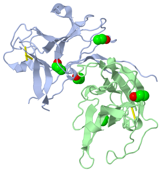
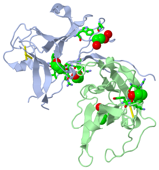
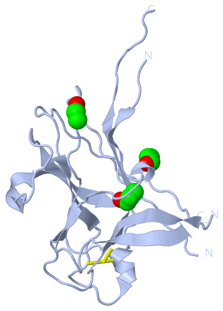
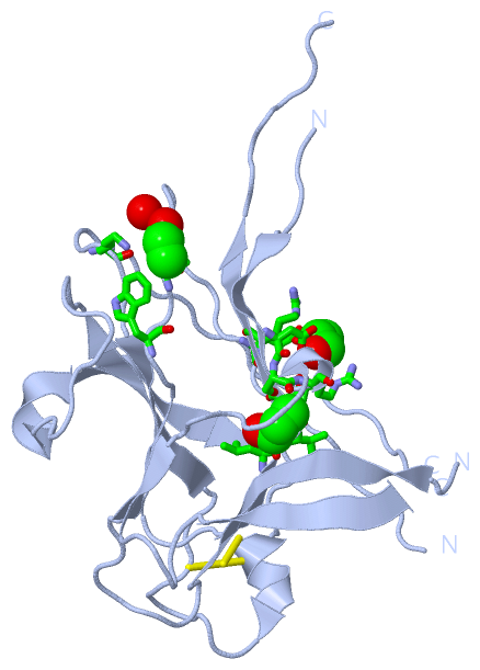
 Description