| molecular function |
|---|
| | GO:0034185 | | apolipoprotein binding | | Interacting selectively and non-covalently with an apolipoprotein, the protein component of a lipoprotein complex. |
| | GO:0016787 | | hydrolase activity | | Catalysis of the hydrolysis of various bonds, e.g. C-O, C-N, C-C, phosphoric anhydride bonds, etc. Hydrolase is the systematic name for any enzyme of EC class 3. |
| | GO:0008233 | | peptidase activity | | Catalysis of the hydrolysis of a peptide bond. A peptide bond is a covalent bond formed when the carbon atom from the carboxyl group of one amino acid shares electrons with the nitrogen atom from the amino group of a second amino acid. |
| | GO:0005515 | | protein binding | | Interacting selectively and non-covalently with any protein or protein complex (a complex of two or more proteins that may include other nonprotein molecules). |
| | GO:0019904 | | protein domain specific binding | | Interacting selectively and non-covalently with a specific domain of a protein. |
| | GO:0005102 | | receptor binding | | Interacting selectively and non-covalently with one or more specific sites on a receptor molecule, a macromolecule that undergoes combination with a hormone, neurotransmitter, drug or intracellular messenger to initiate a change in cell function. |
| | GO:0004252 | | serine-type endopeptidase activity | | Catalysis of the hydrolysis of internal, alpha-peptide bonds in a polypeptide chain by a catalytic mechanism that involves a catalytic triad consisting of a serine nucleophile that is activated by a proton relay involving an acidic residue (e.g. aspartate or glutamate) and a basic residue (usually histidine). |
| | GO:0008236 | | serine-type peptidase activity | | Catalysis of the hydrolysis of peptide bonds in a polypeptide chain by a catalytic mechanism that involves a catalytic triad consisting of a serine nucleophile that is activated by a proton relay involving an acidic residue (e.g. aspartate or glutamate) and a basic residue (usually histidine). |
| biological process |
|---|
| | GO:0007596 | | blood coagulation | | The sequential process in which the multiple coagulation factors of the blood interact, ultimately resulting in the formation of an insoluble fibrin clot; it may be divided into three stages: stage 1, the formation of intrinsic and extrinsic prothrombin converting principle; stage 2, the formation of thrombin; stage 3, the formation of stable fibrin polymers. |
| | GO:0044267 | | cellular protein metabolic process | | The chemical reactions and pathways involving a specific protein, rather than of proteins in general, occurring at the level of an individual cell. Includes cellular protein modification. |
| | GO:0022617 | | extracellular matrix disassembly | | A process that results in the breakdown of the extracellular matrix. |
| | GO:0042730 | | fibrinolysis | | A process that solubilizes fibrin in the bloodstream of a multicellular organism, chiefly by the proteolytic action of plasmin. |
| | GO:0007599 | | hemostasis | | The stopping of bleeding (loss of body fluid) or the arrest of the circulation to an organ or part. |
| | GO:0008285 | | negative regulation of cell proliferation | | Any process that stops, prevents or reduces the rate or extent of cell proliferation. |
| | GO:2000048 | | negative regulation of cell-cell adhesion mediated by cadherin | | Any process that stops, prevents, or reduces the frequency, rate or extent of cell-cell adhesion mediated by cadherin. |
| | GO:0010812 | | negative regulation of cell-substrate adhesion | | Any process that decreases the frequency, rate or extent of cell-substrate adhesion. Cell-substrate adhesion is the attachment of a cell to the underlying substrate via adhesion molecules. |
| | GO:0051918 | | negative regulation of fibrinolysis | | Any process that stops, prevents, or reduces the frequency, rate or extent of fibrinolysis, an ongoing process that solubilizes fibrin, resulting in the removal of small blood clots. |
| | GO:0002576 | | platelet degranulation | | The regulated exocytosis of secretory granules containing preformed mediators such as histamine and serotonin by a platelet. |
| | GO:0051919 | | positive regulation of fibrinolysis | | Any process that activates, maintains or increases the frequency, rate or extent of fibrinolysis, an ongoing process that solubilizes fibrin, resulting in the removal of small blood clots. |
| | GO:0006508 | | proteolysis | | The hydrolysis of proteins into smaller polypeptides and/or amino acids by cleavage of their peptide bonds. |
| | GO:0048771 | | tissue remodeling | | The reorganization or renovation of existing tissues. This process can either change the characteristics of a tissue such as in blood vessel remodeling, or result in the dynamic equilibrium of a tissue such as in bone remodeling. |
| cellular component |
|---|
| | GO:0072562 | | blood microparticle | | A phospholipid microvesicle that is derived from any of several cell types, such as platelets, blood cells, endothelial cells, or others, and contains membrane receptors as well as other proteins characteristic of the parental cell. Microparticles are heterogeneous in size, and are characterized as microvesicles free of nucleic acids. |
| | GO:0009986 | | cell surface | | The external part of the cell wall and/or plasma membrane. |
| | GO:0070062 | | extracellular exosome | | A vesicle that is released into the extracellular region by fusion of the limiting endosomal membrane of a multivesicular body with the plasma membrane. Extracellular exosomes, also simply called exosomes, have a diameter of about 40-100 nm. |
| | GO:0005576 | | extracellular region | | The space external to the outermost structure of a cell. For cells without external protective or external encapsulating structures this refers to space outside of the plasma membrane. This term covers the host cell environment outside an intracellular parasite. |
| | GO:0005615 | | extracellular space | | That part of a multicellular organism outside the cells proper, usually taken to be outside the plasma membranes, and occupied by fluid. |
| | GO:0031232 | | extrinsic component of external side of plasma membrane | | The component of a plasma membrane consisting of gene products and protein complexes that are loosely bound to its external surface, but not integrated into the hydrophobic region. |
| | GO:0044218 | | other organism cell membrane | | The cell membrane of a secondary organism with which the first organism is interacting. |
| | GO:0005886 | | plasma membrane | | The membrane surrounding a cell that separates the cell from its external environment. It consists of a phospholipid bilayer and associated proteins. |
| | GO:0031093 | | platelet alpha granule lumen | | The volume enclosed by the membrane of the platelet alpha granule. |
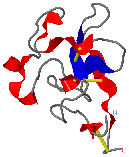
 Description
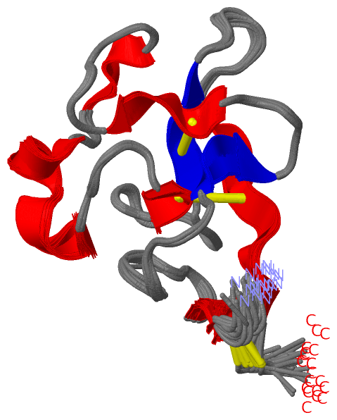