Show PDB file:   
         Plain Text   HTML   (compressed file size)
QuickSearch:   
by PDB,NDB,UniProt,PROSITE Code or Search Term(s)  
(-)Asym./Biol. Unit - manually
(-)Asym./Biol. Unit
(-)Asym./Biol. Unit - sites
collapse expand < >
Image Asym./Biol. Unit - manually
Asym./Biol. Unit - manually  (Jmol Viewer)
Image Asym./Biol. Unit
Asym./Biol. Unit  (Jmol Viewer)
Image Asym./Biol. Unit - sites
Asym./Biol. Unit - sites  (Jmol Viewer)

(-) Description

Title :  0.82A RESOLUTION CRYSTAL STRUCTURE OF ALPHA-LYTIC PROTEASE AT PH 5
 
Authors :  C. N. Fuhrmann, M. D. Daugherty, D. A. Agard
Date :  25 May 06  (Deposition) - 26 Sep 06  (Release) - 13 Jul 11  (Revision)
Method :  X-RAY DIFFRACTION
Resolution :  0.82
Chains :  Asym./Biol. Unit :  A
Keywords :  A-Lytic Protease, Serine Protease, Acylation Transition State, Catalysis, Protein Folding, Protein Stability, Packing Distortion, Hydrolase (Keyword Search: [Gene Ontology, PubMed, Web (Google))
 
Reference :  C. N. Fuhrmann, M. D. Daugherty, D. A. Agard
Subangstrom Crystallography Reveals That Short Ionic Hydrogen Bonds, And Not A His-Asp Low-Barrier Hydrogen Bond Stabilize The Transition State In Serine Protease Catalysis
J. Am. Chem. Soc. V. 128 9086 2006
PubMed-ID: 16834383  |  Reference-DOI: 10.1021/JA057721O

(-) Compounds

Molecule 1 - ALPHA-LYTIC PROTEASE
    ChainsA
    EC Number3.4.21.12
    FragmentMATURE PROTEASE DOMAIN (RESIDUES 200-397)
    Organism ScientificLYSOBACTER ENZYMOGENES
    Organism Taxid69
    Other DetailsGENE ALPHA-LP
    SecretionSECRETED BY THE NATIVE BACTERIUM
    SynonymALPHA-LYTIC ENDOPEPTIDASE

 Structural Features

(-) Chains, Units

  1
Asymmetric/Biological Unit A

Summary Information (see also Sequences/Alignments below)

(-) Ligands, Modified Residues, Ions  (2, 13)

Asymmetric/Biological Unit (2, 13)
No.NameCountTypeFull Name
1GOL2Ligand/IonGLYCEROL
2SO411Ligand/IonSULFATE ION

(-) Sites  (13, 13)

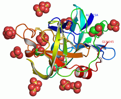
Asymmetric Unit (13, 13)
No.NameEvidenceResiduesDescription
01AC1SOFTWAREARG A:48A , ARG A:120A , GLN A:237 , TYR A:238 , HOH A:389 , HOH A:390 , HOH A:391 , HOH A:485 , HOH A:534 , HOH A:559 , HOH A:568 , HOH A:596 , HOH A:635 , HOH A:698 , HOH A:707 , HOH A:718 , HOH A:719BINDING SITE FOR RESIDUE SO4 A 246
02AC2SOFTWAREALA A:15A , ASN A:15B , ARG A:230 , PRO A:233 , HOH A:333 , HOH A:403 , HOH A:434 , HOH A:460 , HOH A:488 , HOH A:509 , HOH A:633 , HOH A:634 , HOH A:643BINDING SITE FOR RESIDUE SO4 A 247
03AC3SOFTWAREHIS A:57 , ARG A:122 , ARG A:192 , GLY A:193 , SER A:195 , GOL A:253 , HOH A:424 , HOH A:459 , HOH A:491 , HOH A:668 , HOH A:669 , HOH A:670 , HOH A:671 , HOH A:676BINDING SITE FOR RESIDUE SO4 A 248
04AC4SOFTWARETHR A:59A , VAL A:59B , ARG A:90 , HOH A:386 , HOH A:393 , HOH A:468 , HOH A:562 , HOH A:626 , HOH A:627 , HOH A:708BINDING SITE FOR RESIDUE SO4 A 203
05AC5SOFTWAREALA A:48C , THR A:49 , SER A:110 , ALA A:176 , ARG A:178 , HOH A:309 , HOH A:349 , HOH A:461 , HOH A:555 , HOH A:573 , HOH A:661 , HOH A:662 , HOH A:730BINDING SITE FOR RESIDUE SO4 A 204
06AC6SOFTWARETHR A:168 , ARG A:178 , HOH A:417 , HOH A:549 , HOH A:709 , HOH A:731BINDING SITE FOR RESIDUE SO4 A 205
07AC7SOFTWAREGLN A:219 , SER A:219A , HOH A:353 , HOH A:355 , HOH A:630 , HOH A:700BINDING SITE FOR RESIDUE SO4 A 206
08AC8SOFTWAREASN A:36 , THR A:62 , ARG A:65 , HOH A:365 , HOH A:599 , HOH A:701 , HOH A:702BINDING SITE FOR RESIDUE SO4 A 249
09AC9SOFTWAREALA A:131 , VAL A:132 , LYS A:165 , HOH A:566 , HOH A:646 , HOH A:703 , HOH A:711BINDING SITE FOR RESIDUE SO4 A 250
10BC1SOFTWAREARG A:141 , HOH A:314 , HOH A:380 , HOH A:432 , HOH A:527 , HOH A:528 , HOH A:529 , HOH A:548 , HOH A:567 , HOH A:590 , HOH A:611 , HOH A:623 , HOH A:665 , HOH A:674 , HOH A:712BINDING SITE FOR RESIDUE SO4 A 251
11BC2SOFTWAREASN A:217 , GLN A:219 , HOH A:569 , HOH A:679 , HOH A:720 , HOH A:721 , HOH A:722BINDING SITE FOR RESIDUE SO4 A 252
12BC3SOFTWAREHIS A:57 , ARG A:122 , TYR A:171 , GLU A:174 , ARG A:192 , SER A:214 , GLY A:215 , SO4 A:248 , HOH A:337 , HOH A:474 , HOH A:477 , HOH A:491 , HOH A:629 , HOH A:671 , HOH A:745BINDING SITE FOR RESIDUE GOL A 253
13BC4SOFTWAREPHE A:120I , VAL A:120J , THR A:120K , ALA A:173 , GLU A:174 , HOH A:489 , HOH A:531 , HOH A:532 , HOH A:576BINDING SITE FOR RESIDUE GOL A 254

(-) SS Bonds  (3, 3)

Asymmetric/Biological Unit
No.Residues
1A:42 -A:58
2A:137 -A:159
3A:189 -A:220A

(-) Cis Peptide Bonds  (1, 1)

Asymmetric/Biological Unit
No.Residues
1Phe A:94 -Pro A:95

 Sequence-Structure Mapping

(-) SAPs(SNPs)/Variants  (0, 0)

(no "SAP(SNP)/Variant" information available for 2H5C)

(-) PROSITE Motifs  (2, 2)

Asymmetric/Biological Unit (2, 2)
 PROSITEUniProtKBPDB
No.IDACDescriptionIDLocationCountLocation
1TRYPSIN_HISPS00134 Serine proteases, trypsin family, histidine active site.PRLA_LYSEN231-236  1A:53-58
2TRYPSIN_SERPS00135 Serine proteases, trypsin family, serine active site.PRLA_LYSEN336-347  1A:189-200

(-) Exons   (0, 0)

(no "Exon" information available for 2H5C)

(-) Sequences/Alignments

Asymmetric/Biological Unit
   Reformat: Number of residues per line =  ('0' or empty: single-line sequence representation)
  Number of residues per labelling interval =   
  UniProt sequence: complete  aligned part    
   Show mapping: SCOP domains CATH domains Pfam domains Secondary structure (by author)
SAPs(SNPs) PROSITE motifs Exons
(details for a mapped element are shown in a popup box when the mouse pointer rests over it)
Chain A from PDB  Type:PROTEIN  Length:198
 aligned with PRLA_LYSEN | P00778 from UniProtKB/Swiss-Prot  Length:397

    Alignment length:198
                                   209       219       229       239       249       259       269       279       289       299       309       319       329       339       349       359       369       379       389        
          PRLA_LYSEN    200 ANIVGGIEYSINNASLCSVGFSVTRGATKGFVTAGHCGTVNATARIGGAVVGTFAARVFPGNDRAWVSLTSAQTLLPRVANGSSFVTVRGSTEAAVGAAVCRSGRTTGYQCGTITAKNVTANYAEGAVRGLTQGNACMGRGDSGGSWITSAGQAQGVMSGGNVQSNGNNCGIPASQRSSLFERLQPILSQYGLSLVTG  397
               SCOP domains d2h5ca_ A: alpha-Lytic protease                                                                                                                                                                        SCOP domains
               CATH domains --2h5cA01 A:16-119,A:231-243 Trypsin-like serine proteases                  2h5cA02 A:120-230 Trypsin-like serine proteases                                                            2h5cA01      -- CATH domains
               Pfam domains ------------------------------------------------------------------------------------------------------------------------------------------------------------------------------------------------------ Pfam domains
         Sec.struct. author .eeee..eeee...eeee..eeeee..eeeeeehhhhh....eeee..eeeeeeeeee.....eeeeee....eeeeeeee..eeee...........eeeeee...eeeeeeeeeeeeeeee..eeeeeeeee...........eee.....eeeeeeee...........hhhhh.eeeeehhhhhhhhh.ee... Sec.struct. author
                 SAPs(SNPs) ------------------------------------------------------------------------------------------------------------------------------------------------------------------------------------------------------ SAPs(SNPs)
                    PROSITE -------------------------------TRYPSI---------------------------------------------------------------------------------------------------TRYPSIN_SER -------------------------------------------------- PROSITE
                 Transcript ------------------------------------------------------------------------------------------------------------------------------------------------------------------------------------------------------ Transcript
                2h5c A  15A ANIVGGIEYSINNASLCSVGFSVTRGATKGFVTAGHCGTVNATARIGGAVVGTFAARVFPGNDRAWVSLTSAQTLLPRVANGSSFVTVRGSTEAAVGAAVCRSGRTTGYQCGTITAKNVTANYAEGAVRGLTQGNACMGRGDSGGSWITSAGQAQGVMSGGNVQSNGNNCGIPASQRSSLFERLQPILSQYGLSLVTG  245
                            ||   || 34 ||    44A    ||| 51       59B  ||  ||83 || |  |95|      109    ||120C|||||||123 ||    136      |158       168  ||   179     ||192      201A||     215    ||220A  |||  227       237        
                            ||  19|   36|    44A  48A||         59A| 62| 67|  85| | 91|||           114| ||||120I||  125|           143|            171|         185|         201A||         219A||| |  |||                       
                          15A|   31    38          48B|          59B  64  81   87 |  94||            119 |||||120J|   129            156             173          189           202|          219B|| |  |||                       
                           15B                      48C                         88A   95|             120A|||||120K                                                              207           219C| |  |||                       
                                                                                      100              120B|||||                                                                                219D |  |||                       
                                                                                                        120C||||                                                                                  220A  |||                       
                                                                                                         120D|||                                                                                     222A||                       
                                                                                                          120E||                                                                                      222B|                       
                                                                                                           120G|                                                                                       222C                       
                                                                                                            120H                                                                                                                  

   Legend:   → Mismatch (orange background)
  - → Gap (green background, '-', border residues have a numbering label)
    → Modified Residue (blue background, lower-case, 'x' indicates undefined single-letter code, labelled with number + name)
  x → Chemical Group (purple background, 'x', labelled with number + name, e.g. ACE or NH2)
  extra numbering lines below/above indicate numbering irregularities and modified residue names etc., number ends below/above '|'

 Classification and Annotation

(-) SCOP Domains  (1, 1)

Asymmetric/Biological Unit

(-) CATH Domains  (1, 2)

Asymmetric/Biological Unit
(-)
Class: Mainly Beta (13760)

(-) Pfam Domains  (0, 0)

(no "Pfam Domain" information available for 2H5C)

(-) Gene Ontology  (6, 6)

Asymmetric/Biological Unit(hide GO term definitions)
Chain A   (PRLA_LYSEN | P00778)
molecular function
    GO:0016787    hydrolase activity    Catalysis of the hydrolysis of various bonds, e.g. C-O, C-N, C-C, phosphoric anhydride bonds, etc. Hydrolase is the systematic name for any enzyme of EC class 3.
    GO:0008233    peptidase activity    Catalysis of the hydrolysis of a peptide bond. A peptide bond is a covalent bond formed when the carbon atom from the carboxyl group of one amino acid shares electrons with the nitrogen atom from the amino group of a second amino acid.
    GO:0004252    serine-type endopeptidase activity    Catalysis of the hydrolysis of internal, alpha-peptide bonds in a polypeptide chain by a catalytic mechanism that involves a catalytic triad consisting of a serine nucleophile that is activated by a proton relay involving an acidic residue (e.g. aspartate or glutamate) and a basic residue (usually histidine).
    GO:0008236    serine-type peptidase activity    Catalysis of the hydrolysis of peptide bonds in a polypeptide chain by a catalytic mechanism that involves a catalytic triad consisting of a serine nucleophile that is activated by a proton relay involving an acidic residue (e.g. aspartate or glutamate) and a basic residue (usually histidine).
biological process
    GO:0006508    proteolysis    The hydrolysis of proteins into smaller polypeptides and/or amino acids by cleavage of their peptide bonds.
cellular component
    GO:0005576    extracellular region    The space external to the outermost structure of a cell. For cells without external protective or external encapsulating structures this refers to space outside of the plasma membrane. This term covers the host cell environment outside an intracellular parasite.

 Visualization

(-) Interactive Views

Asymmetric/Biological Unit
  Complete Structure
    Jena3D(integrated viewing of ligand, site, SAP, PROSITE, SCOP information)
    WebMol | AstexViewer[tm]@PDBe
(Java Applets, require no local installation except for Java; loading may be slow)
    STRAP
(Java WebStart application, automatic local installation, requires Java; full application with system access!)
    RasMol
(require local installation)
    Molscript (VRML)
(requires installation of a VRML viewer; select preferred view via VRML and generate a mono or stereo PDF format file)
 
  Ligands, Modified Residues, Ions
    GOL  [ RasMol | Jena3D ]  +environment [ RasMol | Jena3D ]
    SO4  [ RasMol | Jena3D ]  +environment [ RasMol | Jena3D ]
 
  Sites
    AC1  [ RasMol ]  +environment [ RasMol ]
    AC2  [ RasMol ]  +environment [ RasMol ]
    AC3  [ RasMol ]  +environment [ RasMol ]
    AC4  [ RasMol ]  +environment [ RasMol ]
    AC5  [ RasMol ]  +environment [ RasMol ]
    AC6  [ RasMol ]  +environment [ RasMol ]
    AC7  [ RasMol ]  +environment [ RasMol ]
    AC8  [ RasMol ]  +environment [ RasMol ]
    AC9  [ RasMol ]  +environment [ RasMol ]
    BC1  [ RasMol ]  +environment [ RasMol ]
    BC2  [ RasMol ]  +environment [ RasMol ]
    BC3  [ RasMol ]  +environment [ RasMol ]
    BC4  [ RasMol ]  +environment [ RasMol ]
 
  Cis Peptide Bonds
    Phe A:94 - Pro A:95   [ RasMol ]  
 

(-) Still Images

Jmol
  protein: cartoon or spacefill or dots and stick; nucleic acid: cartoon and stick; ligands: spacefill; active site: stick
Molscript
  protein, nucleic acid: cartoon; ligands: spacefill; active site: ball and stick

 Databases and Analysis Tools

(-) Databases

Access by PDB/NDB ID
  2h5c
    Family and Domain InformationProDom | SYSTERS
    General Structural InformationGlycoscienceDB | MMDB | NDB | OCA | PDB | PDBe | PDBj | PDBsum | PDBWiki | PQS | PROTEOPEDIA
    Orientation in MembranesOPM
    Protein SurfaceSURFACE
    Secondary StructureDSSP (structure derived) | HSSP (homology derived)
    Structural GenomicsGeneCensus
    Structural NeighboursCE | VAST
    Structure ClassificationCATH | Dali | SCOP
    Validation and Original DataBMRB Data View | BMRB Restraints Grid | EDS | PROCHECK | RECOORD | WHAT_CHECK
 
Access by UniProt ID/Accession number
  PRLA_LYSEN | P00778
    Comparative Protein Structure ModelsModBase
    Genomic InformationEnsembl
    Protein-protein InteractionDIP
    Sequence, Family and Domain InformationInterPro | Pfam | SMART | UniProtKB/SwissProt
 
Access by Enzyme Classificator   (EC Number)
  3.4.21.12
    General Enzyme InformationBRENDA | EC-PDB | Enzyme | IntEnz
    PathwayKEGG | MetaCyc
 
Access by Disease Identifier   (MIM ID)
  (no 'MIM ID' available)
    Disease InformationOMIM
 
Access by GenAge ID
  (no 'GenAge ID' available)
    Age Related InformationGenAge

(-) Analysis Tools

Access by PDB/NDB ID
    Domain InformationXDom
    Interatomic Contacts of Structural UnitsCSU
    Ligand-protein ContactsLPC
    Protein CavitiescastP
    Sequence and Secondary StructurePDBCartoon
    Structure AlignmentSTRAP(Java WebStart application, automatic local installation, requires Java; full application with system access!)
    Structure and Sequence BrowserSTING
 
Access by UniProt ID/Accession number
  PRLA_LYSEN | P00778
    Protein Disorder PredictionDisEMBL | FoldIndex | GLOBPLOT (for more information see DisProt)

 Related Entries

(-) Entries Sharing at Least One Protein Chain (UniProt ID)

UniProtKB/Swiss-Prot
        PRLA_LYSEN | P007781boq 1gba 1gbb 1gbc 1gbd 1gbe 1gbf 1gbh 1gbi 1gbj 1gbk 1gbl 1gbm 1p01 1p02 1p03 1p04 1p05 1p06 1p09 1p10 1p11 1p12 1qq4 1qrw 1qrx 1ssx 1tal 2alp 2h5d 2lpr 2pro 2ull 3lpr 3m7t 3m7u 3pro 3qgj 3urc 3urd 3ure 4pro 5lpr 6lpr 7lpr 8lpr 9lpr

(-) Related Entries Specified in the PDB File

1qrx SAME PROTEIN AT PH 5.14 (1.6A RESOLUTION)
1ssx SAME PROTEIN AT PH 8 (0.83A RESOLUTION)
1tal SAME PROTEIN AT PH 8 (1.5A RESOLUTION)
2h5d
2ull DATA FROM 1TAL WERE MODELED HERE WITH 16 CONFORMATIONS OF THE ENTIRE PROTEIN