|
|
|
|
 Description
|
|
Compounds
|
||||||||||||||||||||||||||||||||||||||||||||||||
Chains, Units
Summary Information (see also Sequences/Alignments below) |
Ligands, Modified Residues, Ions (0, 0)| (no "Ligand,Modified Residues,Ions" information available for 2FO8) |
Sites (0, 0)| (no "Site" information available for 2FO8) |
SS Bonds (0, 0)| (no "SS Bond" information available for 2FO8) |
Cis Peptide Bonds (0, 0)| (no "Cis Peptide Bond" information available for 2FO8) |
SAPs(SNPs)/Variants (0, 0)| (no "SAP(SNP)/Variant" information available for 2FO8) |
PROSITE Motifs (0, 0)| (no "PROSITE Motif" information available for 2FO8) |
Exons (0, 0)| (no "Exon" information available for 2FO8) |
Sequences/Alignments
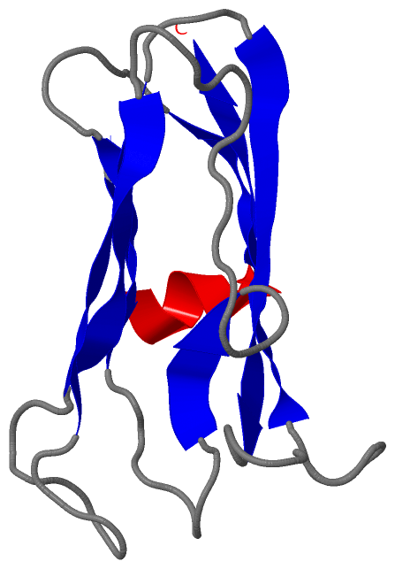
NMR StructureChain A from PDB Type:PROTEIN Length:108 aligned with CHAG_TRYCR | Q966X9 from UniProtKB/Swiss-Prot Length:110 Alignment length:108 12 22 32 42 52 62 72 82 92 102 CHAG_TRYCR 3 HKVTKAHNGATLTVAVGELVEIQLPSNPTTGFAWYFEGGTKESPNESMFTVENKYFPPDSKLLGAGGTEHFHVTVKAAGTHAVNLTYMRPWTGPSHDSERFTVYLKAN 110 SCOP domains d2fo8a1 A:3-110 Chagasin SCOP domains CATH domains ------------------------------------------------------------------------------------------------------------ CATH domains Pfam domains ------------------------------------------------------------------------------------------------------------ Pfam domains SAPs(SNPs) ------------------------------------------------------------------------------------------------------------ SAPs(SNPs) PROSITE ------------------------------------------------------------------------------------------------------------ PROSITE Transcript ------------------------------------------------------------------------------------------------------------ Transcript 2fo8 A 3 HKVTKAHNGATLTVAVGELVEIQLPSNPTTGFAWYFEGGTKESPNESMFTVENKYFPPDSKLLGAGGTEHFHVTVKAAGTHAVNLTYMRPWTGPSHDSERFTVYLKAN 110 12 22 32 42 52 62 72 82 92 102
|
||||||||||||||||||||
SCOP Domains (1, 1)
NMR Structure

|
CATH Domains (0, 0)| (no "CATH Domain" information available for 2FO8) |
Pfam Domains (0, 0)| (no "Pfam Domain" information available for 2FO8) |
Gene Ontology (7, 7)|
NMR Structure(hide GO term definitions) Chain A (CHAG_TRYCR | Q966X9)
|
||||||||||||||||||||||||||||||||||||||||||||||||||||||||||||
Interactive Views
|
||||||||||||||||||||||||||||||||||||||||||||||||||||||||||||||||||||||||||||||||||||||||||||||||||||||||||||||||||||
Still Images
|
||||||||||||||||
Databases
|
||||||||||||||||||||||||||||||||||||||||||||||||||||||||||||||||||||||||||||||||||||||||||||||||||||||||||||||||||||||||||||||||||||||||||||||||||||||||||||||||
Analysis Tools
|
|||||||||||||||||||||||||||||||||||||||||||||||||||||||||||||
Entries Sharing at Least One Protein Chain (UniProt ID)
Related Entries Specified in the PDB File
|
|