Show PDB file:   
         Plain Text   HTML   (compressed file size)
QuickSearch:   
by PDB,NDB,UniProt,PROSITE Code or Search Term(s)  
(-)Asym.Unit - manually
(-)Asymmetric Unit
(-)Biological Unit 1
(-)Biological Unit 2
collapse expand < >
Image Asym.Unit - manually
Asym.Unit - manually  (Jmol Viewer)
Image Asymmetric Unit
Asymmetric Unit  (Jmol Viewer)
Image Biological Unit 1
Biological Unit 1  (Jmol Viewer)
Image Biological Unit 2
Biological Unit 2  (Jmol Viewer)

(-) Description

Title :  CRYSTAL STRUCTURE OF CHAGASIN, THE ENDOGENOUS CYSTEINE-PROTEASE INHIBITOR FROM TRYPANOSOMA CRUZI
 
Authors :  A. A. Figueiredo Da Silva, B. G. Guimaraes
Date :  05 Jun 06  (Deposition) - 10 Oct 06  (Release) - 24 Feb 09  (Revision)
Method :  X-RAY DIFFRACTION
Resolution :  1.70
Chains :  Asym. Unit :  A,B
Biol. Unit 1:  A  (1x)
Biol. Unit 2:  B  (1x)
Keywords :  Beta-Strands, Immunoglobulin-Fold, Hydrolase Inhibitor (Keyword Search: [Gene Ontology, PubMed, Web (Google))
 
Reference :  A. A. Figueiredo Da Silva, L. C. Vieira, M. A. Krieger, S. Goldenberg, N. I. Zanchin, B. G. Guimaraes
Crystal Structure Of Chagasin, The Endogenous Cysteine-Protease Inhibitor From Trypanosoma Cruzi.
J. Struct. Biol. V. 157 416 2007
PubMed-ID: 17011790  |  Reference-DOI: 10.1016/J.JSB.2006.07.017
(for further references see the PDB file header)

(-) Compounds

Molecule 1 - CHAGASIN
    ChainsA, B
    EngineeredYES
    Expression SystemESCHERICHIA COLI
    Expression System PlasmidPET
    Expression System StrainBL21(DE3) SLYD-
    Expression System Taxid562
    Expression System Vector TypePLASMID
    GeneCHA
    Organism ScientificTRYPANOSOMA CRUZI
    Organism Taxid5693
    StrainDM28C

 Structural Features

(-) Chains, Units

  12
Asymmetric Unit AB
Biological Unit 1 (1x)A 
Biological Unit 2 (1x) B

Summary Information (see also Sequences/Alignments below)

(-) Ligands, Modified Residues, Ions  (0, 0)

(no "Ligand,Modified Residues,Ions" information available for 2H7W)

(-) Sites  (0, 0)

(no "Site" information available for 2H7W)

(-) SS Bonds  (0, 0)

(no "SS Bond" information available for 2H7W)

(-) Cis Peptide Bonds  (2, 2)
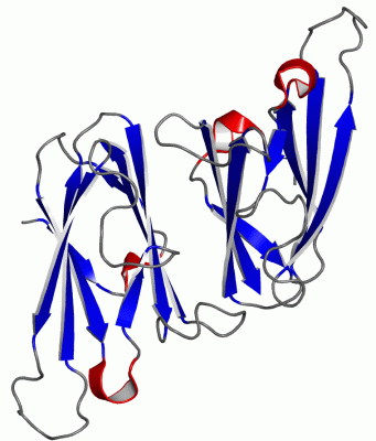
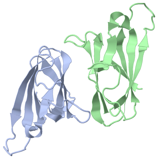
Asymmetric Unit
No.Residues
1Glu B:39 -Gly B:40
2Gly B:40 -Gly B:41

 Sequence-Structure Mapping

(-) SAPs(SNPs)/Variants  (0, 0)

(no "SAP(SNP)/Variant" information available for 2H7W)

(-) PROSITE Motifs  (0, 0)

(no "PROSITE Motif" information available for 2H7W)

(-) Exons   (0, 0)

(no "Exon" information available for 2H7W)

(-) Sequences/Alignments

Asymmetric Unit
   Reformat: Number of residues per line =  ('0' or empty: single-line sequence representation)
  Number of residues per labelling interval =   
  UniProt sequence: complete  aligned part    
   Show mapping: SCOP domains CATH domains Pfam domains Secondary structure (by author)
SAPs(SNPs) PROSITE motifs Exons
(details for a mapped element are shown in a popup box when the mouse pointer rests over it)
Chain A from PDB  Type:PROTEIN  Length:108
 aligned with CHAG_TRYCR | Q966X9 from UniProtKB/Swiss-Prot  Length:110

    Alignment length:108
                                    12        22        32        42        52        62        72        82        92       102        
           CHAG_TRYCR     3 HKVTKAHNGATLTVAVGELVEIQLPSNPTTGFAWYFEGGTKESPNESMFTVENKYFPPDSKLLGAGGTEHFHVTVKAAGTHAVNLTYMRPWTGPSHDSERFTVYLKAN 110
               SCOP domains d2h7wa1 A:3-110 Chagasin                                                                                     SCOP domains
               CATH domains ------------------------------------------------------------------------------------------------------------ CATH domains
               Pfam domains ------------------------------------------------------------------------------------------------------------ Pfam domains
         Sec.struct. author .eehhhhh..eeeee...eeeeeeeehhhhh.eee.............eeeeeeee..........eeeeeeeee...eeeeeeeeee..........eeeeeeeeee Sec.struct. author
                 SAPs(SNPs) ------------------------------------------------------------------------------------------------------------ SAPs(SNPs)
                    PROSITE ------------------------------------------------------------------------------------------------------------ PROSITE
                 Transcript ------------------------------------------------------------------------------------------------------------ Transcript
                 2h7w A   3 HKVTKAHNGATLTVAVGELVEIQLPSNPTTGFAWYFEGGTKESPNESMFTVENKYFPPDSKLLGAGGTEHFHVTVKAAGTHAVNLTYMRPWTGPSHDSERFIVYLKAN 110
                                    12        22        32        42        52        62        72        82        92       102        

Chain B from PDB  Type:PROTEIN  Length:108
 aligned with CHAG_TRYCR | Q966X9 from UniProtKB/Swiss-Prot  Length:110

    Alignment length:108
                                    11        21        31        41        51        61        71        81        91       101        
           CHAG_TRYCR     2 SHKVTKAHNGATLTVAVGELVEIQLPSNPTTGFAWYFEGGTKESPNESMFTVENKYFPPDSKLLGAGGTEHFHVTVKAAGTHAVNLTYMRPWTGPSHDSERFTVYLKA 109
               SCOP domains d2h7wb_ B: Chagasin                                                                                          SCOP domains
               CATH domains ------------------------------------------------------------------------------------------------------------ CATH domains
               Pfam domains ------------------------------------------------------------------------------------------------------------ Pfam domains
         Sec.struct. author .eeee.hhh..eee.....eeeeeeeehhhhh.eeee............eeeeeeee..........eeeeeeeee...eeeeeeeeee..........eeeeeeeee Sec.struct. author
                 SAPs(SNPs) ------------------------------------------------------------------------------------------------------------ SAPs(SNPs)
                    PROSITE ------------------------------------------------------------------------------------------------------------ PROSITE
                 Transcript ------------------------------------------------------------------------------------------------------------ Transcript
                 2h7w B   2 SHKVTKAHNGATLTVAVGELVEIQLPSNPTTGFAWYFEGGTKESPNESMFTVENKYFPPDSKLLGAGGTEHFHVTVKAAGTHAVNLTYMRPWTGPSHDSERFIVYLKA 109
                                    11        21        31        41        51        61        71        81        91       101        

   Legend:   → Mismatch (orange background)
  - → Gap (green background, '-', border residues have a numbering label)
    → Modified Residue (blue background, lower-case, 'x' indicates undefined single-letter code, labelled with number + name)
  x → Chemical Group (purple background, 'x', labelled with number + name, e.g. ACE or NH2)
  extra numbering lines below/above indicate numbering irregularities and modified residue names etc., number ends below/above '|'

 Classification and Annotation

(-) SCOP Domains  (1, 2)

Asymmetric Unit

(-) CATH Domains  (0, 0)

(no "CATH Domain" information available for 2H7W)

(-) Pfam Domains  (0, 0)

(no "Pfam Domain" information available for 2H7W)

(-) Gene Ontology  (7, 7)

Asymmetric Unit(hide GO term definitions)
Chain A,B   (CHAG_TRYCR | Q966X9)
molecular function
    GO:0004869    cysteine-type endopeptidase inhibitor activity    Stops, prevents or reduces the activity of a cysteine-type endopeptidase, any enzyme that hydrolyzes peptide bonds in polypeptides by a mechanism in which the sulfhydryl group of a cysteine residue at the active center acts as a nucleophile.
    GO:0030414    peptidase inhibitor activity    Stops, prevents or reduces the activity of a peptidase, any enzyme that catalyzes the hydrolysis peptide bonds.
biological process
    GO:0010951    negative regulation of endopeptidase activity    Any process that decreases the frequency, rate or extent of endopeptidase activity, the endohydrolysis of peptide bonds within proteins.
    GO:0010466    negative regulation of peptidase activity    Any process that stops or reduces the rate of peptidase activity, the hydrolysis of peptide bonds within proteins.
cellular component
    GO:0009986    cell surface    The external part of the cell wall and/or plasma membrane.
    GO:0020016    ciliary pocket    Invagination of the plasma membrane from which a cilium (also called flagellum) protrudes.
    GO:0031410    cytoplasmic vesicle    A vesicle found in the cytoplasm of a cell.

 Visualization

(-) Interactive Views

Asymmetric Unit
  Complete Structure
    Jena3D(integrated viewing of ligand, site, SAP, PROSITE, SCOP information)
    WebMol | AstexViewer[tm]@PDBe
(Java Applets, require no local installation except for Java; loading may be slow)
    STRAP
(Java WebStart application, automatic local installation, requires Java; full application with system access!)
    RasMol
(require local installation)
    Molscript (VRML)
(requires installation of a VRML viewer; select preferred view via VRML and generate a mono or stereo PDF format file)
 
  Ligands, Modified Residues, Ions
(no "Ligands, Modified Residues, Ions" information available for 2h7w)
 
  Sites
(no "Sites" information available for 2h7w)
 
  Cis Peptide Bonds
    Glu B:39 - Gly B:40   [ RasMol ]  
    Gly B:40 - Gly B:41   [ RasMol ]  
 
Biological Units
  Complete Structure
    Biological Unit 1  [ Jena3D ]
    Biological Unit 2  [ Jena3D ]

(-) Still Images

Jmol
  protein: cartoon or spacefill or dots and stick; nucleic acid: cartoon and stick; ligands: spacefill; active site: stick
Molscript
  protein, nucleic acid: cartoon; ligands: spacefill; active site: ball and stick

 Databases and Analysis Tools

(-) Databases

Access by PDB/NDB ID
  2h7w
    Family and Domain InformationProDom | SYSTERS
    General Structural InformationGlycoscienceDB | MMDB | NDB | OCA | PDB | PDBe | PDBj | PDBsum | PDBWiki | PQS | PROTEOPEDIA
    Orientation in MembranesOPM
    Protein SurfaceSURFACE
    Secondary StructureDSSP (structure derived) | HSSP (homology derived)
    Structural GenomicsGeneCensus
    Structural NeighboursCE | VAST
    Structure ClassificationCATH | Dali | SCOP
    Validation and Original DataBMRB Data View | BMRB Restraints Grid | EDS | PROCHECK | RECOORD | WHAT_CHECK
 
Access by UniProt ID/Accession number
  CHAG_TRYCR | Q966X9
    Comparative Protein Structure ModelsModBase
    Genomic InformationEnsembl
    Protein-protein InteractionDIP
    Sequence, Family and Domain InformationInterPro | Pfam | SMART | UniProtKB/SwissProt
 
Access by Enzyme Classificator   (EC Number)
  (no 'Enzyme Classificator' available)
    General Enzyme InformationBRENDA | EC-PDB | Enzyme | IntEnz
    PathwayKEGG | MetaCyc
 
Access by Disease Identifier   (MIM ID)
  (no 'MIM ID' available)
    Disease InformationOMIM
 
Access by GenAge ID
  (no 'GenAge ID' available)
    Age Related InformationGenAge

(-) Analysis Tools

Access by PDB/NDB ID
    Domain InformationXDom
    Interatomic Contacts of Structural UnitsCSU
    Ligand-protein ContactsLPC
    Protein CavitiescastP
    Sequence and Secondary StructurePDBCartoon
    Structure AlignmentSTRAP(Java WebStart application, automatic local installation, requires Java; full application with system access!)
    Structure and Sequence BrowserSTING
 
Access by UniProt ID/Accession number
  CHAG_TRYCR | Q966X9
    Protein Disorder PredictionDisEMBL | FoldIndex | GLOBPLOT (for more information see DisProt)

 Related Entries

(-) Entries Sharing at Least One Protein Chain (UniProt ID)

UniProtKB/Swiss-Prot
        CHAG_TRYCR | Q966X92fo8 2nnr 2nqd 2oul 3cbj 3cbk 3e1z

(-) Related Entries Specified in the PDB File

(no "Related Entries Specified in the PDB File" available for 2H7W)