Show PDB file:   
         Plain Text   HTML   (compressed file size)
QuickSearch:   
by PDB,NDB,UniProt,PROSITE Code or Search Term(s)  
(-)Asymmetric Unit
(-)Asym. Unit - sites
(-)Biological Unit 1
(-)Biol. Unit 1 - sites
(-)Biological Unit 2
collapse expand < >
Image Asymmetric Unit
Asymmetric Unit  (Jmol Viewer)
Image Asym. Unit - sites
Asym. Unit - sites  (Jmol Viewer)
Image Biological Unit 1
Biological Unit 1  (Jmol Viewer)
Image Biol. Unit 1 - sites
Biol. Unit 1 - sites  (Jmol Viewer)
Image Biological Unit 2
Biological Unit 2  (Jmol Viewer)

(-) Description

Title :  CRYSTAL STRUCTURE OF CHAGASIN, CYSTEINE PROTEASE INHIBITOR FROM TRYPANOSOMA CRUZI
 
Authors :  I. Redzynia, G. Bujacz, A. Ljunggren, M. Jaskolski, M. Abrahamson
Date :  24 Oct 06  (Deposition) - 24 Jul 07  (Release) - 13 Jul 11  (Revision)
Method :  X-RAY DIFFRACTION
Resolution :  1.70
Chains :  Asym. Unit :  A,B
Biol. Unit 1:  A  (1x)
Biol. Unit 2:  B  (1x)
Keywords :  Chagasin, Deformed Jelly Roll, Predominately Beta Structure, Hydrolase Inhibitor (Keyword Search: [Gene Ontology, PubMed, Web (Google))
 
Reference :  A. Ljunggren, I. Redzynia, M. Alvarez-Fernandez, M. Abrahamson, J. S. Mort, J. C. Krupa, M. Jaskolski, G. Bujacz
Crystal Structure Of The Parasite Protease Inhibitor Chagasin In Complex With A Host Target Cysteine Protease
J. Mol. Biol. V. 371 137 2007
PubMed-ID: 17561110  |  Reference-DOI: 10.1016/J.JMB.2007.05.005

(-) Compounds

Molecule 1 - CHAGASIN
    ChainsA, B
    EngineeredYES
    Expression SystemESCHERICHIA COLI BL21
    Expression System PlasmidPGEX-6P-1
    Expression System StrainBL21
    Expression System Taxid511693
    Expression System Vector TypePLASMID
    Organism ScientificTRYPANOSOMA CRUZI
    Organism Taxid5693
    SynonymCYSTEINE PROTEASE INHIBITOR

 Structural Features

(-) Chains, Units

  12
Asymmetric Unit AB
Biological Unit 1 (1x)A 
Biological Unit 2 (1x) B

Summary Information (see also Sequences/Alignments below)

(-) Ligands, Modified Residues, Ions  (3, 9)

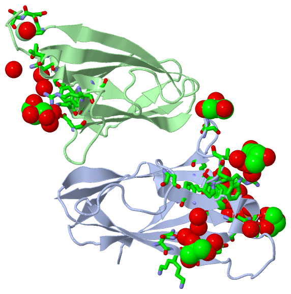
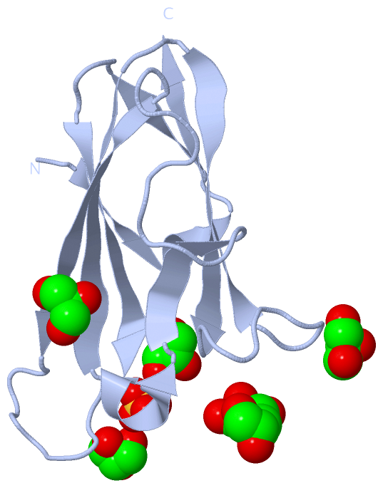
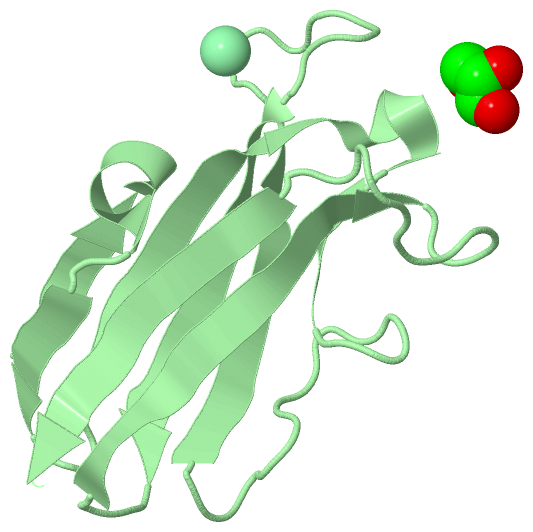
Asymmetric Unit (3, 9)
No.NameCountTypeFull Name
1CL1Ligand/IonCHLORIDE ION
2GOL7Ligand/IonGLYCEROL
3SO41Ligand/IonSULFATE ION
Biological Unit 1 (2, 7)
No.NameCountTypeFull Name
1CL-1Ligand/IonCHLORIDE ION
2GOL6Ligand/IonGLYCEROL
3SO41Ligand/IonSULFATE ION
Biological Unit 2 (1, 1)
No.NameCountTypeFull Name
1CL-1Ligand/IonCHLORIDE ION
2GOL1Ligand/IonGLYCEROL
3SO4-1Ligand/IonSULFATE ION

(-) Sites  (9, 9)

Asymmetric Unit (9, 9)
No.NameEvidenceResiduesDescription
1AC1SOFTWAREASN A:29 , GLY A:68 , GOL A:3001 , GOL A:3002 , HOH A:3052 , HOH A:3074 , HOH A:3107 , ASN B:29 , HOH B:3010 , HOH B:3094BINDING SITE FOR RESIDUE SO4 A 1001
2AC2SOFTWAREPRO B:60 , ASP B:61 , SER B:62BINDING SITE FOR RESIDUE CL B 2001
3AC3SOFTWARESO4 A:1001 , LYS B:7 , PRO B:27 , TRP B:36 , TYR B:89 , GLU B:101 , HOH B:3017 , HOH B:3094BINDING SITE FOR RESIDUE GOL A 3001
4AC4SOFTWARELYS A:7 , PRO A:27 , TRP A:36 , TYR A:89 , GLU A:101 , SO4 A:1001 , HOH A:3025 , HOH A:3107 , HOH B:3012BINDING SITE FOR RESIDUE GOL A 3002
5AC5SOFTWAREARG A:91 , TRP A:93 , THR A:94 , GOL A:3004BINDING SITE FOR RESIDUE GOL A 3003
6AC6SOFTWAREARG A:91 , TRP A:93 , GOL A:3003 , HOH A:3082 , THR B:31 , HOH B:3074BINDING SITE FOR RESIDUE GOL A 3004
7AC7SOFTWARESER A:97 , HIS A:98BINDING SITE FOR RESIDUE GOL A 3005
8AC8SOFTWAREASN A:55 , LYS A:56 , TYR A:57 , HOH A:3024 , HOH A:3041 , HOH A:3060BINDING SITE FOR RESIDUE GOL A 3006
9AC9SOFTWARETHR A:31 , GLY A:41 , PRO A:92 , HOH A:3034 , HOH A:3035 , ARG B:91 , HOH B:3078BINDING SITE FOR RESIDUE GOL B 3007

(-) SS Bonds  (0, 0)

(no "SS Bond" information available for 2NNR)

(-) Cis Peptide Bonds  (0, 0)

(no "Cis Peptide Bond" information available for 2NNR)

 Sequence-Structure Mapping

(-) SAPs(SNPs)/Variants  (0, 0)

(no "SAP(SNP)/Variant" information available for 2NNR)

(-) PROSITE Motifs  (0, 0)

(no "PROSITE Motif" information available for 2NNR)

(-) Exons   (0, 0)

(no "Exon" information available for 2NNR)

(-) Sequences/Alignments

Asymmetric Unit
   Reformat: Number of residues per line =  ('0' or empty: single-line sequence representation)
  Number of residues per labelling interval =   
  UniProt sequence: complete  aligned part    
   Show mapping: SCOP domains CATH domains Pfam domains Secondary structure (by author)
SAPs(SNPs) PROSITE motifs Exons
(details for a mapped element are shown in a popup box when the mouse pointer rests over it)
Chain A from PDB  Type:PROTEIN  Length:110
 aligned with CHAG_TRYCR | Q966X9 from UniProtKB/Swiss-Prot  Length:110

    Alignment length:110
                                    10        20        30        40        50        60        70        80        90       100       110
           CHAG_TRYCR     1 MSHKVTKAHNGATLTVAVGELVEIQLPSNPTTGFAWYFEGGTKESPNESMFTVENKYFPPDSKLLGAGGTEHFHVTVKAAGTHAVNLTYMRPWTGPSHDSERFTVYLKAN 110
               SCOP domains d2nnra_ A: Chagasin                                                                                            SCOP domains
               CATH domains -------------------------------------------------------------------------------------------------------------- CATH domains
               Pfam domains -------------------------------------------------------------------------------------------------------------- Pfam domains
         Sec.struct. author ..eeee.hhh..eeee....eeeeeeeehhhhh.eeee............eeeeeeee..........eeeeeeeee...eeeeeeeeee..........eeeeeeeee. Sec.struct. author
                 SAPs(SNPs) -------------------------------------------------------------------------------------------------------------- SAPs(SNPs)
                    PROSITE -------------------------------------------------------------------------------------------------------------- PROSITE
                 Transcript -------------------------------------------------------------------------------------------------------------- Transcript
                 2nnr A   1 MSHKVTKAHNGATLTVAVGELVEIQLPSNPTTGFAWYFEGGTKESPNESMFTVENKYFPPDSKLLGAGGTEHFHVTVKAAGTHAVNLTYMRPWTGPSHDSERFTVYLKAN 110
                                    10        20        30        40        50        60        70        80        90       100       110

Chain B from PDB  Type:PROTEIN  Length:110
 aligned with CHAG_TRYCR | Q966X9 from UniProtKB/Swiss-Prot  Length:110

    Alignment length:110
                                    10        20        30        40        50        60        70        80        90       100       110
           CHAG_TRYCR     1 MSHKVTKAHNGATLTVAVGELVEIQLPSNPTTGFAWYFEGGTKESPNESMFTVENKYFPPDSKLLGAGGTEHFHVTVKAAGTHAVNLTYMRPWTGPSHDSERFTVYLKAN 110
               SCOP domains d2nnrb_ B: Chagasin                                                                                            SCOP domains
               CATH domains -------------------------------------------------------------------------------------------------------------- CATH domains
           Pfam domains (1) -------------Inhibitor_I42-2nnrB01 B:14-109                                                                  - Pfam domains (1)
           Pfam domains (2) -------------Inhibitor_I42-2nnrB02 B:14-109                                                                  - Pfam domains (2)
         Sec.struct. author ..eeehhhhh..eeee....eeeeeeeehhhhh.eee.............eeeeeeee..........eeeeeeeee...eeeeeeeeee...........eeeeeeee. Sec.struct. author
                 SAPs(SNPs) -------------------------------------------------------------------------------------------------------------- SAPs(SNPs)
                    PROSITE -------------------------------------------------------------------------------------------------------------- PROSITE
                 Transcript -------------------------------------------------------------------------------------------------------------- Transcript
                 2nnr B   1 MSHKVTKAHNGATLTVAVGELVEIQLPSNPTTGFAWYFEGGTKESPNESMFTVENKYFPPDSKLLGAGGTEHFHVTVKAAGTHAVNLTYMRPWTGPSHDSERFTVYLKAN 110
                                    10        20        30        40        50        60        70        80        90       100       110

   Legend:   → Mismatch (orange background)
  - → Gap (green background, '-', border residues have a numbering label)
    → Modified Residue (blue background, lower-case, 'x' indicates undefined single-letter code, labelled with number + name)
  x → Chemical Group (purple background, 'x', labelled with number + name, e.g. ACE or NH2)
  extra numbering lines below/above indicate numbering irregularities and modified residue names etc., number ends below/above '|'

 Classification and Annotation

(-) SCOP Domains  (1, 2)

Asymmetric Unit

(-) CATH Domains  (0, 0)

(no "CATH Domain" information available for 2NNR)

(-) Pfam Domains  (1, 2)

Asymmetric Unit

(-) Gene Ontology  (7, 7)

Asymmetric Unit(hide GO term definitions)
Chain A,B   (CHAG_TRYCR | Q966X9)
molecular function
    GO:0004869    cysteine-type endopeptidase inhibitor activity    Stops, prevents or reduces the activity of a cysteine-type endopeptidase, any enzyme that hydrolyzes peptide bonds in polypeptides by a mechanism in which the sulfhydryl group of a cysteine residue at the active center acts as a nucleophile.
    GO:0030414    peptidase inhibitor activity    Stops, prevents or reduces the activity of a peptidase, any enzyme that catalyzes the hydrolysis peptide bonds.
biological process
    GO:0010951    negative regulation of endopeptidase activity    Any process that decreases the frequency, rate or extent of endopeptidase activity, the endohydrolysis of peptide bonds within proteins.
    GO:0010466    negative regulation of peptidase activity    Any process that stops or reduces the rate of peptidase activity, the hydrolysis of peptide bonds within proteins.
cellular component
    GO:0009986    cell surface    The external part of the cell wall and/or plasma membrane.
    GO:0020016    ciliary pocket    Invagination of the plasma membrane from which a cilium (also called flagellum) protrudes.
    GO:0031410    cytoplasmic vesicle    A vesicle found in the cytoplasm of a cell.

 Visualization

(-) Interactive Views

Asymmetric Unit
  Complete Structure
    Jena3D(integrated viewing of ligand, site, SAP, PROSITE, SCOP information)
    WebMol | AstexViewer[tm]@PDBe
(Java Applets, require no local installation except for Java; loading may be slow)
    STRAP
(Java WebStart application, automatic local installation, requires Java; full application with system access!)
    RasMol
(require local installation)
    Molscript (VRML)
(requires installation of a VRML viewer; select preferred view via VRML and generate a mono or stereo PDF format file)
 
  Ligands, Modified Residues, Ions
    CL  [ RasMol | Jena3D ]  +environment [ RasMol | Jena3D ]
    GOL  [ RasMol | Jena3D ]  +environment [ RasMol | Jena3D ]
    SO4  [ RasMol | Jena3D ]  +environment [ RasMol | Jena3D ]
 
  Sites
    AC1  [ RasMol ]  +environment [ RasMol ]
    AC2  [ RasMol ]  +environment [ RasMol ]
    AC3  [ RasMol ]  +environment [ RasMol ]
    AC4  [ RasMol ]  +environment [ RasMol ]
    AC5  [ RasMol ]  +environment [ RasMol ]
    AC6  [ RasMol ]  +environment [ RasMol ]
    AC7  [ RasMol ]  +environment [ RasMol ]
    AC8  [ RasMol ]  +environment [ RasMol ]
    AC9  [ RasMol ]  +environment [ RasMol ]
 
  Cis Peptide Bonds
(no "Cis Peptide Bonds" information available for 2nnr)
 
Biological Units
  Complete Structure
    Biological Unit 1  [ Jena3D ]
    Biological Unit 2  [ Jena3D ]

(-) Still Images

Jmol
  protein: cartoon or spacefill or dots and stick; nucleic acid: cartoon and stick; ligands: spacefill; active site: stick
Molscript
  protein, nucleic acid: cartoon; ligands: spacefill; active site: ball and stick

 Databases and Analysis Tools

(-) Databases

Access by PDB/NDB ID
  2nnr
    Family and Domain InformationProDom | SYSTERS
    General Structural InformationGlycoscienceDB | MMDB | NDB | OCA | PDB | PDBe | PDBj | PDBsum | PDBWiki | PQS | PROTEOPEDIA
    Orientation in MembranesOPM
    Protein SurfaceSURFACE
    Secondary StructureDSSP (structure derived) | HSSP (homology derived)
    Structural GenomicsGeneCensus
    Structural NeighboursCE | VAST
    Structure ClassificationCATH | Dali | SCOP
    Validation and Original DataBMRB Data View | BMRB Restraints Grid | EDS | PROCHECK | RECOORD | WHAT_CHECK
 
Access by UniProt ID/Accession number
  CHAG_TRYCR | Q966X9
    Comparative Protein Structure ModelsModBase
    Genomic InformationEnsembl
    Protein-protein InteractionDIP
    Sequence, Family and Domain InformationInterPro | Pfam | SMART | UniProtKB/SwissProt
 
Access by Enzyme Classificator   (EC Number)
  (no 'Enzyme Classificator' available)
    General Enzyme InformationBRENDA | EC-PDB | Enzyme | IntEnz
    PathwayKEGG | MetaCyc
 
Access by Disease Identifier   (MIM ID)
  (no 'MIM ID' available)
    Disease InformationOMIM
 
Access by GenAge ID
  (no 'GenAge ID' available)
    Age Related InformationGenAge

(-) Analysis Tools

Access by PDB/NDB ID
    Domain InformationXDom
    Interatomic Contacts of Structural UnitsCSU
    Ligand-protein ContactsLPC
    Protein CavitiescastP
    Sequence and Secondary StructurePDBCartoon
    Structure AlignmentSTRAP(Java WebStart application, automatic local installation, requires Java; full application with system access!)
    Structure and Sequence BrowserSTING
 
Access by UniProt ID/Accession number
  CHAG_TRYCR | Q966X9
    Protein Disorder PredictionDisEMBL | FoldIndex | GLOBPLOT (for more information see DisProt)

 Related Entries

(-) Entries Sharing at Least One Protein Chain (UniProt ID)

UniProtKB/Swiss-Prot
        CHAG_TRYCR | Q966X92fo8 2h7w 2nqd 2oul 3cbj 3cbk 3e1z

(-) Related Entries Specified in the PDB File

2fo8 NMR STRUCTURE OF THE SAME CYSTEINE PROTEASE INHIBITOR
2h7w THE SAME CYSTEINE PROTEASE INHIBIOTOR BASICALLY ISOSTRUCTURAL STRUCTURE CRYSTALLIZED FROM PEG