|
|
|
|
 Description
|
|
Compounds
|
||||||||||||||||||||||||||||||||||||||||||||||||
Chains, Units
Summary Information (see also Sequences/Alignments below) |
Ligands, Modified Residues, Ions (3, 4)| Asymmetric/Biological Unit (3, 4) |
Sites (4, 4)
Asymmetric Unit (4, 4)
|
SS Bonds (0, 0)| (no "SS Bond" information available for 2IYI) |
Cis Peptide Bonds (0, 0)| (no "Cis Peptide Bond" information available for 2IYI) |
SAPs(SNPs)/Variants (0, 0)| (no "SAP(SNP)/Variant" information available for 2IYI) |
PROSITE Motifs (0, 0)| (no "PROSITE Motif" information available for 2IYI) |
Exons (0, 0)| (no "Exon" information available for 2IYI) |
Sequences/Alignments
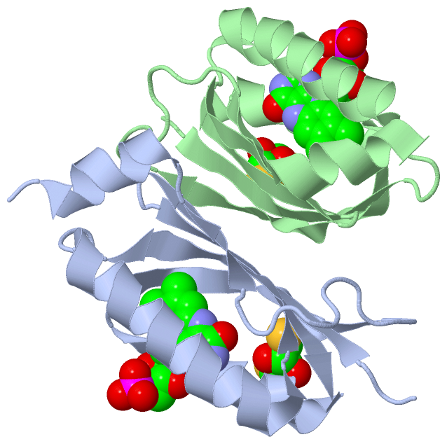
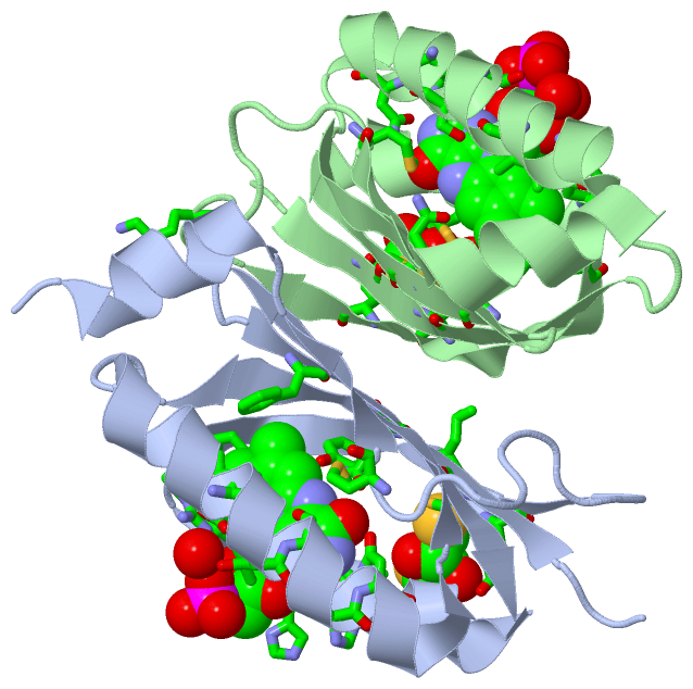
Asymmetric/Biological UnitChain A from PDB Type:PROTEIN Length:108 aligned with Q3J677_RHOS4 | Q3J677 from UniProtKB/TrEMBL Length:450 Alignment length:108 23 33 43 53 63 73 83 93 103 113 Q3J677_RHOS4 14 SDLVSCCYRSLAAPDLTLRDLLDIVETSQAHNARAQLTGALFYSQGVFFQWLEGRPAAVAEVMTHIQRDRRHSNVEILAEEPIAKRRFAGWHMQLSCSEADMRSLGLA 121 SCOP domains d2iyia_ A: automated matches SCOP domains CATH domains ------------------------------------------------------------------------------------------------------------ CATH domains Pfam domains ------------------------------------------------------------------------------------------------------------ Pfam domains SAPs(SNPs) ------------------------------------------------------------------------------------------------------------ SAPs(SNPs) PROSITE ------------------------------------------------------------------------------------------------------------ PROSITE Transcript ------------------------------------------------------------------------------------------------------------ Transcript 2iyi A 14 SDLVSCSYRSLAAPDLTLRDLLDIVETSQAHNARAQLTGALFYSQGVFFQWLEGRPAAVAEVMTHIQRDRRHSNVEILAEEPIAKRRFAGWHMQLSCSEADMRSLGLA 121 23 33 43 53 63 73 83 93 103 113 Chain B from PDB Type:PROTEIN Length:106 aligned with Q3J677_RHOS4 | Q3J677 from UniProtKB/TrEMBL Length:450 Alignment length:106 24 34 44 54 64 74 84 94 104 114 Q3J677_RHOS4 15 DLVSCCYRSLAAPDLTLRDLLDIVETSQAHNARAQLTGALFYSQGVFFQWLEGRPAAVAEVMTHIQRDRRHSNVEILAEEPIAKRRFAGWHMQLSCSEADMRSLGL 120 SCOP domains d2iyib_ B: automated matches SCOP domains CATH domains ---------------------------------------------------------------------------------------------------------- CATH domains Pfam domains ---------------------------------------------------------------------------------------------------------- Pfam domains SAPs(SNPs) ---------------------------------------------------------------------------------------------------------- SAPs(SNPs) PROSITE ---------------------------------------------------------------------------------------------------------- PROSITE Transcript ---------------------------------------------------------------------------------------------------------- Transcript 2iyi B 15 DLVSCSYRSLAAPDLTLRDLLDIVETSQAHNARAQLTGALFYSQGVFFQWLEGRPAAVAEVMTHIQRDRRHSNVEILAEEPIAKRRFAGWHMQLSCSEADMRSLGL 120 24 34 44 54 64 74 84 94 104 114
|
||||||||||||||||||||
SCOP Domains (1, 2)
Asymmetric/Biological Unit
 |
CATH Domains (0, 0)| (no "CATH Domain" information available for 2IYI) |
Pfam Domains (0, 0)| (no "Pfam Domain" information available for 2IYI) |
Gene Ontology (4, 4)|
Asymmetric/Biological Unit(hide GO term definitions) Chain A,B (Q3J677_RHOS4 | Q3J677)
|
||||||||||||||||||||||||||||||||||||
Interactive Views
|
|||||||||||||||||||||||||||||||||||||||||||||||||||||||||||||||||||||||||||||||||||||||||||||||||||||||||||||||||||||||||||||||||||||||||||||||||||||||||
Still Images
|
||||||||||||||||
Databases
|
||||||||||||||||||||||||||||||||||||||||||||||||||||||||||||||||||||||||||||||||||||||||||||||||||||||||||||||||||||||||||||||||||||||||||||||||||||||||||||||||
Analysis Tools
|
|||||||||||||||||||||||||||||||||||||||||||||||||||||||||||||
Entries Sharing at Least One Protein Chain (UniProt ID)
Related Entries Specified in the PDB File
|
|