|
|
|
|
 Description
|
|
Compounds
|
||||||||||||||||||||||||||||||||||||||||||||||||
Chains, Units
Summary Information (see also Sequences/Alignments below) |
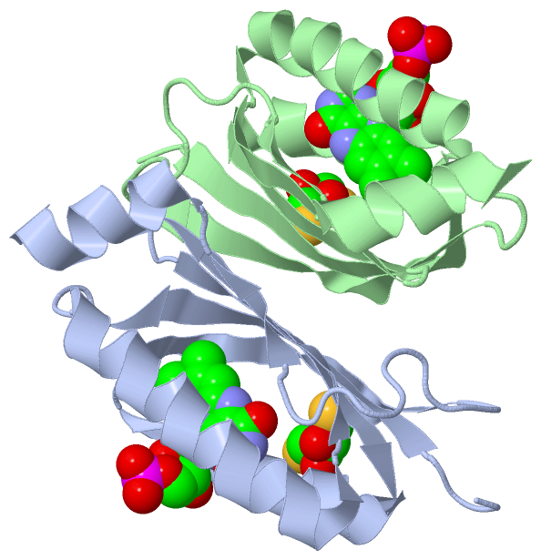
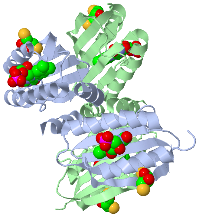
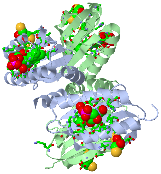
Ligands, Modified Residues, Ions (3, 4)| Asymmetric Unit (3, 4) Biological Unit 1 (3, 8) |
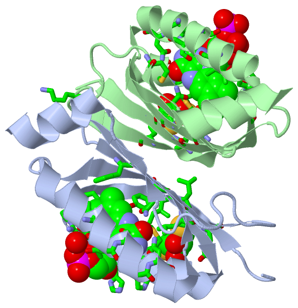
Sites (4, 4)
Asymmetric Unit (4, 4)
|
SS Bonds (0, 0)| (no "SS Bond" information available for 2IYG) |
Cis Peptide Bonds (0, 0)| (no "Cis Peptide Bond" information available for 2IYG) |
SAPs(SNPs)/Variants (0, 0)| (no "SAP(SNP)/Variant" information available for 2IYG) |
PROSITE Motifs (0, 0)| (no "PROSITE Motif" information available for 2IYG) |
Exons (0, 0)| (no "Exon" information available for 2IYG) |
Sequences/Alignments
Asymmetric UnitChain A from PDB Type:PROTEIN Length:109 aligned with Q3J677_RHOS4 | Q3J677 from UniProtKB/TrEMBL Length:450 Alignment length:109 22 32 42 52 62 72 82 92 102 112 Q3J677_RHOS4 13 GSDLVSCCYRSLAAPDLTLRDLLDIVETSQAHNARAQLTGALFYSQGVFFQWLEGRPAAVAEVMTHIQRDRRHSNVEILAEEPIAKRRFAGWHMQLSCSEADMRSLGLA 121 SCOP domains d2iyga_ A: automated matches SCOP domains CATH domains ------------------------------------------------------------------------------------------------------------- CATH domains Pfam domains ------------------------------------------------------------------------------------------------------------- Pfam domains SAPs(SNPs) ------------------------------------------------------------------------------------------------------------- SAPs(SNPs) PROSITE ------------------------------------------------------------------------------------------------------------- PROSITE Transcript ------------------------------------------------------------------------------------------------------------- Transcript 2iyg A 13 GSDLVSCSYRSLAAPDLTLRDLLDIVETSQAHNARAQLTGALFYSQGVFFQWLEGRPAAVAEVMTHIQRDRRHSNVEILAEEPIAKRRFAGWHMQLSCSEADMRSLGLA 121 22 32 42 52 62 72 82 92 102 112 Chain B from PDB Type:PROTEIN Length:108 aligned with Q3J677_RHOS4 | Q3J677 from UniProtKB/TrEMBL Length:450 Alignment length:108 23 33 43 53 63 73 83 93 103 113 Q3J677_RHOS4 14 SDLVSCCYRSLAAPDLTLRDLLDIVETSQAHNARAQLTGALFYSQGVFFQWLEGRPAAVAEVMTHIQRDRRHSNVEILAEEPIAKRRFAGWHMQLSCSEADMRSLGLA 121 SCOP domains d2iygb_ B: automated matches SCOP domains CATH domains ------------------------------------------------------------------------------------------------------------ CATH domains Pfam domains ------------------------------------------------------------------------------------------------------------ Pfam domains SAPs(SNPs) ------------------------------------------------------------------------------------------------------------ SAPs(SNPs) PROSITE ------------------------------------------------------------------------------------------------------------ PROSITE Transcript ------------------------------------------------------------------------------------------------------------ Transcript 2iyg B 14 SDLVSCSYRSLAAPDLTLRDLLDIVETSQAHNARAQLTGALFYSQGVFFQWLEGRPAAVAEVMTHIQRDRRHSNVEILAEEPIAKRRFAGWHMQLSCSEADMRSLGLA 121 23 33 43 53 63 73 83 93 103 113
|
||||||||||||||||||||
SCOP Domains (1, 2)
Asymmetric Unit
 |
CATH Domains (0, 0)| (no "CATH Domain" information available for 2IYG) |
Pfam Domains (0, 0)| (no "Pfam Domain" information available for 2IYG) |
Gene Ontology (4, 4)|
Asymmetric Unit(hide GO term definitions) Chain A,B (Q3J677_RHOS4 | Q3J677)
|
||||||||||||||||||||||||||||||||||||
Interactive Views
|
|||||||||||||||||||||||||||||||||||||||||||||||||||||||||||||||||||||||||||||||||||||||||||||||||||||||||||||||||||||||||||||||||||||||||||||||||||||||||||||||||||||||||||
Still Images
|
||||||||||||||||
Databases
|
||||||||||||||||||||||||||||||||||||||||||||||||||||||||||||||||||||||||||||||||||||||||||||||||||||||||||||||||||||||||||||||||||||||||||||||||||||||||||||||||
Analysis Tools
|
|||||||||||||||||||||||||||||||||||||||||||||||||||||||||||||
Entries Sharing at Least One Protein Chain (UniProt ID)
Related Entries Specified in the PDB File
|
|