|
|
|
|
 Description
|
|
Compounds
|
||||||||||||||||||||||||||||||||||||||||||||
Chains, Units
Summary Information (see also Sequences/Alignments below) |
Ligands, Modified Residues, Ions (1, 1)
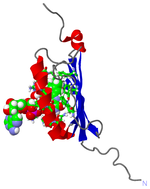
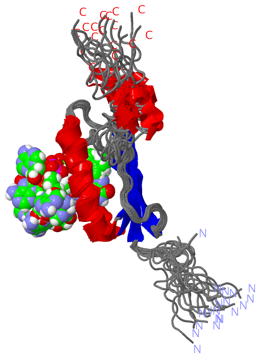
NMR Structure (1, 1)
|
Sites (1, 1)
NMR Structure (1, 1)
|
SS Bonds (0, 0)| (no "SS Bond" information available for 2BUN) |
Cis Peptide Bonds (0, 0)| (no "Cis Peptide Bond" information available for 2BUN) |
SAPs(SNPs)/Variants (0, 0)| (no "SAP(SNP)/Variant" information available for 2BUN) |
PROSITE Motifs (0, 0)| (no "PROSITE Motif" information available for 2BUN) |
Exons (0, 0)| (no "Exon" information available for 2BUN) |
Sequences/Alignments
NMR StructureChain A from PDB Type:PROTEIN Length:121 aligned with Q53119_RHOSH | Q53119 from UniProtKB/TrEMBL Length:450 Alignment length:121 14 24 34 44 54 64 74 84 94 104 114 124 Q53119_RHOSH 5 LEADVTMTGSDLVSCCYRSLAAPDLTLRDLLDIVETSQAHNARAQLTGALFYSQGVFFQWLEGRPAAVAEVMTHIQRDRRHSNVEILAEEPIAKRRFAGWHMQLSCSEADMRSLGLAESRQ 125 SCOP domains d2buna1 A:5-125 Sensor of blue light AppA SCOP domains CATH domains ------------------------------------------------------------------------------------------------------------------------- CATH domains Pfam domains ------------------------------------------------------------------------------------------------------------------------- Pfam domains SAPs(SNPs) ------------------------------------------------------------------------------------------------------------------------- SAPs(SNPs) PROSITE ------------------------------------------------------------------------------------------------------------------------- PROSITE Transcript ------------------------------------------------------------------------------------------------------------------------- Transcript 2bun A 5 LEADVTMTGSDLVSCCYRSLAAPDLTLRDLLDIVETSQAHNARAQLTGALFYSQGVFFQWLEGHPAAVAEVMSHIQRDRRHSNVEILAEESIAKRRFAGWHMQLSCSEADMRSLGLAESRQ 125 14 24 34 44 54 64 74 84 94 104 114 124
|
||||||||||||||||||||
SCOP Domains (1, 1)
NMR Structure

|
CATH Domains (0, 0)| (no "CATH Domain" information available for 2BUN) |
Pfam Domains (0, 0)| (no "Pfam Domain" information available for 2BUN) |
Gene Ontology (4, 4)|
NMR Structure(hide GO term definitions) Chain A (Q53119_RHOSH | Q53119)
|
||||||||||||||||||||||||||||||||||||
Interactive Views
|
||||||||||||||||||||||||||||||||||||||||||||||||||||||||||||||||||||||||||||||||||||||||||||||||||||||||||||||||||||||
Still Images
|
||||||||||||||||
Databases
|
||||||||||||||||||||||||||||||||||||||||||||||||||||||||||||||||||||||||||||||||||||||||||||||||||||||||||||||||||||||||||||||||||||||||||||||||||||||||||||||||
Analysis Tools
|
|||||||||||||||||||||||||||||||||||||||||||||||||||||||||||||
Entries Sharing at Least One Protein Chain (UniProt ID)
Related Entries Specified in the PDB File
|
|