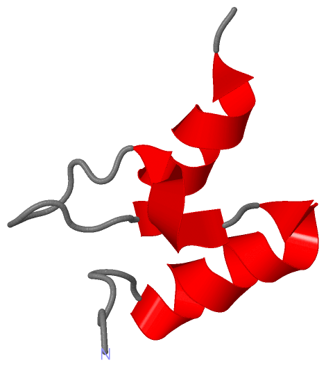
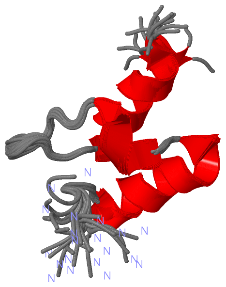
Show PDB file:   
         Plain Text   HTML   (compressed file size)
QuickSearch:   
by PDB,NDB,UniProt,PROSITE Code or Search Term(s)  
(-)NMR Structure - manually
(-)NMR Structure - model 1
(-)NMR Structure - all models
collapse expand < >
Image NMR Structure - manually
NMR Structure - manually  (Jmol Viewer)
Image NMR Structure - model 1
NMR Structure - model 1  (Jmol Viewer)
Image NMR Structure - all models
NMR Structure - all models  (Jmol Viewer)

(-) Description

Title :  NMR STRUCTURE OF THE PERIPHERAL-SUBUNIT BINDING DOMAIN OF BACILLUS STEAROTHERMOPHILUS E2P
 
Authors :  M. D. Allen, R. W. Broadhurst, R. G. Solomon, R. N. Perham
Date :  14 Jul 04  (Deposition) - 20 Jul 04  (Release) - 24 Feb 09  (Revision)
Method :  SOLUTION NMR
Resolution :  NOT APPLICABLE
Chains :  NMR Structure  :  A  (20x)
Keywords :  Transferase, Peripheral-Subunit Binding Domain, Dihydrolipoamide Acetyltransferase, Dihydrolipoamide Dehydrogenase, Protein- Protein Interaction, Nmr, Protein Structure, Multienzyme Complex, Bacillus Sterothermophilus, Glycolysis, Acyltransferase, Lipoyl (Keyword Search: [Gene Ontology, PubMed, Web (Google))
 
Reference :  M. D. Allen, R. W. Broadhurst, R. G. Solomon, R. N. Perham
Interaction Of The E2 And E3 Components Of The Pyruvate Dehydrogenase Multienzyme Complex Of Bacillus Stearothermophilus. Use Of A Truncated Protein Domain In Nmr Spectroscopy
Febs J. V. 272 259 2005
PubMed-ID: 15634348  |  Reference-DOI: 10.1111/J.1432-1033.2004.04405.X

(-) Compounds

Molecule 1 - DIHYDROLIPOYLLYSINE-RESIDUE ACETYLTRANSFERASE COMPONENT OF PYRUVATE DEHYDROGENASE
    ChainsA
    EC Number2.3.1.12
    EngineeredYES
    Expression SystemESCHERICHIA COLI
    Expression System StrainC41
    Expression System Taxid562
    Expression System VectorPET11C
    FragmentRESIDUES 118-170
    Organism ScientificGEOBACILLUS STEAROTHERMOPHILUS
    Organism Taxid1422
    SynonymCOMPLEX DIHYDROPOAMIDE ACETYLTRANSFERASE

 Structural Features

(-) Chains, Units

  
NMR Structure (20x)

Summary Information (see also Sequences/Alignments below)

(-) Ligands, Modified Residues, Ions  (0, 0)

(no "Ligand,Modified Residues,Ions" information available for 1W3D)

(-) Sites  (0, 0)

(no "Site" information available for 1W3D)

(-) SS Bonds  (0, 0)

(no "SS Bond" information available for 1W3D)

(-) Cis Peptide Bonds  (0, 0)

(no "Cis Peptide Bond" information available for 1W3D)

 Sequence-Structure Mapping

(-) SAPs(SNPs)/Variants  (0, 0)

(no "SAP(SNP)/Variant" information available for 1W3D)

(-) PROSITE Motifs  (1, 1)

NMR Structure (1, 1)
 PROSITEUniProtKBPDB
No.IDACDescriptionIDLocationCountLocation
1PSBDPS51826 Peripheral subunit-binding (PSBD) domain profile.ODP2_GEOSE130-167  1A:130-167

(-) Exons   (0, 0)

(no "Exon" information available for 1W3D)

(-) Sequences/Alignments

NMR Structure
   Reformat: Number of residues per line =  ('0' or empty: single-line sequence representation)
  Number of residues per labelling interval =   
  UniProt sequence: complete  aligned part    
   Show mapping: SCOP domains CATH domains Pfam domains Secondary structure (by author)
SAPs(SNPs) PROSITE motifs Exons
(details for a mapped element are shown in a popup box when the mouse pointer rests over it)
Chain A from PDB  Type:PROTEIN  Length:46
 aligned with ODP2_GEOSE | P11961 from UniProtKB/Swiss-Prot  Length:428

    Alignment length:46
                                   135       145       155       165      
           ODP2_GEOSE   126 NRRVIAMPSVRKYAREKGVDIRLVQGTGKNGRVLKEDIDAFLAGGA 171
               SCOP domains d1w3da_ A:                                     SCOP domains
               CATH domains 1w3dA00 A:126-171 Dihydrolipoamide Transferase CATH domains
               Pfam domains -E3_binding-1w3dA01 A:127-165           ------ Pfam domains
         Sec.struct. author ......hhhhhhhhhhhh.hhhhh.........hhhhhhhhhh... Sec.struct. author
                 SAPs(SNPs) ---------------------------------------------- SAPs(SNPs)
                    PROSITE ----PSBD  PDB: A:130-167 UniProt: 130-167 ---- PROSITE
                 Transcript ---------------------------------------------- Transcript
                 1w3d A 126 NRRVIAMPSVRKYAREKGVDIRLVQGTGKNGRVLKEDIDAFLAGGA 171
                                   135       145       155       165      

   Legend:   → Mismatch (orange background)
  - → Gap (green background, '-', border residues have a numbering label)
    → Modified Residue (blue background, lower-case, 'x' indicates undefined single-letter code, labelled with number + name)
  x → Chemical Group (purple background, 'x', labelled with number + name, e.g. ACE or NH2)
  extra numbering lines below/above indicate numbering irregularities and modified residue names etc., number ends below/above '|'

 Classification and Annotation

(-) SCOP Domains  (1, 1)

NMR Structure

(-) CATH Domains  (1, 1)

NMR Structure

(-) Pfam Domains  (1, 1)

NMR Structure

(-) Gene Ontology  (5, 5)

NMR Structure(hide GO term definitions)
Chain A   (ODP2_GEOSE | P11961)
molecular function
    GO:0004742    dihydrolipoyllysine-residue acetyltransferase activity    Catalysis of the reaction: acetyl-CoA + dihydrolipoamide = CoA + S-acetyldihydrolipoamide.
    GO:0016740    transferase activity    Catalysis of the transfer of a group, e.g. a methyl group, glycosyl group, acyl group, phosphorus-containing, or other groups, from one compound (generally regarded as the donor) to another compound (generally regarded as the acceptor). Transferase is the systematic name for any enzyme of EC class 2.
    GO:0016746    transferase activity, transferring acyl groups    Catalysis of the transfer of an acyl group from one compound (donor) to another (acceptor).
biological process
    GO:0006096    glycolytic process    The chemical reactions and pathways resulting in the breakdown of a carbohydrate into pyruvate, with the concomitant production of a small amount of ATP and the reduction of NAD(P) to NAD(P)H. Glycolysis begins with the metabolism of a carbohydrate to generate products that can enter the pathway and ends with the production of pyruvate. Pyruvate may be converted to acetyl-coenzyme A, ethanol, lactate, or other small molecules.
    GO:0008152    metabolic process    The chemical reactions and pathways, including anabolism and catabolism, by which living organisms transform chemical substances. Metabolic processes typically transform small molecules, but also include macromolecular processes such as DNA repair and replication, and protein synthesis and degradation.

 Visualization

(-) Interactive Views

NMR Structure
  Complete Structure
    Jena3D(integrated viewing of ligand, site, SAP, PROSITE, SCOP information)
    WebMol | AstexViewer[tm]@PDBe
(Java Applets, require no local installation except for Java; loading may be slow)
    STRAP
(Java WebStart application, automatic local installation, requires Java; full application with system access!)
    RasMol
(require local installation)
    Molscript (VRML)
(requires installation of a VRML viewer; select preferred view via VRML and generate a mono or stereo PDF format file)
 
  Ligands, Modified Residues, Ions
(no "Ligands, Modified Residues, Ions" information available for 1w3d)
 
  Sites
(no "Sites" information available for 1w3d)
 
  Cis Peptide Bonds
(no "Cis Peptide Bonds" information available for 1w3d)
 

(-) Still Images

Jmol
  protein: cartoon or spacefill or dots and stick; nucleic acid: cartoon and stick; ligands: spacefill; active site: stick
Molscript
  protein, nucleic acid: cartoon; ligands: spacefill; active site: ball and stick

 Databases and Analysis Tools

(-) Databases

Access by PDB/NDB ID
  1w3d
    Family and Domain InformationProDom | SYSTERS
    General Structural InformationGlycoscienceDB | MMDB | NDB | OCA | PDB | PDBe | PDBj | PDBsum | PDBWiki | PQS | PROTEOPEDIA
    Orientation in MembranesOPM
    Protein SurfaceSURFACE
    Secondary StructureDSSP (structure derived) | HSSP (homology derived)
    Structural GenomicsGeneCensus
    Structural NeighboursCE | VAST
    Structure ClassificationCATH | Dali | SCOP
    Validation and Original DataBMRB Data View | BMRB Restraints Grid | EDS | PROCHECK | RECOORD | WHAT_CHECK
 
Access by UniProt ID/Accession number
  ODP2_GEOSE | P11961
    Comparative Protein Structure ModelsModBase
    Genomic InformationEnsembl
    Protein-protein InteractionDIP
    Sequence, Family and Domain InformationInterPro | Pfam | SMART | UniProtKB/SwissProt
 
Access by Enzyme Classificator   (EC Number)
  2.3.1.12
    General Enzyme InformationBRENDA | EC-PDB | Enzyme | IntEnz
    PathwayKEGG | MetaCyc
 
Access by Disease Identifier   (MIM ID)
  (no 'MIM ID' available)
    Disease InformationOMIM
 
Access by GenAge ID
  (no 'GenAge ID' available)
    Age Related InformationGenAge

(-) Analysis Tools

Access by PDB/NDB ID
    Domain InformationXDom
    Interatomic Contacts of Structural UnitsCSU
    Ligand-protein ContactsLPC
    Protein CavitiescastP
    Sequence and Secondary StructurePDBCartoon
    Structure AlignmentSTRAP(Java WebStart application, automatic local installation, requires Java; full application with system access!)
    Structure and Sequence BrowserSTING
 
Access by UniProt ID/Accession number
  ODP2_GEOSE | P11961
    Protein Disorder PredictionDisEMBL | FoldIndex | GLOBPLOT (for more information see DisProt)

 Related Entries

(-) Entries Sharing at Least One Protein Chain (UniProt ID)

UniProtKB/Swiss-Prot
        ODP2_GEOSE | P119611b5s 1ebd 1lab 1lac 1w4e 1w4f 1w4g 1w4h 1w85 1w88 2pdd 2pde 3duf 3dv0 3dva

(-) Related Entries Specified in the PDB File

1b5s DIHYDROLIPOYL TRANSACETYLASE CATALYTIC DOMAIN ( RESIDUES 184-425) FROM BACILLUS STEAROTHERMOPHILUS
1ebd DIHYDROLIPOAMIDE DEHYDROGENASE COMPLEXED WITH THE BINDING DOMAIN OF THE DIHYDROLIPOAMIDE ACETYLASE
1lab DIHYDROLIPOAMIDE ACETYLTRANSFERASE (E2P) SUBUNIT OF THE PYRUVATE DEHYDROGENASE (PDH) MULTIENZYME COMPLEX (LIPOYLATED DOMAIN, RESIDUES 1 - 80) (NMR, 11 STRUCTURES)
1lac DIHYDROLIPOAMIDE ACETYLTRANSFERASE (E2P) SUBUNIT OF THE PYRUVATE DEHYDROGENASE (PDH) MULTIENZYME COMPLEX (LIPOYLATED DOMAIN, RESIDUES 1 - 80) (NMR, AVERAGE STRUCTURE)
2pdd DIHYDROLIPOAMIDE ACETYLTRANSFERASE (E2P) SUBUNIT OF THE PYRUVATE DEHYDROGENASE (PDH) MULTIENZYME COMPLEX (PYRUVATE DECARBOXYLASE (E1P ) / DIHYDROLIPOAMIDE DEHYDROGENASE (E3) 43 RESIDUE BINDING DOMAIN) (NMR, 35 STRUCTURES)
2pde DIHYDROLIPOAMIDE ACETYLTRANSFERASE (E2P) SUBUNIT OF THE PYRUVATE DEHYDROGENASE (PDH) MULTIENZYME COMPLEX (PYRUVATE DECARBOXYLASE (E1P ) / DIHYDROLIPOAMIDE DEHYDROGENASE (E3) 43 RESIDUE BINDING DOMAIN) (NMR, AVERAGE STRUCTURE)