|
|
|
|
 Description
|
|
Compounds
|
||||||||||||||||||||||||||||||||||||||||||||||||
Chains, Units
Summary Information (see also Sequences/Alignments below) |
Ligands, Modified Residues, Ions (0, 0)| (no "Ligand,Modified Residues,Ions" information available for 1TNW) |
Sites (4, 4)
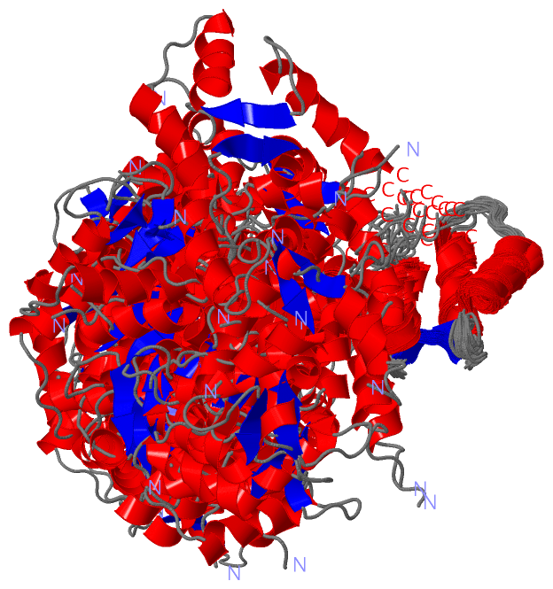
NMR Structure (4, 4)
|
SS Bonds (0, 0)| (no "SS Bond" information available for 1TNW) |
Cis Peptide Bonds (0, 0)| (no "Cis Peptide Bond" information available for 1TNW) |
SAPs(SNPs)/Variants (0, 0)| (no "SAP(SNP)/Variant" information available for 1TNW) |
PROSITE Motifs (2, 8)
NMR Structure (2, 8)
|
||||||||||||||||||||||||||||||||
Exons (4, 4)
NMR Structure (4, 4)
|
||||||||||||||||||||||||||||||||||||||||||||||||||||||||||||||||||||||||
Sequences/Alignments
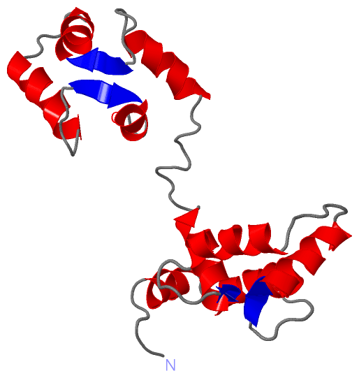
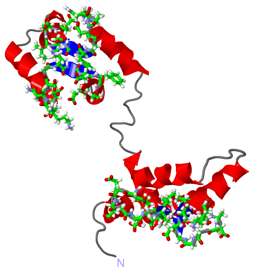
NMR StructureChain A from PDB Type:PROTEIN Length:162 aligned with TNNC2_CHICK | P02588 from UniProtKB/Swiss-Prot Length:163 Alignment length:162 11 21 31 41 51 61 71 81 91 101 111 121 131 141 151 161 TNNC2_CHICK 2 ASMTDQQAEARAFLSEEMIAEFKAAFDMFDADGGGDISTKELGTVMRMLGQNPTKEELDAIIEEVDEDGSGTIDFEEFLVMMVRQMKEDAKGKSEEELANCFRIFDKNADGFIDIEELGEILRATGEHVTEEDIEDLMKDSDKNNDGRIDFDEFLKMMEGVQ 163 SCOP domains d1tnwa_ A: Troponin C SCOP domains CATH domains 1tnwA01 A:1-88 EF-hand 1tnwA02 A:89-162 EF-hand CATH domains Pfam domains ------------------------------------------------------------------------------------------------------------------------------------------------------------------ Pfam domains SAPs(SNPs) ------------------------------------------------------------------------------------------------------------------------------------------------------------------ SAPs(SNPs) PROSITE (1) ----------------EF_HAND_2 PDB: A:17-52 EF_HAND_2 PDB: A:53-88 ----EF_HAND_2 PDB: A:93-128 EF_HAND_2 PDB: A:129-162 PROSITE (1) PROSITE (2) -----------------------------EF_HAND_1 -----------------------EF_HAND_1 ---------------------------EF_HAND_1 -----------------------EF_HAND_1 -------- PROSITE (2) Transcript 1 (1) Exon 1.1 PDB: A:1-49 UniProt: 1-50 [INCOMPLETE] -------------------------------------Exon 1.3 PDB: A:87-133 UniProt: 88-134 ----------------------------- Transcript 1 (1) Transcript 1 (2) ------------------------------------------------Exon 1.2 PDB: A:49-87 UniProt: 50-88 ---------------------------------------------Exon 1.4 -------------------- Transcript 1 (2) 1tnw A 1 ASMTDQQAEARAFLSEEMIAEFKAAFDMFDADGGGDISTKELGTVMRMLGQNPTKEELDAIIEEVDEDGSGTIDFEEFLVMMVRQMKEDAKGKSEEELANCFRIFDKNADGFIDIEELGEILRATGEHVIEEDIEDLMKDSDKNNDGRIDFDEFLKMMEGVQ 162 10 20 30 40 50 60 70 80 90 100 110 120 130 140 150 160
|
||||||||||||||||||||
SCOP Domains (1, 1)
NMR Structure
 |
CATH Domains (1, 2)
NMR Structure
|
Pfam Domains (0, 0)| (no "Pfam Domain" information available for 1TNW) |
Gene Ontology (5, 5)|
NMR Structure(hide GO term definitions) Chain A (TNNC2_CHICK | P02588)
|
||||||||||||||||||||||||||||||||||||||||||||||||
Interactive Views
|
||||||||||||||||||||||||||||||||||||||||||||||||||||||||||||||||||||||||||||||||||||||||||||||||||||||||||||||||||||||||||||||||||||||||||
Still Images
|
||||||||||||||||
Databases
|
||||||||||||||||||||||||||||||||||||||||||||||||||||||||||||||||||||||||||||||||||||||||||||||||||||||||||||||||||||||||||||||||||||||||||||||||||||||||||||||||
Analysis Tools
|
|||||||||||||||||||||||||||||||||||||||||||||||||||||||||||||
Entries Sharing at Least One Protein Chain (UniProt ID)
Related Entries Specified in the PDB File
|
|