Show PDB file:   
         Plain Text   HTML   (compressed file size)
QuickSearch:   
by PDB,NDB,UniProt,PROSITE Code or Search Term(s)  
(-)NMR Structure - model 1
(-)NMR Structure - model 1, sites
(-)NMR Structure - all models
collapse expand < >
Image NMR Structure - model 1
NMR Structure - model 1  (Jmol Viewer)
Image NMR Structure - model 1, sites
NMR Structure - model 1, sites  (Jmol Viewer)
Image NMR Structure - all models
NMR Structure - all models  (Jmol Viewer)

(-) Description

Title :  STRUCTURE OF A RHODAMINE-LABELED N-DOMAIN TROPONIN C MUTANT (CA2+ SATURATED) IN COMPLEX WITH SKELETAL TROPONIN I 115-131
 
Authors :  P. Mercier, R. E. Ferguson, M. Irving, J. E. T. Corrie, D. R. Trentham, B. D. Sykes
Date :  18 Jan 03  (Deposition) - 29 Apr 03  (Release) - 24 Feb 09  (Revision)
Method :  SOLUTION NMR
Resolution :  NOT APPLICABLE
Chains :  NMR Structure  :  A,B  (21x)
Keywords :  Troponin C- Troponin I Complex, Bifunctional Rhodamine Labeled Toponin C, Structural Protein (Keyword Search: [Gene Ontology, PubMed, Web (Google))
 
Reference :  P. Mercier, R. E. Ferguson, M. Irving, J. E. T. Corrie, D. R. Trentham, B. D. Sykes
Nmr Structure Of A Bifunctional Rhodamine Labeled N-Domain Of Troponin C Complexed With The Regulatory "Switch" Peptide From Troponin I: Implications For In Situ Fluorescence Studies In Muscle Fibers
Biochemistry V. 42 4333 2003
PubMed-ID: 12693929  |  Reference-DOI: 10.1021/BI027041N
(for further references see the PDB file header)

(-) Compounds

Molecule 1 - TROPONIN C
    ChainsA
    EngineeredYES
    Expression SystemESCHERICHIA COLI
    Expression System PlasmidPET3ASNTNC
    Expression System StrainBL21(DE3)PLYSS
    Expression System Taxid562
    Expression System Vector TypePET3A
    FragmentTNC, RESIDUES 1-90
    GeneTNNC2
    MutationYES
    Organism CommonCHICKEN
    Organism ScientificGALLUS GALLUS
    Organism Taxid9031
    SynonymTROPONIN C, SKELETAL MUSCLE
 
Molecule 2 - TROPONIN I
    ChainsB
    EngineeredYES
    FragmentSWITCH PEPTIDE, RESIDUES 115-131
    Other DetailsTHIS SEQUENCE OCCURS NATURALLY IN ORYCTOLAGUS CUNICULUS (RABIT)
    SynonymTROPONIN I, FAST SKELETAL MUSCLE
    SyntheticYES

 Structural Features

(-) Chains, Units

  
NMR Structure (21x)

Summary Information (see also Sequences/Alignments below)

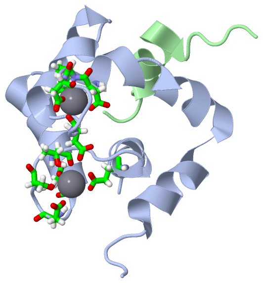
(-) Ligands, Modified Residues, Ions  (1, 2)

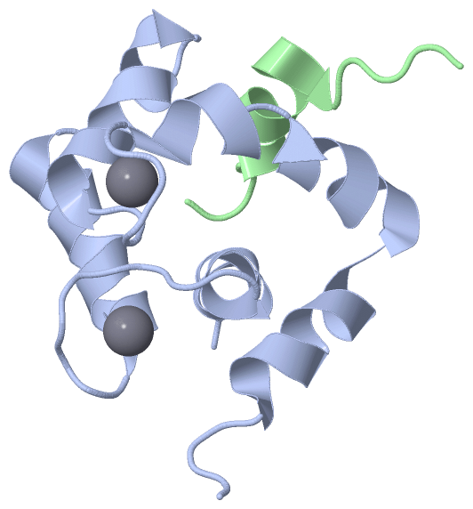
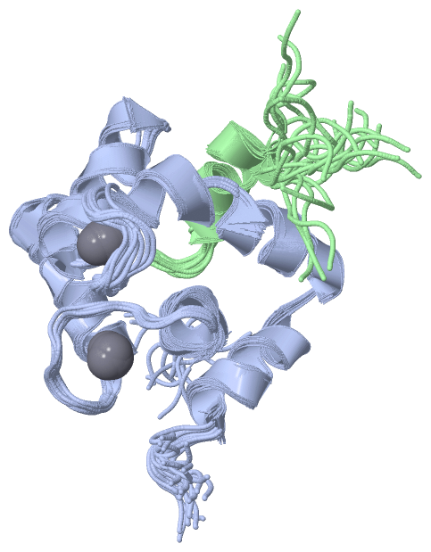
NMR Structure (1, 2)
No.NameCountTypeFull Name
1CA2Ligand/IonCALCIUM ION

(-) Sites  (2, 2)

NMR Structure (2, 2)
No.NameEvidenceResiduesDescription
1AC1SOFTWAREASP A:66 , ASP A:68 , SER A:70 , THR A:72 , GLU A:77BINDING SITE FOR RESIDUE CA A 132
2AC2SOFTWAREASP A:30 , ALA A:31 , ASP A:32 , ASP A:36 , GLU A:41BINDING SITE FOR RESIDUE CA A 133

(-) SS Bonds  (0, 0)

(no "SS Bond" information available for 1NPQ)

(-) Cis Peptide Bonds  (0, 0)

(no "Cis Peptide Bond" information available for 1NPQ)

 Sequence-Structure Mapping

(-) SAPs(SNPs)/Variants  (0, 0)

(no "SAP(SNP)/Variant" information available for 1NPQ)

(-) PROSITE Motifs  (2, 4)

NMR Structure (2, 4)
 PROSITEUniProtKBPDB
No.IDACDescriptionIDLocationCountLocation
1EF_HAND_2PS50222 EF-hand calcium-binding domain profile.TNNC2_CHICK18-53
54-89
94-129
130-163
  2A:17-52
A:53-88
-
-
2EF_HAND_1PS00018 EF-hand calcium-binding domain.TNNC2_CHICK31-43
67-79
107-119
143-155
  2A:30-42
A:66-78
-
-

(-) Exons   (3, 3)

NMR Structure (3, 3)
 ENSEMBLUniProtKBPDB
No.Transcript IDExonExon IDGenome LocationLengthIDLocationLengthCountLocationLength
1.1ENSGALT000000110601ENSGALE00000073521Un_random:20732110-20732258149TNNC2_CHICK1-50501A:1-4949
1.2ENSGALT000000110602ENSGALE00000073519Un_random:20732332-20732446115TNNC2_CHICK50-88391A:49-8739
1.3ENSGALT000000110603ENSGALE00000073522Un_random:20732522-20732658137TNNC2_CHICK88-134471A:87-904
1.4ENSGALT000000110604ENSGALE00000073520Un_random:20732749-20732979231TNNC2_CHICK134-143100--

(-) Sequences/Alignments

NMR Structure
   Reformat: Number of residues per line =  ('0' or empty: single-line sequence representation)
  Number of residues per labelling interval =   
  UniProt sequence: complete  aligned part    
   Show mapping: SCOP domains CATH domains Pfam domains Secondary structure (by author)
SAPs(SNPs) PROSITE motifs Exons
(details for a mapped element are shown in a popup box when the mouse pointer rests over it)
Chain A from PDB  Type:PROTEIN  Length:90
 aligned with TNNC2_CHICK | P02588 from UniProtKB/Swiss-Prot  Length:163

    Alignment length:90
                                    11        21        31        41        51        61        71        81        91
          TNNC2_CHICK     2 ASMTDQQAEARAFLSEEMIAEFKAAFDMFDADGGGDISTKELGTVMRMLGQNPTKEELDAIIEEVDEDGSGTIDFEEFLVMMVRQMKEDA  91
               SCOP domains d1npqa_ A: Troponin C                                                                      SCOP domains
               CATH domains 1npqA00 A:1-90 EF-hand                                                                     CATH domains
           Pfam domains (1) -------------EF_hand_5-1npqA01 A:14-82                                            -------- Pfam domains (1)
           Pfam domains (2) --------------------------------EF_hand_6-1npqA02 A:33-85                            ----- Pfam domains (2)
         Sec.struct. author ....hhhhhhhhhhhhhhhhhhh.hhhhhh.......hhhhhhhhhhh.....hhhhhhhhhhhhh.........hhhhhhhhhhhh... Sec.struct. author
                 SAPs(SNPs) ------------------------------------------------------------------------------------------ SAPs(SNPs)
                PROSITE (1) ----------------EF_HAND_2  PDB: A:17-52             EF_HAND_2  PDB: A:53-88             -- PROSITE (1)
                PROSITE (2) -----------------------------EF_HAND_1    -----------------------EF_HAND_1    ------------ PROSITE (2)
           Transcript 1 (1) Exon 1.1  PDB: A:1-49 UniProt: 1-50 [INCOMPLETE] -------------------------------------1.3  Transcript 1 (1)
           Transcript 1 (2) ------------------------------------------------Exon 1.2  PDB: A:49-87 UniProt: 50-88  --- Transcript 1 (2)
                 1npq A   1 ASMTDQQAEARAFLSEEMIAEFKAAFDMFDADGGGDISTKELGTVMRMLGQNPTKCELDAIICEVDEDGSGTIDFEEFLVMMVRQMKEDA  90
                                    10        20        30        40        50        60        70        80        90

Chain B from PDB  Type:PROTEIN  Length:17
 aligned with TNNI2_RABIT | P02643 from UniProtKB/Swiss-Prot  Length:182

    Alignment length:17
                                   125       
          TNNI2_RABIT   116 RMSADAMLKALLGSKHK 132
               SCOP domains ----------------- SCOP domains
               CATH domains ----------------- CATH domains
               Pfam domains Troponin-1npqB01  Pfam domains
         Sec.struct. author ...hhhhhhhhh..... Sec.struct. author
                 SAPs(SNPs) ----------------- SAPs(SNPs)
                    PROSITE ----------------- PROSITE
                 Transcript ----------------- Transcript
                 1npq B 115 RMSADAMLRALLGSKHK 131
                                   124       

   Legend:   → Mismatch (orange background)
  - → Gap (green background, '-', border residues have a numbering label)
    → Modified Residue (blue background, lower-case, 'x' indicates undefined single-letter code, labelled with number + name)
  x → Chemical Group (purple background, 'x', labelled with number + name, e.g. ACE or NH2)
  extra numbering lines below/above indicate numbering irregularities and modified residue names etc., number ends below/above '|'

 Classification and Annotation

(-) SCOP Domains  (1, 1)

NMR Structure

(-) CATH Domains  (1, 1)

NMR Structure

(-) Pfam Domains  (3, 3)

NMR Structure
(-)
Clan: EF_hand (270)

(-) Gene Ontology  (6, 9)

NMR Structure(hide GO term definitions)
Chain A   (TNNC2_CHICK | P02588)
molecular function
    GO:0003779    actin binding    Interacting selectively and non-covalently with monomeric or multimeric forms of actin, including actin filaments.
    GO:0005509    calcium ion binding    Interacting selectively and non-covalently with calcium ions (Ca2+).
    GO:0046872    metal ion binding    Interacting selectively and non-covalently with any metal ion.
biological process
    GO:0003009    skeletal muscle contraction    A process in which force is generated within skeletal muscle tissue, resulting in a change in muscle geometry. Force generation involves a chemo-mechanical energy conversion step that is carried out by the actin/myosin complex activity, which generates force through ATP hydrolysis. In the skeletal muscle, the muscle contraction takes advantage of an ordered sarcomeric structure and in most cases it is under voluntary control.
cellular component
    GO:0005861    troponin complex    A complex of accessory proteins (typically troponin T, troponin I and troponin C) found associated with actin in muscle thin filaments; involved in calcium regulation of muscle contraction.

Chain B   (TNNI2_RABIT | P02643)
molecular function
    GO:0003779    actin binding    Interacting selectively and non-covalently with monomeric or multimeric forms of actin, including actin filaments.
    GO:0046872    metal ion binding    Interacting selectively and non-covalently with any metal ion.
    GO:0031014    troponin T binding    Interacting selectively and non-covalently with troponin T, the tropomyosin-binding subunit of the troponin complex.
cellular component
    GO:0005861    troponin complex    A complex of accessory proteins (typically troponin T, troponin I and troponin C) found associated with actin in muscle thin filaments; involved in calcium regulation of muscle contraction.

 Visualization

(-) Interactive Views

NMR Structure
  Complete Structure
    Jena3D(integrated viewing of ligand, site, SAP, PROSITE, SCOP information)
    WebMol | AstexViewer[tm]@PDBe
(Java Applets, require no local installation except for Java; loading may be slow)
    STRAP
(Java WebStart application, automatic local installation, requires Java; full application with system access!)
    RasMol
(require local installation)
    Molscript (VRML)
(requires installation of a VRML viewer; select preferred view via VRML and generate a mono or stereo PDF format file)
 
  Ligands, Modified Residues, Ions
    CA  [ RasMol | Jena3D ]  +environment [ RasMol | Jena3D ]
 
  Sites
    AC1  [ RasMol ]  +environment [ RasMol ]
    AC2  [ RasMol ]  +environment [ RasMol ]
 
  Cis Peptide Bonds
(no "Cis Peptide Bonds" information available for 1npq)
 

(-) Still Images

Jmol
  protein: cartoon or spacefill or dots and stick; nucleic acid: cartoon and stick; ligands: spacefill; active site: stick
Molscript
  protein, nucleic acid: cartoon; ligands: spacefill; active site: ball and stick

 Databases and Analysis Tools

(-) Databases

Access by PDB/NDB ID
  1npq
    Family and Domain InformationProDom | SYSTERS
    General Structural InformationGlycoscienceDB | MMDB | NDB | OCA | PDB | PDBe | PDBj | PDBsum | PDBWiki | PQS | PROTEOPEDIA
    Orientation in MembranesOPM
    Protein SurfaceSURFACE
    Secondary StructureDSSP (structure derived) | HSSP (homology derived)
    Structural GenomicsGeneCensus
    Structural NeighboursCE | VAST
    Structure ClassificationCATH | Dali | SCOP
    Validation and Original DataBMRB Data View | BMRB Restraints Grid | EDS | PROCHECK | RECOORD | WHAT_CHECK
 
Access by UniProt ID/Accession number
  TNNC2_CHICK | P02588
    Comparative Protein Structure ModelsModBase
    Genomic InformationEnsembl
    Protein-protein InteractionDIP
    Sequence, Family and Domain InformationInterPro | Pfam | SMART | UniProtKB/SwissProt
  TNNI2_RABIT | P02643
    Comparative Protein Structure ModelsModBase
    Genomic InformationEnsembl
    Protein-protein InteractionDIP
    Sequence, Family and Domain InformationInterPro | Pfam | SMART | UniProtKB/SwissProt
 
Access by Enzyme Classificator   (EC Number)
  (no 'Enzyme Classificator' available)
    General Enzyme InformationBRENDA | EC-PDB | Enzyme | IntEnz
    PathwayKEGG | MetaCyc
 
Access by Disease Identifier   (MIM ID)
  (no 'MIM ID' available)
    Disease InformationOMIM
 
Access by GenAge ID
  (no 'GenAge ID' available)
    Age Related InformationGenAge

(-) Analysis Tools

Access by PDB/NDB ID
    Domain InformationXDom
    Interatomic Contacts of Structural UnitsCSU
    Ligand-protein ContactsLPC
    Protein CavitiescastP
    Sequence and Secondary StructurePDBCartoon
    Structure AlignmentSTRAP(Java WebStart application, automatic local installation, requires Java; full application with system access!)
    Structure and Sequence BrowserSTING
 
Access by UniProt ID/Accession number
  TNNC2_CHICK | P02588
    Protein Disorder PredictionDisEMBL | FoldIndex | GLOBPLOT (for more information see DisProt)
  TNNI2_RABIT | P02643
    Protein Disorder PredictionDisEMBL | FoldIndex | GLOBPLOT (for more information see DisProt)

 Related Entries

(-) Entries Sharing at Least One Protein Chain (UniProt ID)

UniProtKB/Swiss-Prot
        TNNC2_CHICK | P025881avs 1blq 1cta 1ctd 1ew7 1jc2 1ncx 1ncy 1ncz 1pon 1skt 1smg 1tnp 1tnq 1tnw 1tnx 1top 1ytz 1yv0 1zac 2w49 2w4u 4tnc
        TNNI2_RABIT | P026431a2x 1ew7

(-) Related Entries Specified in the PDB File

(no "Related Entries Specified in the PDB File" available for 1NPQ)