Show PDB file:   
         Plain Text   HTML   (compressed file size)
QuickSearch:   
by PDB,NDB,UniProt,PROSITE Code or Search Term(s)  
(-)Asym.Unit - manually
(-)Asymmetric Unit
(-)Asym. Unit - sites
(-)Biological Unit 1
(-)Biol. Unit 1 - sites
(-)Biological Unit 2
collapse expand < >
Image Asym.Unit - manually
Asym.Unit - manually  (Jmol Viewer)
Image Asymmetric Unit
Asymmetric Unit  (Jmol Viewer)
Image Asym. Unit - sites
Asym. Unit - sites  (Jmol Viewer)
Image Biological Unit 1
Biological Unit 1  (Jmol Viewer)
Image Biol. Unit 1 - sites
Biol. Unit 1 - sites  (Jmol Viewer)
Image Biological Unit 2
Biological Unit 2  (Jmol Viewer)

(-) Description

Title :  CALCIUM-LOADED APO-AEQUORIN FROM AEQUOREA VICTORIA
 
Authors :  L. Deng, S. V. Markova, E. S. Vysotski, Z. J. Liu, J. Lee, J. Rose, B. C. Wan Southeast Collaboratory For Structural Genomics (Secsg)
Date :  05 Mar 04  (Deposition) - 28 Dec 04  (Release) - 13 Jul 11  (Revision)
Method :  X-RAY DIFFRACTION
Resolution :  1.70
Chains :  Asym. Unit :  A
Biol. Unit 1:  A  (1x)
Biol. Unit 2:  A  (2x)
Keywords :  Photoprotein, Obelin, Bioluminescence, Calcium Binding, Ef-Hand, Aequorin, Structural Genomics, Psi, Protein Structure Initiative, Southeast Collaboratory For Structural Genomics, Secsg, Luminescent Protein (Keyword Search: [Gene Ontology, PubMed, Web (Google))
 
Reference :  L. Deng, E. S. Vysotski, S. V. Markova, Z. J. Liu, J. Lee, J. Rose, B. C. Wang
All Three Ca2+-Binding Loops Of Photoproteins Bind Calcium Ions: The Crystal Structures Of Calcium-Loaded Apo-Aequorin And Apo-Obelin.
Protein Sci. V. 14 663 2005
PubMed-ID: 15689515  |  Reference-DOI: 10.1110/PS.041142905
(for further references see the PDB file header)

(-) Compounds

Molecule 1 - AEQUORIN 1
    ChainsA
    EngineeredYES
    Expression SystemESCHERICHIA COLI
    Expression System PlasmidPET19B+
    Expression System StrainBL21-GOLD(DE3)
    Expression System Taxid562
    Expression System Vector TypePLASMID
    Organism ScientificAEQUOREA VICTORIA
    Organism Taxid6100

 Structural Features

(-) Chains, Units

  1
Asymmetric Unit A
Biological Unit 1 (1x)A
Biological Unit 2 (2x)A

Summary Information (see also Sequences/Alignments below)

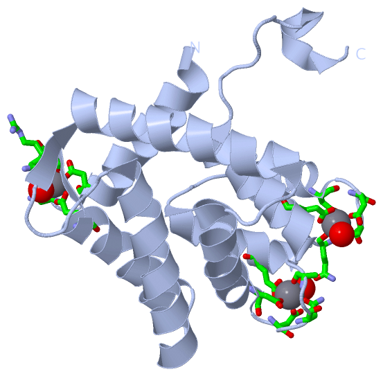
(-) Ligands, Modified Residues, Ions  (1, 3)

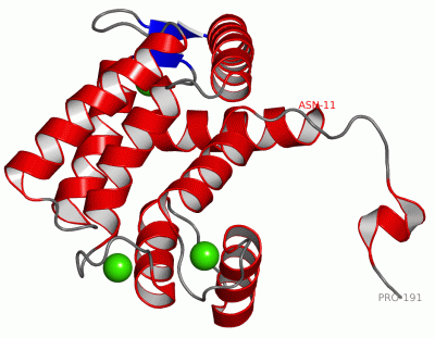
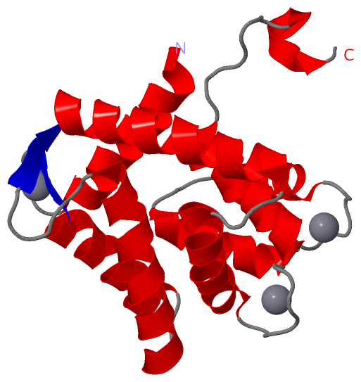
Asymmetric Unit (1, 3)
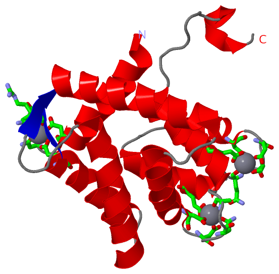
No.NameCountTypeFull Name
1CA3Ligand/IonCALCIUM ION
Biological Unit 1 (0, 0)
No.NameCountTypeFull Name
1CA-1Ligand/IonCALCIUM ION
Biological Unit 2 (0, 0)
No.NameCountTypeFull Name
1CA-1Ligand/IonCALCIUM ION

(-) Sites  (3, 3)

Asymmetric Unit (3, 3)
No.NameEvidenceResiduesDescription
1AC1SOFTWAREASP A:155 , ASP A:157 , SER A:159 , GLN A:161 , GLU A:166 , HOH A:680BINDING SITE FOR RESIDUE CA A 669
2AC2SOFTWAREASP A:26 , ASN A:28 , ASN A:30 , ARG A:32 , GLU A:37 , HOH A:716BINDING SITE FOR RESIDUE CA A 670
3AC3SOFTWAREASP A:119 , ASP A:121 , ASN A:123 , ALA A:125 , GLU A:130 , HOH A:698BINDING SITE FOR RESIDUE CA A 671

(-) SS Bonds  (0, 0)

(no "SS Bond" information available for 1SL8)

(-) Cis Peptide Bonds  (0, 0)

(no "Cis Peptide Bond" information available for 1SL8)

 Sequence-Structure Mapping

(-) SAPs(SNPs)/Variants  (0, 0)

(no "SAP(SNP)/Variant" information available for 1SL8)

(-) PROSITE Motifs  (2, 6)

Asymmetric Unit (2, 6)
 PROSITEUniProtKBPDB
No.IDACDescriptionIDLocationCountLocation
1EF_HAND_2PS50222 EF-hand calcium-binding domain profile.AEQ1_AEQVI18-53
117-146
147-182
  3A:13-48
A:112-141
A:142-177
2EF_HAND_1PS00018 EF-hand calcium-binding domain.AEQ1_AEQVI31-43
124-136
160-172
  3A:26-38
A:119-131
A:155-167
Biological Unit 1 (2, 6)
 PROSITEUniProtKBPDB
No.IDACDescriptionIDLocationCountLocation
1EF_HAND_2PS50222 EF-hand calcium-binding domain profile.AEQ1_AEQVI18-53
117-146
147-182
  3A:13-48
A:112-141
A:142-177
2EF_HAND_1PS00018 EF-hand calcium-binding domain.AEQ1_AEQVI31-43
124-136
160-172
  3A:26-38
A:119-131
A:155-167
Biological Unit 2 (2, 12)
 PROSITEUniProtKBPDB
No.IDACDescriptionIDLocationCountLocation
1EF_HAND_2PS50222 EF-hand calcium-binding domain profile.AEQ1_AEQVI18-53
117-146
147-182
  6A:13-48
A:112-141
A:142-177
2EF_HAND_1PS00018 EF-hand calcium-binding domain.AEQ1_AEQVI31-43
124-136
160-172
  6A:26-38
A:119-131
A:155-167

(-) Exons   (0, 0)

(no "Exon" information available for 1SL8)

(-) Sequences/Alignments

Asymmetric Unit
   Reformat: Number of residues per line =  ('0' or empty: single-line sequence representation)
  Number of residues per labelling interval =   
  UniProt sequence: complete  aligned part    
   Show mapping: SCOP domains CATH domains Pfam domains Secondary structure (by author)
SAPs(SNPs) PROSITE motifs Exons
(details for a mapped element are shown in a popup box when the mouse pointer rests over it)
Chain A from PDB  Type:PROTEIN  Length:181
 aligned with AEQ1_AEQVI | P07164 from UniProtKB/Swiss-Prot  Length:196

    Alignment length:181
                                    25        35        45        55        65        75        85        95       105       115       125       135       145       155       165       175       185       195 
           AEQ1_AEQVI    16 NPKWIGRHKHMFNFLDVNHNGRISLDEMVYKASDIVINNLGATPEQAKRHKDAVEAFFGGAGMKYGVETEWPEYIEGWKRLASEELKRYSKNQITLIRLWGDALFDIIDKDQNGAISLDEWKAYTKSDGIIQSSEDCEETFRVCDIDESGQLDVDEMTRQHLGFWYTMDPACEKLYGGAVP 196
               SCOP domains d1sl8a_ A: Calcium-regulated photoprotein                                                                                                                                             SCOP domains
               CATH domains 1sl8A00 A:11-191 EF-hand                                                                                                                                                              CATH domains
               Pfam domains -----------------------------------------------------------------------------------------------------EF_hand_6-1sl8A01 A:112-138----------------------------------------------------- Pfam domains
         Sec.struct. author hhhhhhhhhhhhhhhh.....eeehhhhhhhhhhhhhhh...hhhhhhhhhhhhhhhhhhh......eeehhhhhhhhhhhhhhhhhhhhh...hhhhhhhhhhhhhhh.......hhhhhhhhhhhh....hhhhhhhhhhhhh.......hhhhhhhhhhhhhh.......hhhhhh.. Sec.struct. author
                 SAPs(SNPs) ------------------------------------------------------------------------------------------------------------------------------------------------------------------------------------- SAPs(SNPs)
                PROSITE (1) --EF_HAND_2  PDB: A:13-48             ---------------------------------------------------------------EF_HAND_2  PDB: A:112-141     EF_HAND_2  PDB: A:142-177           -------------- PROSITE (1)
                PROSITE (2) ---------------EF_HAND_1    --------------------------------------------------------------------------------EF_HAND_1    -----------------------EF_HAND_1    ------------------------ PROSITE (2)
                 Transcript ------------------------------------------------------------------------------------------------------------------------------------------------------------------------------------- Transcript
                 1sl8 A  11 NPKWIGRHKHMFNFLDVNHNGRISLDEMVYKASDIVINNLGATPEQAKRHKDAVEAFFGGAGMKYGVETEWPEYIEGWKRLASEELKRYSKNQITLIRLWGDALFDIIDKDQNGAISLDEWKAYTKSAGIIQSSEDCEETFRVCDIDESGQLDVDEMTRQHLGFWYTMDPACEKLYGGAVP 191
                                    20        30        40        50        60        70        80        90       100       110       120       130       140       150       160       170       180       190 
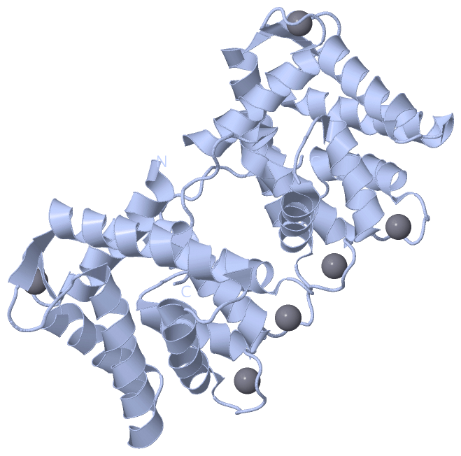
   Legend:   → Mismatch (orange background)
  - → Gap (green background, '-', border residues have a numbering label)
    → Modified Residue (blue background, lower-case, 'x' indicates undefined single-letter code, labelled with number + name)
  x → Chemical Group (purple background, 'x', labelled with number + name, e.g. ACE or NH2)
  extra numbering lines below/above indicate numbering irregularities and modified residue names etc., number ends below/above '|'

 Classification and Annotation

(-) SCOP Domains  (1, 1)

Asymmetric Unit

(-) CATH Domains  (1, 1)

Asymmetric Unit

(-) Pfam Domains  (1, 1)

Asymmetric Unit
(-)
Clan: EF_hand (270)

(-) Gene Ontology  (3, 3)

Asymmetric Unit(hide GO term definitions)
Chain A   (AEQ1_AEQVI | P07164)
molecular function
    GO:0005509    calcium ion binding    Interacting selectively and non-covalently with calcium ions (Ca2+).
    GO:0046872    metal ion binding    Interacting selectively and non-covalently with any metal ion.
biological process
    GO:0008218    bioluminescence    The production of light by certain enzyme-catalyzed reactions in cells.

 Visualization

(-) Interactive Views

Asymmetric Unit
  Complete Structure
    Jena3D(integrated viewing of ligand, site, SAP, PROSITE, SCOP information)
    WebMol | AstexViewer[tm]@PDBe
(Java Applets, require no local installation except for Java; loading may be slow)
    STRAP
(Java WebStart application, automatic local installation, requires Java; full application with system access!)
    RasMol
(require local installation)
    Molscript (VRML)
(requires installation of a VRML viewer; select preferred view via VRML and generate a mono or stereo PDF format file)
 
  Ligands, Modified Residues, Ions
    CA  [ RasMol | Jena3D ]  +environment [ RasMol | Jena3D ]
 
  Sites
    AC1  [ RasMol ]  +environment [ RasMol ]
    AC2  [ RasMol ]  +environment [ RasMol ]
    AC3  [ RasMol ]  +environment [ RasMol ]
 
  Cis Peptide Bonds
(no "Cis Peptide Bonds" information available for 1sl8)
 
Biological Units
  Complete Structure
    Biological Unit 1  [ Jena3D ]
    Biological Unit 2  [ Jena3D ]

(-) Still Images

Jmol
  protein: cartoon or spacefill or dots and stick; nucleic acid: cartoon and stick; ligands: spacefill; active site: stick
Molscript
  protein, nucleic acid: cartoon; ligands: spacefill; active site: ball and stick

 Databases and Analysis Tools

(-) Databases

Access by PDB/NDB ID
  1sl8
    Family and Domain InformationProDom | SYSTERS
    General Structural InformationGlycoscienceDB | MMDB | NDB | OCA | PDB | PDBe | PDBj | PDBsum | PDBWiki | PQS | PROTEOPEDIA
    Orientation in MembranesOPM
    Protein SurfaceSURFACE
    Secondary StructureDSSP (structure derived) | HSSP (homology derived)
    Structural GenomicsGeneCensus
    Structural NeighboursCE | VAST
    Structure ClassificationCATH | Dali | SCOP
    Validation and Original DataBMRB Data View | BMRB Restraints Grid | EDS | PROCHECK | RECOORD | WHAT_CHECK
 
Access by UniProt ID/Accession number
  AEQ1_AEQVI | P07164
    Comparative Protein Structure ModelsModBase
    Genomic InformationEnsembl
    Protein-protein InteractionDIP
    Sequence, Family and Domain InformationInterPro | Pfam | SMART | UniProtKB/SwissProt
 
Access by Enzyme Classificator   (EC Number)
  (no 'Enzyme Classificator' available)
    General Enzyme InformationBRENDA | EC-PDB | Enzyme | IntEnz
    PathwayKEGG | MetaCyc
 
Access by Disease Identifier   (MIM ID)
  (no 'MIM ID' available)
    Disease InformationOMIM
 
Access by GenAge ID
  (no 'GenAge ID' available)
    Age Related InformationGenAge

(-) Analysis Tools

Access by PDB/NDB ID
    Domain InformationXDom
    Interatomic Contacts of Structural UnitsCSU
    Ligand-protein ContactsLPC
    Protein CavitiescastP
    Sequence and Secondary StructurePDBCartoon
    Structure AlignmentSTRAP(Java WebStart application, automatic local installation, requires Java; full application with system access!)
    Structure and Sequence BrowserSTING
 
Access by UniProt ID/Accession number
  AEQ1_AEQVI | P07164
    Protein Disorder PredictionDisEMBL | FoldIndex | GLOBPLOT (for more information see DisProt)

 Related Entries

(-) Entries Sharing at Least One Protein Chain (UniProt ID)

(no "Entries Sharing at Least One Protein Chain" available for 1SL8)

(-) Related Entries Specified in the PDB File

1qv1 CONTAINS CALCIUM-REGULATED PHOTOPROTEIN OBELIN
1ej3 CONTAINS CALCIUM-REGULATED PHOTOPROTEIN AEQUORIN
1el4 CONTAINS CALCIUM-REGULATED PHOTOPROTEIN OBELIN
1jf0 CONTAINS CALCIUM-REGULATED PHOTOPROTEIN OBELIN
1jf2 CONTAINS CALCIUM-REGULATED PHOTOPROTEIN OBELIN W92F MUTANT
1qv0 CONTAINS CALCIUM-REGULATED PHOTOPROTEIN OBELIN
1s36 CONTAINS CALCIUM-DISCHARGED PHOTOPROTEIN W92F OBELIN RELATED ID: AAE-AEQ RELATED DB: TARGETDB