|
|
|
|
 Description
|
|
Compounds
|
||||||||||||||||||||||||||||||||||||||||||||||||
Chains, Units
Summary Information (see also Sequences/Alignments below) |
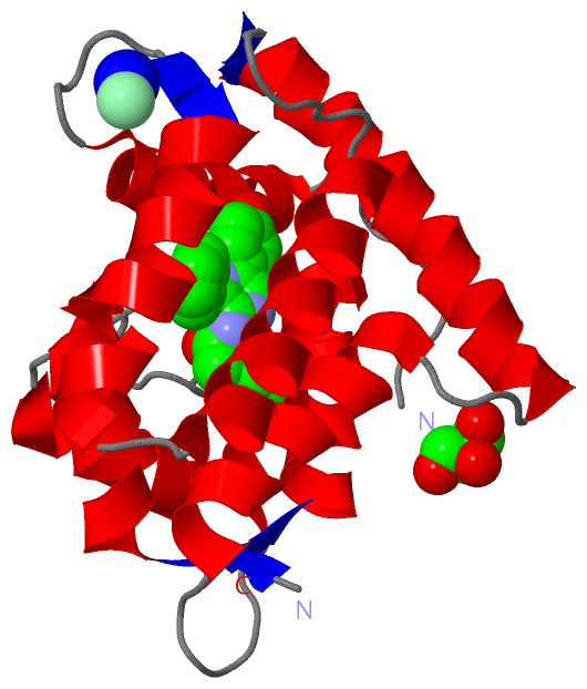
Ligands, Modified Residues, Ions (4, 4)| Asymmetric/Biological Unit (4, 4) |
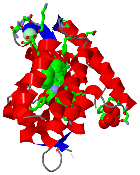
Sites (4, 4)
Asymmetric Unit (4, 4)
|
SS Bonds (0, 0)| (no "SS Bond" information available for 1S36) |
Cis Peptide Bonds (0, 0)| (no "Cis Peptide Bond" information available for 1S36) |
SAPs(SNPs)/Variants (0, 0)| (no "SAP(SNP)/Variant" information available for 1S36) |
PROSITE Motifs (2, 6)| Asymmetric/Biological Unit (2, 6) |
Exons (0, 0)| (no "Exon" information available for 1S36) |
Sequences/Alignments
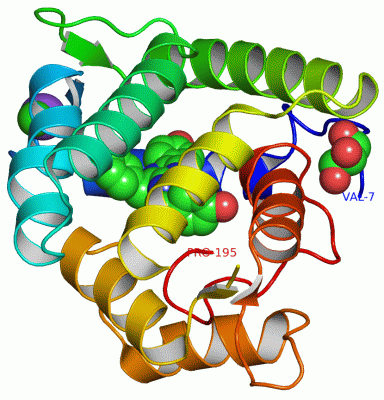
Asymmetric/Biological UnitChain A from PDB Type:PROTEIN Length:185 aligned with OBL_OBELO | Q27709 from UniProtKB/Swiss-Prot Length:195 Alignment length:189 16 26 36 46 56 66 76 86 96 106 116 126 136 146 156 166 176 186 OBL_OBELO 7 VKLKTDFDNPRWIKRHKHMFDFLDINGNGKITLDEIVSKASDDICAKLEATPEQTKRHQVCVEAFFRGCGMEYGKEIAFPQFLDGWKQLATSELKKWARNEPTLIREWGDAVFDIFDKDGSGTITLDEWKAYGKISGISPSQEDCEATFRHCDLDNSGDLDVDEMTRQHLGFWYTLDPEADGLYGNGVP 195 SCOP domains d1s36a_ A: Calcium-regulated photoprotein SCOP domains CATH domains 1s36A00 A:7-195 EF-hand CATH domains Pfam domains ----------------EF_hand_3-1s36A01 -----------------------------------------------EF_hand_5-1s36A02 A:93-173 ---------------------- Pfam domains SAPs(SNPs) --------------------------------------------------------------------------------------------------------------------------------------------------------------------------------------------- SAPs(SNPs) PROSITE (1) ----------EF_HAND_2 PDB: A:17-52 ---------------------------------------------------------EF_HAND_2 PDB: A:110-145 EF_HAND_2 PDB: A:146-181 -------------- PROSITE (1) PROSITE (2) -----------------------EF_HAND_1 --------------------------------------------------------------------------------EF_HAND_1 -----------------------EF_HAND_1 ------------------------ PROSITE (2) Transcript --------------------------------------------------------------------------------------------------------------------------------------------------------------------------------------------- Transcript 1s36 A 7 VKLKTDFDNPRWIKRHKHMFDFLDINGNGKITLDEIVSKASDDICAKLEATPEQTKRHQVCVEAFFRGCGMEYGKEIAFPQFLDGFKQLATSELKKWARNEPTLIREWGDAVFDIFD----GTITLDEWKAYGKISGISPSQEDCEATFRHCDLDNSGDLDVDEMTRQHLGFWYTLDPEADGLYGNGVP 195 16 26 36 46 56 66 76 86 96 106 116 | - | 136 146 156 166 176 186 123 128
|
||||||||||||||||||||
SCOP Domains (1, 1)
Asymmetric/Biological Unit
 |
CATH Domains (1, 1)
Asymmetric/Biological Unit
|
Pfam Domains (2, 2)
Asymmetric/Biological Unit
|
Gene Ontology (3, 3)|
Asymmetric/Biological Unit(hide GO term definitions) Chain A (OBL_OBELO | Q27709)
|
||||||||||||||||||||||||||||||
Interactive Views
|
||||||||||||||||||||||||||||||||||||||||||||||||||||||||||||||||||||||||||||||||||||||||||||||||||||||||||||||||||||||||||||||||||||||||||||||||||||||||||||||||
Still Images
|
||||||||||||||||
Databases
|
||||||||||||||||||||||||||||||||||||||||||||||||||||||||||||||||||||||||||||||||||||||||||||||||||||||||||||||||||||||||||||||||||||||||||||||||||||||||||||||||
Analysis Tools
|
|||||||||||||||||||||||||||||||||||||||||||||||||||||||||||||
Entries Sharing at Least One Protein Chain (UniProt ID)
Related Entries Specified in the PDB File
|
|