|
|
|
|
 Description
|
|
Compounds
|
||||||||||||||||||||||||||||||||||||||||
Chains, Units
Summary Information (see also Sequences/Alignments below) |
Ligands, Modified Residues, Ions (2, 2)| Asymmetric/Biological Unit (2, 2) |
Sites (2, 2)
Asymmetric Unit (2, 2)
|
SS Bonds (0, 0)| (no "SS Bond" information available for 1EL4) |
Cis Peptide Bonds (0, 0)| (no "Cis Peptide Bond" information available for 1EL4) |
SAPs(SNPs)/Variants (0, 0)| (no "SAP(SNP)/Variant" information available for 1EL4) |
PROSITE Motifs (2, 6)| Asymmetric/Biological Unit (2, 6) |
Exons (0, 0)| (no "Exon" information available for 1EL4) |
Sequences/Alignments
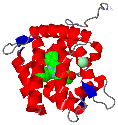
Asymmetric/Biological UnitChain A from PDB Type:PROTEIN Length:194 aligned with OBL_OBELO | Q27709 from UniProtKB/Swiss-Prot Length:195 Alignment length:194 11 21 31 41 51 61 71 81 91 101 111 121 131 141 151 161 171 181 191 OBL_OBELO 2 SSKYAVKLKTDFDNPRWIKRHKHMFDFLDINGNGKITLDEIVSKASDDICAKLEATPEQTKRHQVCVEAFFRGCGMEYGKEIAFPQFLDGWKQLATSELKKWARNEPTLIREWGDAVFDIFDKDGSGTITLDEWKAYGKISGISPSQEDCEATFRHCDLDNSGDLDVDEMTRQHLGFWYTLDPEADGLYGNGVP 195 SCOP domains d1el4a_ A: Calcium-regulated photoprotein SCOP domains CATH domains 1el4A00 A:2-195 EF-hand CATH domains Pfam domains -------------------------------------------------------------------------------------------------------------------------------------------------------------------------------------------------- Pfam domains SAPs(SNPs) -------------------------------------------------------------------------------------------------------------------------------------------------------------------------------------------------- SAPs(SNPs) PROSITE (1) ---------------EF_HAND_2 PDB: A:17-52 ---------------------------------------------------------EF_HAND_2 PDB: A:110-145 EF_HAND_2 PDB: A:146-181 -------------- PROSITE (1) PROSITE (2) ----------------------------EF_HAND_1 --------------------------------------------------------------------------------EF_HAND_1 -----------------------EF_HAND_1 ------------------------ PROSITE (2) Transcript -------------------------------------------------------------------------------------------------------------------------------------------------------------------------------------------------- Transcript 1el4 A 2 SSKYAVKLKTDFDNPRWIKRHKHMFDFLDINGNGKITLDEIVSKASDDICAKLEATPEQTKRHQVCVEAFFRGCGMEYGKEIAFPQFLDGWKQLATSELKKWARNEPTLIREWGDAVFDIFDKDGSGTITLDEWKAYGKISGISPSQEDCEATFRHCDLDNSGDLDVDEMTRQHLGFWYTLDPEADGLYGNGVP 195 11 21 31 41 51 61 71 81 91 101 111 121 131 141 151 161 171 181 191
|
||||||||||||||||||||
SCOP Domains (1, 1)
Asymmetric/Biological Unit
 |
CATH Domains (1, 1)
Asymmetric/Biological Unit
|
Pfam Domains (0, 0)| (no "Pfam Domain" information available for 1EL4) |
Gene Ontology (3, 3)|
Asymmetric/Biological Unit(hide GO term definitions) Chain A (OBL_OBELO | Q27709)
|
||||||||||||||||||||||||||||||
Interactive Views
|
||||||||||||||||||||||||||||||||||||||||||||||||||||||||||||||||||||||||||||||||||||||||||||||||||||||||||||||||||||||||||||||||||||
Still Images
|
||||||||||||||||
Databases
|
||||||||||||||||||||||||||||||||||||||||||||||||||||||||||||||||||||||||||||||||||||||||||||||||||||||||||||||||||||||||||||||||||||||||||||||||||||||||||||||||
Analysis Tools
|
|||||||||||||||||||||||||||||||||||||||||||||||||||||||||||||
Entries Sharing at Least One Protein Chain (UniProt ID)
Related Entries Specified in the PDB File
|
|