| molecular function |
|---|
| | GO:0031683 | | G-protein beta/gamma-subunit complex binding | | Interacting selectively and non-covalently with a complex of G-protein beta/gamma subunits. |
| | GO:0005509 | | calcium ion binding | | Interacting selectively and non-covalently with calcium ions (Ca2+). |
| | GO:0032795 | | heterotrimeric G-protein binding | | Interacting selectively and non-covalently with a heterotrimeric G-protein. |
| | GO:0046872 | | metal ion binding | | Interacting selectively and non-covalently with any metal ion. |
| | GO:0008017 | | microtubule binding | | Interacting selectively and non-covalently with microtubules, filaments composed of tubulin monomers. |
| | GO:0005515 | | protein binding | | Interacting selectively and non-covalently with any protein or protein complex (a complex of two or more proteins that may include other nonprotein molecules). |
| biological process |
|---|
| | GO:0006281 | | DNA repair | | The process of restoring DNA after damage. Genomes are subject to damage by chemical and physical agents in the environment (e.g. UV and ionizing radiations, chemical mutagens, fungal and bacterial toxins, etc.) and by free radicals or alkylating agents endogenously generated in metabolism. DNA is also damaged because of errors during its replication. A variety of different DNA repair pathways have been reported that include direct reversal, base excision repair, nucleotide excision repair, photoreactivation, bypass, double-strand break repair pathway, and mismatch repair pathway. |
| | GO:0000086 | | G2/M transition of mitotic cell cycle | | The mitotic cell cycle transition by which a cell in G2 commits to M phase. The process begins when the kinase activity of M cyclin/CDK complex reaches a threshold high enough for the cell cycle to proceed. This is accomplished by activating a positive feedback loop that results in the accumulation of unphosphorylated and active M cyclin/CDK complex. |
| | GO:0007049 | | cell cycle | | The progression of biochemical and morphological phases and events that occur in a cell during successive cell replication or nuclear replication events. Canonically, the cell cycle comprises the replication and segregation of genetic material followed by the division of the cell, but in endocycles or syncytial cells nuclear replication or nuclear division may not be followed by cell division. |
| | GO:0051301 | | cell division | | The process resulting in division and partitioning of components of a cell to form more cells; may or may not be accompanied by the physical separation of a cell into distinct, individually membrane-bounded daughter cells. |
| | GO:0006974 | | cellular response to DNA damage stimulus | | Any process that results in a change in state or activity of a cell (in terms of movement, secretion, enzyme production, gene expression, etc.) as a result of a stimulus indicating damage to its DNA from environmental insults or errors during metabolism. |
| | GO:0007099 | | centriole replication | | The cell cycle process in which a daughter centriole is formed perpendicular to an existing centriole. An immature centriole contains a ninefold radially symmetric array of single microtubules; mature centrioles consist of a radial array of nine microtubule triplets, doublets, or singlets depending upon the species and cell type. Duplicated centrioles also become the ciliary basal body in cells that form cilia during G0. |
| | GO:0070911 | | global genome nucleotide-excision repair | | The nucleotide-excision repair process in which DNA lesions are removed from nontranscribed strands and from transcriptionally silent regions over the entire genome. |
| | GO:0006289 | | nucleotide-excision repair | | A DNA repair process in which a small region of the strand surrounding the damage is removed from the DNA helix as an oligonucleotide. The small gap left in the DNA helix is filled in by the sequential action of DNA polymerase and DNA ligase. Nucleotide excision repair recognizes a wide range of substrates, including damage caused by UV irradiation (pyrimidine dimers and 6-4 photoproducts) and chemicals (intrastrand cross-links and bulky adducts). |
| | GO:0000715 | | nucleotide-excision repair, DNA damage recognition | | The identification of lesions in DNA, such as pyrimidine-dimers, intrastrand cross-links, and bulky adducts. The wide range of substrate specificity suggests the repair complex recognizes distortions in the DNA helix. |
| | GO:0000717 | | nucleotide-excision repair, DNA duplex unwinding | | The unwinding, or local denaturation, of the DNA duplex to create a bubble around the site of the DNA damage. |
| | GO:0006294 | | nucleotide-excision repair, preincision complex assembly | | The aggregation, arrangement and bonding together of proteins on DNA to form the multiprotein complex involved in damage recognition, DNA helix unwinding, and endonucleolytic cleavage at the site of DNA damage. This assembly occurs before the phosphodiester backbone of the damaged strand is cleaved 3' and 5' of the site of DNA damage. |
| | GO:0016925 | | protein sumoylation | | The process in which a SUMO protein (small ubiquitin-related modifier) is conjugated to a target protein via an isopeptide bond between the carboxyl terminus of SUMO with an epsilon-amino group of a lysine residue of the target protein. |
| | GO:0032465 | | regulation of cytokinesis | | Any process that modulates the frequency, rate or extent of the division of the cytoplasm of a cell and its separation into two daughter cells. |
| | GO:0007283 | | spermatogenesis | | The process of formation of spermatozoa, including spermatocytogenesis and spermiogenesis. |
| cellular component |
|---|
| | GO:0071942 | | XPC complex | | A nucleotide-excision repair complex that is involved in damage sensing during global genome nucleotide excision repair (GG-NER). It is part of the pre-incision (or initial recognition) complex bound to sites of DNA damage. In human, it is composed of XPC, RAD23B and CETN2. |
| | GO:0005814 | | centriole | | A cellular organelle, found close to the nucleus in many eukaryotic cells, consisting of a small cylinder with microtubular walls, 300-500 nm long and 150-250 nm in diameter. It contains nine short, parallel, peripheral microtubular fibrils, each fibril consisting of one complete microtubule fused to two incomplete microtubules. Cells usually have two centrioles, lying at right angles to each other. At division, each pair of centrioles generates another pair and the twin pairs form the pole of the mitotic spindle. |
| | GO:0005813 | | centrosome | | A structure comprised of a core structure (in most organisms, a pair of centrioles) and peripheral material from which a microtubule-based structure, such as a spindle apparatus, is organized. Centrosomes occur close to the nucleus during interphase in many eukaryotic cells, though in animal cells it changes continually during the cell-division cycle. |
| | GO:0036064 | | ciliary basal body | | A membrane-tethered, short cylindrical array of microtubules and associated proteins found at the base of a eukaryotic cilium (also called flagellum) that is similar in structure to a centriole and derives from it. The cilium basal body is the site of assembly and remodelling of the cilium and serves as a nucleation site for axoneme growth. As well as anchoring the cilium, it is thought to provide a selective gateway regulating the entry of ciliary proteins and vesicles by intraflagellar transport. |
| | GO:0005929 | | cilium | | A specialized eukaryotic organelle that consists of a filiform extrusion of the cell surface and of some cytoplasmic parts. Each cilium is largely bounded by an extrusion of the cytoplasmic (plasma) membrane, and contains a regular longitudinal array of microtubules, anchored to a basal body. |
| | GO:0005737 | | cytoplasm | | All of the contents of a cell excluding the plasma membrane and nucleus, but including other subcellular structures. |
| | GO:0005856 | | cytoskeleton | | Any of the various filamentous elements that form the internal framework of cells, and typically remain after treatment of the cells with mild detergent to remove membrane constituents and soluble components of the cytoplasm. The term embraces intermediate filaments, microfilaments, microtubules, the microtrabecular lattice, and other structures characterized by a polymeric filamentous nature and long-range order within the cell. The various elements of the cytoskeleton not only serve in the maintenance of cellular shape but also have roles in other cellular functions, including cellular movement, cell division, endocytosis, and movement of organelles. |
| | GO:0005829 | | cytosol | | The part of the cytoplasm that does not contain organelles but which does contain other particulate matter, such as protein complexes. |
| | GO:0005622 | | intracellular | | The living contents of a cell; the matter contained within (but not including) the plasma membrane, usually taken to exclude large vacuoles and masses of secretory or ingested material. In eukaryotes it includes the nucleus and cytoplasm. |
| | GO:0005654 | | nucleoplasm | | That part of the nuclear content other than the chromosomes or the nucleolus. |
| | GO:0005634 | | nucleus | | A membrane-bounded organelle of eukaryotic cells in which chromosomes are housed and replicated. In most cells, the nucleus contains all of the cell's chromosomes except the organellar chromosomes, and is the site of RNA synthesis and processing. In some species, or in specialized cell types, RNA metabolism or DNA replication may be absent. |
| | GO:0032391 | | photoreceptor connecting cilium | | The portion of the photoreceptor cell cilium linking the photoreceptor inner and outer segments. It's considered to be equivalent to the ciliary transition zone. |
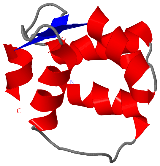
 Description
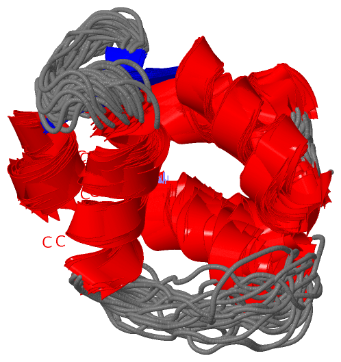