|
|
|
|
 Description
|
|
Compounds
|
||||||||||||||||||||||||
Chains, Units
Summary Information (see also Sequences/Alignments below) |
Ligands, Modified Residues, Ions (3, 9)
Asymmetric Unit (3, 9)
|
Sites (9, 9)
Asymmetric Unit (9, 9)
|
SS Bonds (6, 6)
Asymmetric Unit
|
||||||||||||||||||||||||||||
Cis Peptide Bonds (0, 0)| (no "Cis Peptide Bond" information available for 1IJV) |
SAPs(SNPs)/Variants (3, 6)
Asymmetric Unit (3, 6)
|
|||||||||||||||||||||||||||||||||||||||||||||||||||||||||||||||||||||||||||||||||||||||||||||||||||||||||||||||||||||||||||||||||||||||||||||||||||||||||||||||||||||||||||||||||||||||||||||||||||||||||||||||||||||||||||||||||||||||||||||
PROSITE Motifs (0, 0)| (no "PROSITE Motif" information available for 1IJV) |
Exons (1, 2)
Asymmetric Unit (1, 2)
|
||||||||||||||||||||||||||||||||||||||||||||||||
Sequences/Alignments
Asymmetric UnitChain A from PDB Type:PROTEIN Length:36 aligned with DEFB1_HUMAN | P60022 from UniProtKB/Swiss-Prot Length:68 Alignment length:36 42 52 62 DEFB1_HUMAN 33 DHYNCVSSGGQCLYSACPIFTKIQGTCYRGKAKCCK 68 SCOP domains d1ijva_ A: Beta-defensin, BD SCOP domains CATH domains ------------------------------------ CATH domains Pfam domains ------------------------------------ Pfam domains SAPs(SNPs) -----I---------V------------------S- SAPs(SNPs) PROSITE ------------------------------------ PROSITE Transcript 1 Exon 1.2 PDB: A:1-36 UniProt: 21-68 Transcript 1 1ijv A 1 DHYNCVSSGGQCLYSACPIFTKIQGTCYRGKAKCCK 36 10 20 30 Chain B from PDB Type:PROTEIN Length:36 aligned with DEFB1_HUMAN | P60022 from UniProtKB/Swiss-Prot Length:68 Alignment length:36 42 52 62 DEFB1_HUMAN 33 DHYNCVSSGGQCLYSACPIFTKIQGTCYRGKAKCCK 68 SCOP domains d1ijvb_ B: Beta-defensin, BD SCOP domains CATH domains ------------------------------------ CATH domains Pfam domains ------------------------------------ Pfam domains SAPs(SNPs) -----I---------V------------------S- SAPs(SNPs) PROSITE ------------------------------------ PROSITE Transcript 1 Exon 1.2 PDB: B:1-36 UniProt: 21-68 Transcript 1 1ijv B 1 DHYNCVSSGGQCLYSACPIFTKIQGTCYRGKAKCCK 36 10 20 30
|
||||||||||||||||||||
SCOP Domains (1, 2)
Asymmetric Unit
 |
CATH Domains (0, 0)| (no "CATH Domain" information available for 1IJV) |
Pfam Domains (0, 0)| (no "Pfam Domain" information available for 1IJV) |
Gene Ontology (16, 16)|
Asymmetric Unit(hide GO term definitions) Chain A,B (DEFB1_HUMAN | P60022)
|
||||||||||||||||||||||||||||||||||||||||||||||||||||||||||||||||||||||||||||||||||||||||||||||||||||||||||||
Interactive Views
|
|||||||||||||||||||||||||||||||||||||||||||||||||||||||||||||||||||||||||||||||||||||||||||||||||||||||||||||||||||||||||||||||||||||||||||||||||||||||||||||||||||||||||||||||||||||||||||||||||||||||||||||||||||
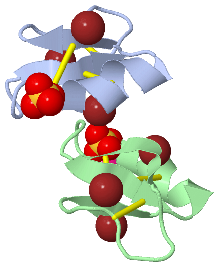
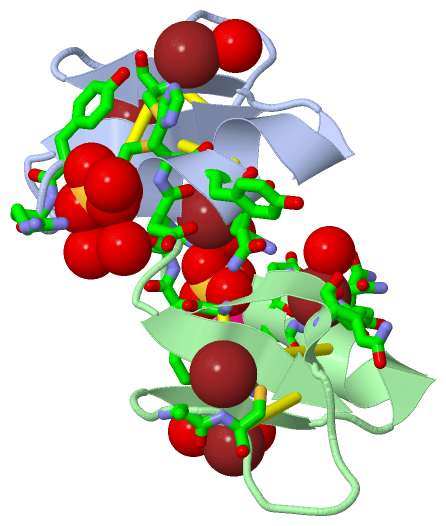
Still Images
|
||||||||||||||||
Databases
|
||||||||||||||||||||||||||||||||||||||||||||||||||||||||||||||||||||||||||||||||||||||||||||||||||||||||||||||||||||||||||||||||||||||||||||||||||||||||||||||||
Analysis Tools
|
|||||||||||||||||||||||||||||||||||||||||||||||||||||||||||||
Entries Sharing at Least One Protein Chain (UniProt ID)
Related Entries Specified in the PDB File
|
|