|
|
|
|
 Description
|
|
Compounds
|
||||||||||||||||||||||||||||||||||||||||||||||||||||||||||||||||||||||||||||||||||
Chains, Units
Summary Information (see also Sequences/Alignments below) |
Ligands, Modified Residues, Ions (4, 29)
Asymmetric Unit (4, 29)
|
Sites (21, 21)
Asymmetric Unit (21, 21)
|
SS Bonds (0, 0)| (no "SS Bond" information available for 1HC8) |
Cis Peptide Bonds (0, 0)| (no "Cis Peptide Bond" information available for 1HC8) |
SAPs(SNPs)/Variants (0, 0)| (no "SAP(SNP)/Variant" information available for 1HC8) |
PROSITE Motifs (1, 2)
Asymmetric Unit (1, 2)
|
||||||||||||||||||||||||||||||||||||||||||||||||||||||||||||||||||||||||
Exons (0, 0)| (no "Exon" information available for 1HC8) |
Sequences/Alignments
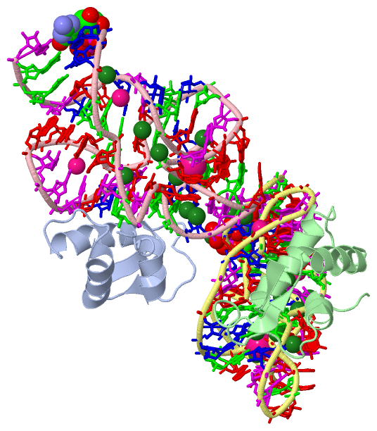
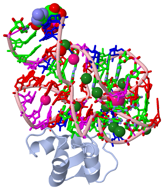
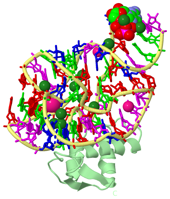
Asymmetric UnitChain A from PDB Type:PROTEIN Length:74 aligned with RL11_GEOSE | P56210 from UniProtKB/Swiss-Prot Length:133 Alignment length:74 68 78 88 98 108 118 128 RL11_GEOSE 59 TFITKTPPAAVLLKKAAGIESGSGEPNRNKVATIKRDKVREIAELKMPDLNAASIEAAMRMIEGTARSMGIVVE 132 SCOP domains d1hc8a_ A: Ribosomal protein L11, C-terminal domain SCOP domains CATH domains 1hc8A00 A:2-75 [code=1.10.10.250, no name defined] CATH domains Pfam domains -------------------------------------------------------------------------- Pfam domains SAPs(SNPs) -------------------------------------------------------------------------- SAPs(SNPs) PROSITE -----------------------------------------------------------RIBOSOMAL_L11 PROSITE Transcript -------------------------------------------------------------------------- Transcript 1hc8 A 2 TFITKTPPAAVLLKKAAGIESGSGEPNRNKVATIKRDKVREIAELKMPDLNAASIEAAMRMIEGTARSMGIVVE 75 11 21 31 41 51 61 71 Chain B from PDB Type:PROTEIN Length:70 aligned with RL11_GEOSE | P56210 from UniProtKB/Swiss-Prot Length:133 Alignment length:70 72 82 92 102 112 122 132 RL11_GEOSE 63 KTPPAAVLLKKAAGIESGSGEPNRNKVATIKRDKVREIAELKMPDLNAASIEAAMRMIEGTARSMGIVVE 132 SCOP domains d1hc8b_ B: Ribosomal protein L11, C-terminal domain SCOP domains CATH domains 1hc8B00 B:206-275 [code=1.10.10.250, no name defined] CATH domains Pfam domains ---------------------------------------------------------------------- Pfam domains SAPs(SNPs) ---------------------------------------------------------------------- SAPs(SNPs) PROSITE -------------------------------------------------------RIBOSOMAL_L11 PROSITE Transcript ---------------------------------------------------------------------- Transcript 1hc8 B 206 KTPPAAVLLKKAAGIESGSGEPNRNKVATIKRDKVREIAELKMPDLNAASIEAAMRMIEGTARSMGIVVE 275 215 225 235 245 255 265 275
Chain C from PDB  Type:RNA  Length:58

                 1hc8 C 101 xCCAGGAUGUAGGCUUAGAAGCAGCCAUCAUUUAAAGAAAGCGUAAUAGCUCACUGGU 158
                            |      110       120       130       140       150
                          101-GTP
Chain D from PDB  Type:RNA  Length:58

                 1hc8 D 301 xCCAGGAUGUAGGCUUAGAAGCAGCCAUCAUUUAAAGAAAGCGUAAUAGCUCACUGGU 358
                            |      310       320       330       340       350
                          301-GTP
|
||||||||||||||||||||
SCOP Domains (1, 2)
Asymmetric Unit
 |
CATH Domains (1, 2)
Asymmetric Unit
|
Pfam Domains (0, 0)| (no "Pfam Domain" information available for 1HC8) |
Gene Ontology (6, 6)|
Asymmetric Unit(hide GO term definitions) Chain A,B (RL11_GEOSE | P56210)
|
||||||||||||||||||||||||||||||||||||||||||||||||||||||
Interactive Views
|
||||||||||||||||||||||||||||||||||||||||||||||||||||||||||||||||||||||||||||||||||||||||||||||||||||||||||||||||||||||||||||||||||||||||||||||||||||||||||||||||||||||||||||||||||||||||||||||||||||||||||||||||||||||||||||||||||||||||||||||||||||||||||||||||||||||||||||||||||||||||||||||||||||||||||||||
Still Images
|
||||||||||||||||
Databases
|
||||||||||||||||||||||||||||||||||||||||||||||||||||||||||||||||||||||||||||||||||||||||||||||||||||||||||||||||||||||||||||||||||||||||||||||||||||||||||||||||
Analysis Tools
|
|||||||||||||||||||||||||||||||||||||||||||||||||||||||||||||
Entries Sharing at Least One Protein Chain (UniProt ID)
Related Entries Specified in the PDB File
|
|