Show PDB file:   
         Plain Text   HTML   (compressed file size)
QuickSearch:   
by PDB,NDB,UniProt,PROSITE Code or Search Term(s)  
(-)NMR Structure - manually
(-)NMR Structure - model 1
(-)NMR Structure - all models
collapse expand < >
Image NMR Structure - manually
NMR Structure - manually  (Jmol Viewer)
Image NMR Structure - model 1
NMR Structure - model 1  (Jmol Viewer)
Image NMR Structure - all models
NMR Structure - all models  (Jmol Viewer)

(-) Description

Title :  PEPTIDE FRAGMENT THR671-LEU690 OF THE RABBIT SKELETAL DIHYDROPYRIDINE RECEPTOR
 
Authors :  M. Casarotto, A. Dulhunty
Date :  13 Jan 00  (Deposition) - 19 Jul 00  (Release) - 24 Feb 09  (Revision)
Method :  SOLUTION NMR
Resolution :  NOT APPLICABLE
Chains :  NMR Structure  :  A
Keywords :  Dihydropyridine Receptor, Ryanodine Receptor, Nmr Structure, Signaling Protein (Keyword Search: [Gene Ontology, PubMed, Web (Google))
 
Reference :  M. G. Casarotto, F. Gibson, S. M. Pace, S. M. Curtis, M. Mulcair, A. F. Dulhunty
A Structural Requirement For Activation Of Skeletal Ryanodine Receptors By Peptides Of The Dihydropyridine Receptor Ii-Iii Loop.
J. Biol. Chem. V. 275 11631 2000
PubMed-ID: 10766780  |  Reference-DOI: 10.1074/JBC.275.16.11631
(for further references see the PDB file header)

(-) Compounds

Molecule 1 - SKELETAL DIHYDROPYRIDINE RECEPTOR
    ChainsA
    EngineeredYES
    FragmentII-III LOOP REGION, FRAGMENT THR671-LEU690 A1
    Other DetailsTHIS PEPTIDE WAS CHEMICALLY SYNTHESIZED. THE SEQUENCE OF THIS PEPTIDE NATURALLY OCCURS IN RABBIT (ORYCTOLAGUS CUNICULUS)
    SyntheticYES

 Structural Features

(-) Chains, Units

  
NMR Structure 

Summary Information (see also Sequences/Alignments below)

(-) Ligands, Modified Residues, Ions  (0, 0)

(no "Ligand,Modified Residues,Ions" information available for 1DU1)

(-) Sites  (0, 0)

(no "Site" information available for 1DU1)

(-) SS Bonds  (0, 0)

(no "SS Bond" information available for 1DU1)

(-) Cis Peptide Bonds  (0, 0)

(no "Cis Peptide Bond" information available for 1DU1)

 Sequence-Structure Mapping

(-) SAPs(SNPs)/Variants  (0, 0)

(no "SAP(SNP)/Variant" information available for 1DU1)

(-) PROSITE Motifs  (0, 0)

(no "PROSITE Motif" information available for 1DU1)

(-) Exons   (0, 0)

(no "Exon" information available for 1DU1)

(-) Sequences/Alignments

NMR Structure
   Reformat: Number of residues per line =  ('0' or empty: single-line sequence representation)
  Number of residues per labelling interval =   
  UniProt sequence: complete  aligned part    
   Show mapping: SCOP domains CATH domains Pfam domains Secondary structure (by author)
SAPs(SNPs) PROSITE motifs Exons
(details for a mapped element are shown in a popup box when the mouse pointer rests over it)
Chain A from PDB  Type:PROTEIN  Length:20
 aligned with CAC1S_RABIT | P07293 from UniProtKB/Swiss-Prot  Length:1873

    Alignment length:20
                                   680       690
          CAC1S_RABIT   671 TSAQKAKAEERKRRKMSRGL 690
               SCOP domains d1du1a_ A:           SCOP domains
               CATH domains -------------------- CATH domains
               Pfam domains -------------------- Pfam domains
         Sec.struct. author hhhhhhhh............ Sec.struct. author
                 SAPs(SNPs) -------------------- SAPs(SNPs)
                    PROSITE -------------------- PROSITE
                 Transcript -------------------- Transcript
                 1du1 A   1 TSAQKAKAEERKRRKMSRGL  20
                                    10        20

   Legend:   → Mismatch (orange background)
  - → Gap (green background, '-', border residues have a numbering label)
    → Modified Residue (blue background, lower-case, 'x' indicates undefined single-letter code, labelled with number + name)
  x → Chemical Group (purple background, 'x', labelled with number + name, e.g. ACE or NH2)
  extra numbering lines below/above indicate numbering irregularities and modified residue names etc., number ends below/above '|'

 Classification and Annotation

(-) SCOP Domains  (1, 1)

NMR Structure
(-)
Class: Peptides (792)

(-) CATH Domains  (0, 0)

(no "CATH Domain" information available for 1DU1)

(-) Pfam Domains  (0, 0)

(no "Pfam Domain" information available for 1DU1)

(-) Gene Ontology  (15, 15)

NMR Structure(hide GO term definitions)
Chain A   (CAC1S_RABIT | P07293)
molecular function
    GO:0005262    calcium channel activity    Enables the facilitated diffusion of a calcium ion (by an energy-independent process) involving passage through a transmembrane aqueous pore or channel without evidence for a carrier-mediated mechanism.
    GO:0005216    ion channel activity    Enables the facilitated diffusion of an ion (by an energy-independent process) by passage through a transmembrane aqueous pore or channel without evidence for a carrier-mediated mechanism. May be either selective (it enables passage of a specific ion only) or non-selective (it enables passage of two or more ions of same charge but different size).
    GO:0046872    metal ion binding    Interacting selectively and non-covalently with any metal ion.
    GO:0005515    protein binding    Interacting selectively and non-covalently with any protein or protein complex (a complex of two or more proteins that may include other nonprotein molecules).
    GO:0005245    voltage-gated calcium channel activity    Enables the transmembrane transfer of a calcium ion by a voltage-gated channel. A voltage-gated channel is a channel whose open state is dependent on the voltage across the membrane in which it is embedded.
    GO:0005244    voltage-gated ion channel activity    Enables the transmembrane transfer of an ion by a voltage-gated channel. An ion is an atom or group of atoms carrying an electric charge by virtue of having gained or lost one or more electrons. A voltage-gated channel is a channel whose open state is dependent on the voltage across the membrane in which it is embedded.
biological process
    GO:0070588    calcium ion transmembrane transport    A process in which a calcium ion is transported from one side of a membrane to the other by means of some agent such as a transporter or pore.
    GO:0006816    calcium ion transport    The directed movement of calcium (Ca) ions into, out of or within a cell, or between cells, by means of some agent such as a transporter or pore.
    GO:0006811    ion transport    The directed movement of charged atoms or small charged molecules into, out of or within a cell, or between cells, by means of some agent such as a transporter or pore.
    GO:0034765    regulation of ion transmembrane transport    Any process that modulates the frequency, rate or extent of the directed movement of ions from one side of a membrane to the other.
    GO:0055085    transmembrane transport    The process in which a solute is transported across a lipid bilayer, from one side of a membrane to the other
    GO:0006810    transport    The directed movement of substances (such as macromolecules, small molecules, ions) or cellular components (such as complexes and organelles) into, out of or within a cell, or between cells, or within a multicellular organism by means of some agent such as a transporter, pore or motor protein.
cellular component
    GO:0016021    integral component of membrane    The component of a membrane consisting of the gene products and protein complexes having at least some part of their peptide sequence embedded in the hydrophobic region of the membrane.
    GO:0016020    membrane    A lipid bilayer along with all the proteins and protein complexes embedded in it an attached to it.
    GO:0005891    voltage-gated calcium channel complex    A protein complex that forms a transmembrane channel through which calcium ions may pass in response to changes in membrane potential.
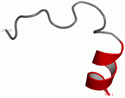
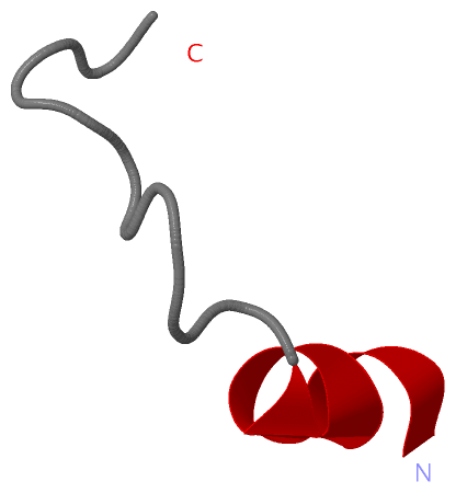
 Visualization

(-) Interactive Views

NMR Structure
  Complete Structure
    Jena3D(integrated viewing of ligand, site, SAP, PROSITE, SCOP information)
    WebMol | AstexViewer[tm]@PDBe
(Java Applets, require no local installation except for Java; loading may be slow)
    STRAP
(Java WebStart application, automatic local installation, requires Java; full application with system access!)
    RasMol
(require local installation)
    Molscript (VRML)
(requires installation of a VRML viewer; select preferred view via VRML and generate a mono or stereo PDF format file)
 
  Ligands, Modified Residues, Ions
(no "Ligands, Modified Residues, Ions" information available for 1du1)
 
  Sites
(no "Sites" information available for 1du1)
 
  Cis Peptide Bonds
(no "Cis Peptide Bonds" information available for 1du1)
 

(-) Still Images

Jmol
  protein: cartoon or spacefill or dots and stick; nucleic acid: cartoon and stick; ligands: spacefill; active site: stick
Molscript
  protein, nucleic acid: cartoon; ligands: spacefill; active site: ball and stick

 Databases and Analysis Tools

(-) Databases

Access by PDB/NDB ID
  1du1
    Family and Domain InformationProDom | SYSTERS
    General Structural InformationGlycoscienceDB | MMDB | NDB | OCA | PDB | PDBe | PDBj | PDBsum | PDBWiki | PQS | PROTEOPEDIA
    Orientation in MembranesOPM
    Protein SurfaceSURFACE
    Secondary StructureDSSP (structure derived) | HSSP (homology derived)
    Structural GenomicsGeneCensus
    Structural NeighboursCE | VAST
    Structure ClassificationCATH | Dali | SCOP
    Validation and Original DataBMRB Data View | BMRB Restraints Grid | EDS | PROCHECK | RECOORD | WHAT_CHECK
 
Access by UniProt ID/Accession number
  CAC1S_RABIT | P07293
    Comparative Protein Structure ModelsModBase
    Genomic InformationEnsembl
    Protein-protein InteractionDIP
    Sequence, Family and Domain InformationInterPro | Pfam | SMART | UniProtKB/SwissProt
 
Access by Enzyme Classificator   (EC Number)
  (no 'Enzyme Classificator' available)
    General Enzyme InformationBRENDA | EC-PDB | Enzyme | IntEnz
    PathwayKEGG | MetaCyc
 
Access by Disease Identifier   (MIM ID)
  (no 'MIM ID' available)
    Disease InformationOMIM
 
Access by GenAge ID
  (no 'GenAge ID' available)
    Age Related InformationGenAge

(-) Analysis Tools

Access by PDB/NDB ID
    Domain InformationXDom
    Interatomic Contacts of Structural UnitsCSU
    Ligand-protein ContactsLPC
    Protein CavitiescastP
    Sequence and Secondary StructurePDBCartoon
    Structure AlignmentSTRAP(Java WebStart application, automatic local installation, requires Java; full application with system access!)
    Structure and Sequence BrowserSTING
 
Access by UniProt ID/Accession number
  CAC1S_RABIT | P07293
    Protein Disorder PredictionDisEMBL | FoldIndex | GLOBPLOT (for more information see DisProt)

 Related Entries

(-) Entries Sharing at Least One Protein Chain (UniProt ID)

UniProtKB/Swiss-Prot
        CAC1S_RABIT | P072931jzp 1t3l 3jbr 5gjv 5gjw

(-) Related Entries Specified in the PDB File

(no "Related Entries Specified in the PDB File" available for 1DU1)