|
|
|
|
 Description
|
|
Compounds
|
||||||||||||||||||||||||
Chains, Units
Summary Information (see also Sequences/Alignments below) |
Ligands, Modified Residues, Ions (3, 12)
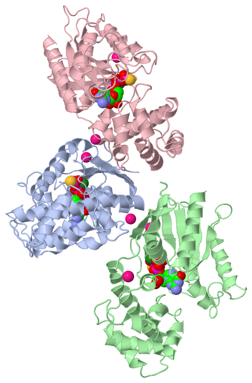
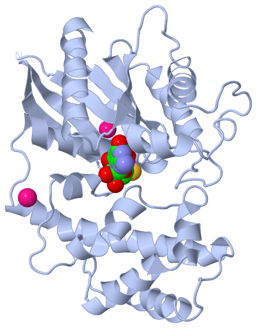
Asymmetric Unit (3, 12)
|
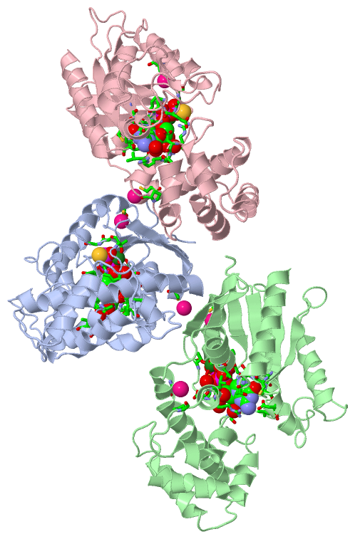
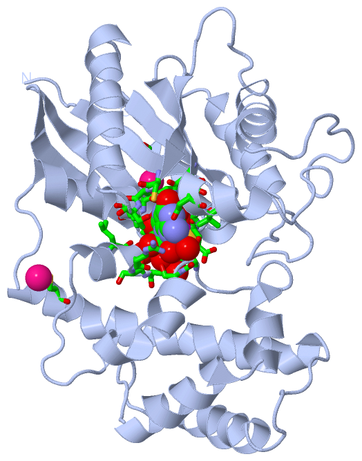
Sites (12, 12)
Asymmetric Unit (12, 12)
|
SS Bonds (0, 0)| (no "SS Bond" information available for 1TND) |
Cis Peptide Bonds (0, 0)| (no "Cis Peptide Bond" information available for 1TND) |
SAPs(SNPs)/Variants (0, 0)| (no "SAP(SNP)/Variant" information available for 1TND) |
PROSITE Motifs (0, 0)| (no "PROSITE Motif" information available for 1TND) |
Exons (8, 24)
Sequences/Alignments
Asymmetric UnitChain A from PDB Type:PROTEIN Length:323 aligned with GNAT1_BOVIN | P04695 from UniProtKB/Swiss-Prot Length:350 Alignment length:323 36 46 56 66 76 86 96 106 116 126 136 146 156 166 176 186 196 206 216 226 236 246 256 266 276 286 296 306 316 326 336 346 GNAT1_BOVIN 27 ARTVKLLLLGAGESGKSTIVKQMKIIHQDGYSLEECLEFIAIIYGNTLQSILAIVRAMTTLNIQYGDSARQDDARKLMHMADTIEEGTMPKEMSDIIQRLWKDSGIQACFDRASEYQLNDSAGYYLSDLERLVTPGYVPTEQDVLRSRVKTTGIIETQFSFKDLNFRMFDVGGQRSERKKWIHCFEGVTCIIFIAALSAYDMVLVEDDEVNRMHESLHLFNSICNHRYFATTSIVLFLNKKDVFSEKIKKAHLSICFPDYNGPNTYEDAGNYIKVQFLELNMRRDVKEIYSHMTCATDTQNVKFVFDAVTDIIIKENLKDCGL 349 SCOP domains d1tnda2 A:27-56,A:178-349 d1tnda1 A:57-177 Transducin (alpha subunit), insertion domain d1tnda2 A:27-56,A:178-349 Transducin (alpha subunit) SCOP domains CATH domains 1tndA01 A:27-57,A:177-347 1tndA02 A:58-176 GI Alpha 1, domain 2-like 1tndA01 A:27-57,A:177-347 P-loop containing nucleotide triphosphate hydrolases -- CATH domains Pfam domains ----------------------------------------------------------------------------------------------------------------------------------------------------------------------------------------------------------------------------------------------------------------------------------------------------------------------------------- Pfam domains SAPs(SNPs) ----------------------------------------------------------------------------------------------------------------------------------------------------------------------------------------------------------------------------------------------------------------------------------------------------------------------------------- SAPs(SNPs) PROSITE ----------------------------------------------------------------------------------------------------------------------------------------------------------------------------------------------------------------------------------------------------------------------------------------------------------------------------------- PROSITE Transcript 1 (1) Exon 1.1 -------------Exon 1.3 PDB: A:50-97 UniProt: 50-97 Exon 1.4 PDB: A:98-150 UniProt: 98-150 ------------------------------------------Exon 1.6 PDB: A:193-236 UniProt: 193-236 Exon 1.7 PDB: A:237-288 UniProt: 237-288 ------------------------------------------------------------- Transcript 1 (1) Transcript 1 (2) ---------Exon 1.2 ---------------------------------------------------------------------------------------------------Exon 1.5 PDB: A:150-193 UniProt: 150-193 ----------------------------------------------------------------------------------------------Exon 1.8 PDB: A:288-349 UniProt: 288-350 [INCOMPLETE] Transcript 1 (2) 1tnd A 27 ARTVKLLLLGAGESGKSTIVKQMKIIHQDGYSLEECLEFIAIIYGNTLQSILAIVRAMTTLNIQYGDSARQDDARKLMHMADTIEEGTMPKEMSDIIQRLWKDSGIQACFDRASEYQLNDSAGYYLSDLERLVTPGYVPTEQDVLRSRVKTTGIIETQFSFKDLNFRMFDVGGQRSERKKWIHCFEGVTCIIFIAALSAYDMVLVEDDEVNRMHESLHLFNSICNHRYFATTSIVLFLNKKDVFSEKIKKAHLSICFPDYNGPNTYEDAGNYIKVQFLELNMRRDVKEIYSHMTCATDTQNVKFVFDAVTDIIIKENLKDCGL 349 36 46 56 66 76 86 96 106 116 126 136 146 156 166 176 186 196 206 216 226 236 246 256 266 276 286 296 306 316 326 336 346 Chain B from PDB Type:PROTEIN Length:316 aligned with GNAT1_BOVIN | P04695 from UniProtKB/Swiss-Prot Length:350 Alignment length:316 36 46 56 66 76 86 96 106 116 126 136 146 156 166 176 186 196 206 216 226 236 246 256 266 276 286 296 306 316 326 336 GNAT1_BOVIN 27 ARTVKLLLLGAGESGKSTIVKQMKIIHQDGYSLEECLEFIAIIYGNTLQSILAIVRAMTTLNIQYGDSARQDDARKLMHMADTIEEGTMPKEMSDIIQRLWKDSGIQACFDRASEYQLNDSAGYYLSDLERLVTPGYVPTEQDVLRSRVKTTGIIETQFSFKDLNFRMFDVGGQRSERKKWIHCFEGVTCIIFIAALSAYDMVLVEDDEVNRMHESLHLFNSICNHRYFATTSIVLFLNKKDVFSEKIKKAHLSICFPDYNGPNTYEDAGNYIKVQFLELNMRRDVKEIYSHMTCATDTQNVKFVFDAVTDIIIKE 342 SCOP domains d1tndb2 B:27-56,B:178-342 d1tndb1 B:57-177 Transducin (alpha subunit), insertion domain d1tndb2 B:27-56,B:178-342 Transducin (alpha subunit) SCOP domains CATH domains 1tndB01 B:27-57,B:177-342 1tndB02 B:58-176 GI Alpha 1, domain 2-like 1tndB01 B:27-57,B:177-342 P-loop containing nucleotide triphosphate hydrolases CATH domains Pfam domains ---------------------------------------------------------------------------------------------------------------------------------------------------------------------------------------------------------------------------------------------------------------------------------------------------------------------------- Pfam domains SAPs(SNPs) ---------------------------------------------------------------------------------------------------------------------------------------------------------------------------------------------------------------------------------------------------------------------------------------------------------------------------- SAPs(SNPs) PROSITE ---------------------------------------------------------------------------------------------------------------------------------------------------------------------------------------------------------------------------------------------------------------------------------------------------------------------------- PROSITE Transcript 1 (1) Exon 1.1 -------------Exon 1.3 PDB: B:50-97 UniProt: 50-97 Exon 1.4 PDB: B:98-150 UniProt: 98-150 ------------------------------------------Exon 1.6 PDB: B:193-236 UniProt: 193-236 Exon 1.7 PDB: B:237-288 UniProt: 237-288 ------------------------------------------------------ Transcript 1 (1) Transcript 1 (2) ---------Exon 1.2 ---------------------------------------------------------------------------------------------------Exon 1.5 PDB: B:150-193 UniProt: 150-193 ----------------------------------------------------------------------------------------------Exon 1.8 PDB: B:288-342 UniProt: 288-350 [INCOMPLETE] Transcript 1 (2) 1tnd B 27 ARTVKLLLLGAGESGKSTIVKQMKIIHQDGYSLEECLEFIAIIYGNTLQSILAIVRAMTTLNIQYGDSARQDDARKLMHMADTIEEGTMPKEMSDIIQRLWKDSGIQACFDRASEYQLNDSAGYYLSDLERLVTPGYVPTEQDVLRSRVKTTGIIETQFSFKDLNFRMFDVGGQRSERKKWIHCFEGVTCIIFIAALSAYDMVLVEDDEVNRMHESLHLFNSICNHRYFATTSIVLFLNKKDVFSEKIKKAHLSICFPDYNGPNTYEDAGNYIKVQFLELNMRRDVKEIYSHMTCATDTQNVKFVFDAVTDIIIKE 342 36 46 56 66 76 86 96 106 116 126 136 146 156 166 176 186 196 206 216 226 236 246 256 266 276 286 296 306 316 326 336 Chain C from PDB Type:PROTEIN Length:316 aligned with GNAT1_BOVIN | P04695 from UniProtKB/Swiss-Prot Length:350 Alignment length:316 36 46 56 66 76 86 96 106 116 126 136 146 156 166 176 186 196 206 216 226 236 246 256 266 276 286 296 306 316 326 336 GNAT1_BOVIN 27 ARTVKLLLLGAGESGKSTIVKQMKIIHQDGYSLEECLEFIAIIYGNTLQSILAIVRAMTTLNIQYGDSARQDDARKLMHMADTIEEGTMPKEMSDIIQRLWKDSGIQACFDRASEYQLNDSAGYYLSDLERLVTPGYVPTEQDVLRSRVKTTGIIETQFSFKDLNFRMFDVGGQRSERKKWIHCFEGVTCIIFIAALSAYDMVLVEDDEVNRMHESLHLFNSICNHRYFATTSIVLFLNKKDVFSEKIKKAHLSICFPDYNGPNTYEDAGNYIKVQFLELNMRRDVKEIYSHMTCATDTQNVKFVFDAVTDIIIKE 342 SCOP domains d1tndc2 C:27-56,C:178-342 d1tndc1 C:57-177 Transducin (alpha subunit), insertion domain d1tndc2 C:27-56,C:178-342 Transducin (alpha subunit) SCOP domains CATH domains 1tndC01 C:27-57,C:177-342 1tndC02 C:58-176 GI Alpha 1, domain 2-like 1tndC01 C:27-57,C:177-342 P-loop containing nucleotide triphosphate hydrolases CATH domains Pfam domains (1) G-alpha-1tndC01 C:27-342 Pfam domains (1) Pfam domains (2) G-alpha-1tndC02 C:27-342 Pfam domains (2) Pfam domains (3) G-alpha-1tndC03 C:27-342 Pfam domains (3) SAPs(SNPs) ---------------------------------------------------------------------------------------------------------------------------------------------------------------------------------------------------------------------------------------------------------------------------------------------------------------------------- SAPs(SNPs) PROSITE ---------------------------------------------------------------------------------------------------------------------------------------------------------------------------------------------------------------------------------------------------------------------------------------------------------------------------- PROSITE Transcript 1 (1) Exon 1.1 -------------Exon 1.3 PDB: C:50-97 UniProt: 50-97 Exon 1.4 PDB: C:98-150 UniProt: 98-150 ------------------------------------------Exon 1.6 PDB: C:193-236 UniProt: 193-236 Exon 1.7 PDB: C:237-288 UniProt: 237-288 ------------------------------------------------------ Transcript 1 (1) Transcript 1 (2) ---------Exon 1.2 ---------------------------------------------------------------------------------------------------Exon 1.5 PDB: C:150-193 UniProt: 150-193 ----------------------------------------------------------------------------------------------Exon 1.8 PDB: C:288-342 UniProt: 288-350 [INCOMPLETE] Transcript 1 (2) 1tnd C 27 ARTVKLLLLGAGESGKSTIVKQMKIIHQDGYSLEECLEFIAIIYGNTLQSILAIVRAMTTLNIQYGDSARQDDARKLMHMADTIEEGTMPKEMSDIIQRLWKDSGIQACFDRASEYQLNDSAGYYLSDLERLVTPGYVPTEQDVLRSRVKTTGIIETQFSFKDLNFRMFDVGGQRSERKKWIHCFEGVTCIIFIAALSAYDMVLVEDDEVNRMHESLHLFNSICNHRYFATTSIVLFLNKKDVFSEKIKKAHLSICFPDYNGPNTYEDAGNYIKVQFLELNMRRDVKEIYSHMTCATDTQNVKFVFDAVTDIIIKE 342 36 46 56 66 76 86 96 106 116 126 136 146 156 166 176 186 196 206 216 226 236 246 256 266 276 286 296 306 316 326 336
|
||||||||||||||||||||
SCOP Domains (2, 6)
Asymmetric Unit
 |
CATH Domains (2, 6)
Asymmetric Unit
|
Pfam Domains (1, 3)
Asymmetric Unit
|
Gene Ontology (34, 34)|
Asymmetric Unit(hide GO term definitions) Chain A,B,C (GNAT1_BOVIN | P04695)
|
||||||||||||||||||||||||||||||||||||||||||||||||||||||||||||||||||||||||||||||||||||||||||||||||||||||||||||||||||||||||||||||||||||||||||||||||||||||||||||||||||||||||||||||||||||||||||||||||||||||||||||||||||||||||||||||
Interactive Views
|
|||||||||||||||||||||||||||||||||||||||||||||||||||||||||||||||||||||||||||||||||||||||||||||||||||||||||||||||||||||||||||||||||||||||||||||||||||||||||||||||||||||||||||||||||||||||||||||||||||||||||||||||||||||||||||||||||||||||||||||
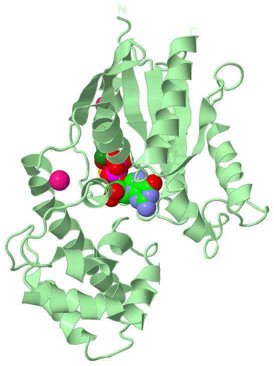
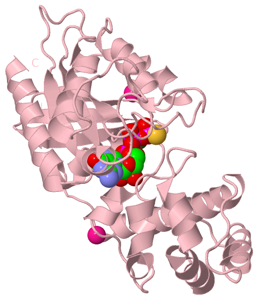
Still Images
|
||||||||||||||||
Databases
|
||||||||||||||||||||||||||||||||||||||||||||||||||||||||||||||||||||||||||||||||||||||||||||||||||||||||||||||||||||||||||||||||||||||||||||||||||||||||||||||||
Analysis Tools
|
|||||||||||||||||||||||||||||||||||||||||||||||||||||||||||||
Entries Sharing at Least One Protein Chain (UniProt ID)
Related Entries Specified in the PDB File
|
|