Show PDB file:   
         Plain Text   HTML   (compressed file size)
QuickSearch:   
by PDB,NDB,UniProt,PROSITE Code or Search Term(s)  
(-)Asym./Biol. Unit
(-)Asym./Biol. Unit - sites
collapse expand < >
Image Asym./Biol. Unit
Asym./Biol. Unit  (Jmol Viewer)
Image Asym./Biol. Unit - sites
Asym./Biol. Unit - sites  (Jmol Viewer)

(-) Description

Title :  CRYSTAL STRUCTURE OF THE COMPLEX FORMED BETWEEN ALKALINE PROTEINASE SAVINASE AND GRAMICIDIN S AT 1.5A RESOLUTION
 
Authors :  V. S. Bhatt, P. Kaur, S. Klupsch, C. Betzel, S. Brenner, T. P. Singh
Date :  08 Jun 04  (Deposition) - 22 Jun 04  (Release) - 27 Jul 11  (Revision)
Method :  X-RAY DIFFRACTION
Resolution :  1.54
Chains :  Asym./Biol. Unit :  A,B
Keywords :  Gramicidin, Antibiotic, Antifungal, Antibacterial, Cyclic Gramicidin, Membrane Ion Channel, Hydrolase-Antibiotic Complex (Keyword Search: [Gene Ontology, PubMed, Web (Google))
 
Reference :  V. S. Bhatt, P. Kaur, S. Klupsch, C. Betzel, S. Brenner, T. P. Singh
Crystal Structure Of The Complex Formed Between Alkaline Proteinase Savinase And Gramicidin S At 1. 5A Resolution.
To Be Published
PubMed: search

(-) Compounds

Molecule 1 - SUBTILISIN SAVINASE
    ChainsA
    EC Number3.4.21.62
    Organism ScientificBACILLUS LENTUS
    Organism Taxid1467
    SynonymALKALINE PROTEASE
 
Molecule 2 - GRAMICIDIN S
    ChainsB
    EngineeredYES
    Organism ScientificBREVIBACILLUS BREVIS
    Organism Taxid1393
    SynonymGRAMICIDIN SOVIET
    SyntheticYES

 Structural Features

(-) Chains, Units

  12
Asymmetric/Biological Unit AB

Summary Information (see also Sequences/Alignments below)
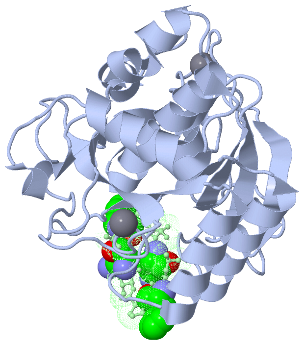
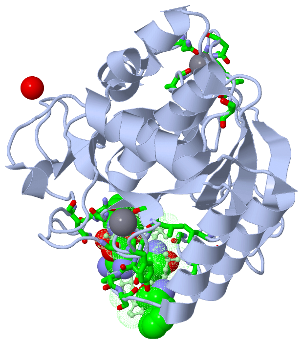
(-) Ligands, Modified Residues, Ions  (3, 6)

Asymmetric/Biological Unit (3, 6)
No.NameCountTypeFull Name
1CA2Ligand/IonCALCIUM ION
2DPN2Mod. Amino AcidD-PHENYLALANINE
3ORN2Mod. Amino AcidL-ORNITHINE

(-) Sites  (3, 3)

Asymmetric Unit (3, 3)
No.NameEvidenceResiduesDescription
1AC1SOFTWAREALA A:169 , TYR A:171 , ALA A:174 , HOH A:2114 , HOH A:2137BINDING SITE FOR RESIDUE CA A 1276
2AC2SOFTWAREGLN A:2 , ASP A:41 , LEU A:75 , ASN A:77 , ILE A:79 , VAL A:81BINDING SITE FOR RESIDUE CA A 1277
3AC3SOFTWARELEU A:96 , GLY A:100 , GLY A:102 , ILE A:107 , LEU A:126 , GLY A:127 , SER A:128 , PRO A:129 , SER A:130 , GLY A:154 , ASN A:155 , SER A:166 , GLN A:191 , SER A:240 , HOH A:2127 , HOH B:2001BINDING SITE FOR CHAIN B OF GRAMICIDIN S

(-) SS Bonds  (0, 0)

(no "SS Bond" information available for 1TK2)

(-) Cis Peptide Bonds  (1, 1)

Asymmetric/Biological Unit
No.Residues
1Tyr A:167 -Pro A:168

 Sequence-Structure Mapping

(-) SAPs(SNPs)/Variants  (0, 0)

(no "SAP(SNP)/Variant" information available for 1TK2)

(-) PROSITE Motifs  (3, 3)

Asymmetric/Biological Unit (3, 3)
 PROSITEUniProtKBPDB
No.IDACDescriptionIDLocationCountLocation
1SUBTILASE_ASPPS00136 Serine proteases, subtilase family, aspartic acid active site.SUBS_BACLE28-38  1A:28-39
2SUBTILASE_HISPS00137 Serine proteases, subtilase family, histidine active site.SUBS_BACLE62-72  1A:64-74
3SUBTILASE_SERPS00138 Serine proteases, subtilase family, serine active site.SUBS_BACLE213-223  1A:219-229

(-) Exons   (0, 0)

(no "Exon" information available for 1TK2)

(-) Sequences/Alignments

Asymmetric/Biological Unit
   Reformat: Number of residues per line =  ('0' or empty: single-line sequence representation)
  Number of residues per labelling interval =   
  UniProt sequence: complete  aligned part    
   Show mapping: SCOP domains CATH domains Pfam domains Secondary structure (by author)
SAPs(SNPs) PROSITE motifs Exons
(details for a mapped element are shown in a popup box when the mouse pointer rests over it)
Chain A from PDB  Type:PROTEIN  Length:269
 aligned with SUBS_BACLE | P29600 from UniProtKB/Swiss-Prot  Length:269

    Alignment length:269
                                    10        20        30        40        50        60        70        80        90       100       110       120       130       140       150       160       170       180       190       200       210       220       230       240       250       260         
           SUBS_BACLE     1 AQSVPWGISRVQAPAAHNRGLTGSGVKVAVLDTGISTHPDLNIRGGASFVPGEPSTQDGNGHGTHVAGTIAALNNSIGVLGVAPSAELYAVKVLGASGSGSVSSIAQGLEWAGNNGMHVANLSLGSPSPSATLEQAVNSATSRGVLVVAASGNSGAGSISYPARYANAMAVGATDQNNNRASFSQYGAGLDIVAPGVNVQSTYPGSTYASLNGTSMATPHVAGAAALVKQKNPSWSNVQIRNHLKNTATSLGSTNLYGSGLVNAEAATR 269
               SCOP domains d1tk2a_ A: Subtilisin                                                                                                                                                                                                                                                         SCOP domains
               CATH domains 1tk2A00 A:1-275  [code=3.40.50.200, no name defined]                                                                                                                                                                                                                          CATH domains
               Pfam domains --------------------------Peptidase_S8-1tk2A01 A:27-269                                                                                                                                                                                                                ------ Pfam domains
         Sec.struct. author ....hhhhhhhhhhhhhh........eeeeee..........eeeeee............hhhhhhhhhhhh..............eeeeee........hhhhhhhhhhhhhhh...eeee.......hhhhhhhhhhhhhh..eeeee.................eeeeeee.................eeee...eeeee...eeeee.hhhhhhhhhhhhhhhhhhhh...hhhhhhhhhhhh.....hhhhhh....hhhhhh. Sec.struct. author
                 SAPs(SNPs) ----------------------------------------------------------------------------------------------------------------------------------------------------------------------------------------------------------------------------------------------------------------------------- SAPs(SNPs)
                    PROSITE ---------------------------SUBTILASE_A-----------------------SUBTILASE_H--------------------------------------------------------------------------------------------------------------------------------------------SUBTILASE_S---------------------------------------------- PROSITE
                 Transcript ----------------------------------------------------------------------------------------------------------------------------------------------------------------------------------------------------------------------------------------------------------------------------- Transcript
                 1tk2 A   1 AQSVPWGISRVQAPAAHNRGLTGSGVKVAVLDTGISTHPDLNIRGGASFVPGEPSTQDGNGHGTHVAGTIAALNNSIGVLGVAPSAELYAVKVLGASGSGSVSSIAQGLEWAGNNGMHVANLSLGSPSPSATLEQAVNSATSRGVLVVAASGNSGAGSISYPARYANAMAVGATDQNNNRASFSQYGAGLDIVAPGVNVQSTYPGSTYASLNGTSMATPHVAGAAALVKQKNPSWSNVQIRNHLKNTATSLGSTNLYGSGLVNAEAATR 275
                                    10        20        30    ||  41        51     || 62        72        82        92       102       112       122       132       142       152    || 166       176       186       196       206       216       226       236       246       256       266         
                                                             35|                  57|                                                                                               157| ||                                                                                                              
                                                              37                   59                                                                                                160 ||                                                                                                              
                                                                                                                                                                                       162|                                                                                                              
                                                                                                                                                                                        165                                                                                                              

Chain B from PDB  Type:PROTEIN  Length:10
                                          
               SCOP domains d1tk2b_ B: SCOP domains
               CATH domains ---------- CATH domains
               Pfam domains ---------- Pfam domains
         Sec.struct. author .ee..ee... Sec.struct. author
                 SAPs(SNPs) ---------- SAPs(SNPs)
                    PROSITE ---------- PROSITE
                 Transcript ---------- Transcript
                 1tk2 B   1 VxLxPVxLxP  10
                             | |  | 10
                             2-ORN| | 
                               4-DPN| 
                                  7-ORN
                                    9-DPN

   Legend:   → Mismatch (orange background)
  - → Gap (green background, '-', border residues have a numbering label)
    → Modified Residue (blue background, lower-case, 'x' indicates undefined single-letter code, labelled with number + name)
  x → Chemical Group (purple background, 'x', labelled with number + name, e.g. ACE or NH2)
  extra numbering lines below/above indicate numbering irregularities and modified residue names etc., number ends below/above '|'

 Classification and Annotation

(-) SCOP Domains  (2, 2)

Asymmetric/Biological Unit
(-)
Class: Peptides (792)

(-) CATH Domains  (1, 1)

Asymmetric/Biological Unit
(-)
Class: Alpha Beta (26913)

(-) Pfam Domains  (1, 1)

Asymmetric/Biological Unit

(-) Gene Ontology  (8, 8)

Asymmetric/Biological Unit(hide GO term definitions)
Chain A   (SUBS_BACLE | P29600)
molecular function
    GO:0016787    hydrolase activity    Catalysis of the hydrolysis of various bonds, e.g. C-O, C-N, C-C, phosphoric anhydride bonds, etc. Hydrolase is the systematic name for any enzyme of EC class 3.
    GO:0046872    metal ion binding    Interacting selectively and non-covalently with any metal ion.
    GO:0008233    peptidase activity    Catalysis of the hydrolysis of a peptide bond. A peptide bond is a covalent bond formed when the carbon atom from the carboxyl group of one amino acid shares electrons with the nitrogen atom from the amino group of a second amino acid.
    GO:0004252    serine-type endopeptidase activity    Catalysis of the hydrolysis of internal, alpha-peptide bonds in a polypeptide chain by a catalytic mechanism that involves a catalytic triad consisting of a serine nucleophile that is activated by a proton relay involving an acidic residue (e.g. aspartate or glutamate) and a basic residue (usually histidine).
    GO:0008236    serine-type peptidase activity    Catalysis of the hydrolysis of peptide bonds in a polypeptide chain by a catalytic mechanism that involves a catalytic triad consisting of a serine nucleophile that is activated by a proton relay involving an acidic residue (e.g. aspartate or glutamate) and a basic residue (usually histidine).
biological process
    GO:0006508    proteolysis    The hydrolysis of proteins into smaller polypeptides and/or amino acids by cleavage of their peptide bonds.
    GO:0030435    sporulation resulting in formation of a cellular spore    The process in which a relatively unspecialized cell acquires the specialized features of a cellular spore, a cell form that can be used for dissemination, for survival of adverse conditions because of its heat and dessication resistance, and/or for reproduction.
cellular component
    GO:0005576    extracellular region    The space external to the outermost structure of a cell. For cells without external protective or external encapsulating structures this refers to space outside of the plasma membrane. This term covers the host cell environment outside an intracellular parasite.

 Visualization

(-) Interactive Views

Asymmetric/Biological Unit
  Complete Structure
    Jena3D(integrated viewing of ligand, site, SAP, PROSITE, SCOP information)
    WebMol | AstexViewer[tm]@PDBe
(Java Applets, require no local installation except for Java; loading may be slow)
    STRAP
(Java WebStart application, automatic local installation, requires Java; full application with system access!)
    RasMol
(require local installation)
    Molscript (VRML)
(requires installation of a VRML viewer; select preferred view via VRML and generate a mono or stereo PDF format file)
 
  Ligands, Modified Residues, Ions
    CA  [ RasMol | Jena3D ]  +environment [ RasMol | Jena3D ]
    DPN  [ RasMol | Jena3D ]  +environment [ RasMol | Jena3D ]
    ORN  [ RasMol | Jena3D ]  +environment [ RasMol | Jena3D ]
 
  Sites
    AC1  [ RasMol ]  +environment [ RasMol ]
    AC2  [ RasMol ]  +environment [ RasMol ]
    AC3  [ RasMol ]  +environment [ RasMol ]
 
  Cis Peptide Bonds
    Tyr A:167 - Pro A:168   [ RasMol ]  
 

(-) Still Images

Jmol
  protein: cartoon or spacefill or dots and stick; nucleic acid: cartoon and stick; ligands: spacefill; active site: stick
Molscript
  protein, nucleic acid: cartoon; ligands: spacefill; active site: ball and stick

 Databases and Analysis Tools

(-) Databases

Access by PDB/NDB ID
  1tk2
    Family and Domain InformationProDom | SYSTERS
    General Structural InformationGlycoscienceDB | MMDB | NDB | OCA | PDB | PDBe | PDBj | PDBsum | PDBWiki | PQS | PROTEOPEDIA
    Orientation in MembranesOPM
    Protein SurfaceSURFACE
    Secondary StructureDSSP (structure derived) | HSSP (homology derived)
    Structural GenomicsGeneCensus
    Structural NeighboursCE | VAST
    Structure ClassificationCATH | Dali | SCOP
    Validation and Original DataBMRB Data View | BMRB Restraints Grid | EDS | PROCHECK | RECOORD | WHAT_CHECK
 
Access by UniProt ID/Accession number
  SUBS_BACLE | P29600
    Comparative Protein Structure ModelsModBase
    Genomic InformationEnsembl
    Protein-protein InteractionDIP
    Sequence, Family and Domain InformationInterPro | Pfam | SMART | UniProtKB/SwissProt
 
Access by Enzyme Classificator   (EC Number)
  3.4.21.62
    General Enzyme InformationBRENDA | EC-PDB | Enzyme | IntEnz
    PathwayKEGG | MetaCyc
 
Access by Disease Identifier   (MIM ID)
  (no 'MIM ID' available)
    Disease InformationOMIM
 
Access by GenAge ID
  (no 'GenAge ID' available)
    Age Related InformationGenAge

(-) Analysis Tools

Access by PDB/NDB ID
    Domain InformationXDom
    Interatomic Contacts of Structural UnitsCSU
    Ligand-protein ContactsLPC
    Protein CavitiescastP
    Sequence and Secondary StructurePDBCartoon
    Structure AlignmentSTRAP(Java WebStart application, automatic local installation, requires Java; full application with system access!)
    Structure and Sequence BrowserSTING
 
Access by UniProt ID/Accession number
  SUBS_BACLE | P29600
    Protein Disorder PredictionDisEMBL | FoldIndex | GLOBPLOT (for more information see DisProt)

 Related Entries

(-) Entries Sharing at Least One Protein Chain (UniProt ID)

UniProtKB/Swiss-Prot
        SUBS_BACLE | P296001c9j 1c9m 1c9n 1gci 1iav 1jea 1ndq 1ndu 1q5p 1svn 3bx1 4cfy 4cfz 4cg0 5aqe 5arb 5arc 5ard

(-) Related Entries Specified in the PDB File

1al4 CRYSTAL STRUCTURE OF GRAMICIDIN D IN N-PROPANOL
1alx CRYSTAL STRUCTURE OF GRAMICIDIN D IN METHANOL
1alz CRYSTAL STRUCTURE OF GRAMICIDIN D IN ETHANOL
1av2 CRYSTAL STRUCTURE OF GRAMICIDIN A COMPLEXED WITH CESIUM CHLORIDE
1bdw CRYSTAL STRUCTURE OF GRAMICIDIN A FROM BACILLUS BREVIS
1c4d CRYSTAL STRUCTURE OF GRAMICIDIN A COMPLEXED WITH CESIUM CHLORIDE
1gmk CRYSTAL STRUCTURE OF GRAMICIDIN A COMPLRXED WITH POTASSIUM THIOCYANATE
1grm SOLUTION STRUCTURE OF THE GRAMICIDIN A
1jno SOLUTION STRUCTURE OF GRAMICIDIN A IN SODIUM DODECYL SULFATE MICELLES
1jo3 SOLUTION STRUCTURE OF GRAMICIDIN B IN SODIUM DODECYL SULFATE MICELLES
1jo4 SOLUTION STRUCTURE OF GRAMICIDIN C IN SODIUM DODECYL SULFATE MICELLES
1kqe SOLUTION STRUCTURE OF A LINKED SHORTENED GRAMICIDIN A IN BENZENE/ACETONE 10:1
1mag SOLID STATE NMR STRUCTURE OF GRAMICIDIN A IN HYDRATED DMPC BILAYERS,
1mic SOLUTION STRUCTURE OF GRAMICIDIN A IN METHANOL IN THE PRESENCE OF CACL
1ng8 SOLUTION STRUCTURE OF GRAMICIDIN A (W15G) IN SODIUM DODECYL SULFATE MICELLES
1nrm SOLUTION STRUCTURE OF GRAMICIDIN A IN DODECYL PHOSPHOCHOLINE MICELLES
1nru SOLUTION STRUCTURE OF GRAMICIDIN A IN DODECYL PHOSPHOCHOLINE MICELLES IN THE PRESENCE OF EXCESS NA+
1nt5 SOLUTION STRUCTURE OF GRAMICIDIN A (V1F) IN SODIUM DODECYL SULFATE MICELLES
1nt6 SOLUTION STRUCTURE OF F1-GRAMICIDIN C IN SODIUM DODECYL SULFATE MICELLES
1tkq SOLUTION STRUCTURE OF A LINKED UNSYMMETRIC GRAMICIDIN A IN A MEMBRANE-ISOELECTRICAL SOLVENTS MIXTURE, IN THE PRESENCE OF CSCL
1w5u CRYSTAL STRUCTURE OF GRAMICIDIN D IN ETHANOL
2izq CRYSTAL STRUCTURE OF GRAMICIDIN D COMPLEX WITH KI IN METHANOL
2xdc CRYSTAL STRUCTURE OF GRAMICIDIN A FROM CRYSTALS GROWN IN A LIPID CUBIC PHASE. RELATED ENTRIES
3l8l CRYSTAL STRUCTURE OF GRAMICIDIN D COMPLEX WITH NAI