Show PDB file:   
         Plain Text   HTML   (compressed file size)
QuickSearch:   
by PDB,NDB,UniProt,PROSITE Code or Search Term(s)  
(-)Asymmetric Unit
(-)Asym. Unit - sites
(-)Biological Unit 1
(-)Biol. Unit 1 - sites
(-)Biological Unit 2
(-)Biological Unit 3
(-)Biological Unit 4
(-)Biological Unit 5
collapse expand < >
Image Asymmetric Unit
Asymmetric Unit  (Jmol Viewer)
Image Asym. Unit - sites
Asym. Unit - sites  (Jmol Viewer)
Image Biological Unit 1
Biological Unit 1  (Jmol Viewer)
Image Biol. Unit 1 - sites
Biol. Unit 1 - sites  (Jmol Viewer)
Image Biological Unit 2
Biological Unit 2  (Jmol Viewer)
Image Biological Unit 3
Biological Unit 3  (Jmol Viewer)
Image Biological Unit 4
Biological Unit 4  (Jmol Viewer)
Image Biological Unit 5
Biological Unit 5  (Jmol Viewer)

(-) Description

Title :  X-RAY STRUCTURE OF CALCIUM-LOADED CALBINDOMODULIN (A CALBINDIN D9K RE-ENGINEERED TO UNDERGO A CONFORMATIONAL OPENING) AT 1.44 A RESOLUTION
 
Authors :  C. G. Bunick, M. R. Nelson, S. Mangahas, L. S. Mizoue, G. J. Bunick, W. J. C
Date :  04 Sep 03  (Deposition) - 25 May 04  (Release) - 13 Jul 11  (Revision)
Method :  X-RAY DIFFRACTION
Resolution :  1.44
Chains :  Asym. Unit :  A,B
Biol. Unit 1:  A  (1x)
Biol. Unit 2:  B  (1x)
Biol. Unit 3:  A (1x),B (1x)
Biol. Unit 4:  A (1x),B (1x)
Biol. Unit 5:  A,B  (1x)
Keywords :  Ef-Hand (Helix-Loop-Helix) Calcium Binding Protein, Four-Helix Domain, Protein Engineering, Calcium-Induced Conformational Response, Calmodulin, Calbindin D9K, Signaling Protein (Keyword Search: [Gene Ontology, PubMed, Web (Google))
 
Reference :  C. G. Bunick, M. R. Nelson, S. Mangahas, M. J. Hunter, J. H. Sheehan, L. S. Mizoue, G. J. Bunick, W. J. Chazin
Designing Sequence To Control Protein Function In An Ef-Han Protein
J. Am. Chem. Soc. V. 126 5990 2004
PubMed-ID: 15137763  |  Reference-DOI: 10.1021/JA0397456

(-) Compounds

Molecule 1 - VITAMIN D-DEPENDENT CALCIUM-BINDING PROTEIN, INTESTINAL
    ChainsA, B
    EngineeredYES
    Expression SystemESCHERICHIA COLI BL21(DE3)
    Expression System PlasmidPSV271
    Expression System StrainBL21(DE3)
    Expression System Taxid469008
    Expression System Vector TypePET27 DERIVATIVE
    GeneCALB3 OR S100D
    MutationYES
    Organism CommonCATTLE
    Organism ScientificBOS TAURUS
    Organism Taxid9913
    SynonymCABP, CALBINDIN D9K

 Structural Features

(-) Chains, Units

  12
Asymmetric Unit AB
Biological Unit 1 (1x)A 
Biological Unit 2 (1x) B
Biological Unit 3 (1x)A (1x)B (1x)
Biological Unit 4 (1x)A (1x)B (1x)
Biological Unit 5 (1x)AB

Summary Information (see also Sequences/Alignments below)

(-) Ligands, Modified Residues, Ions  (3, 8)

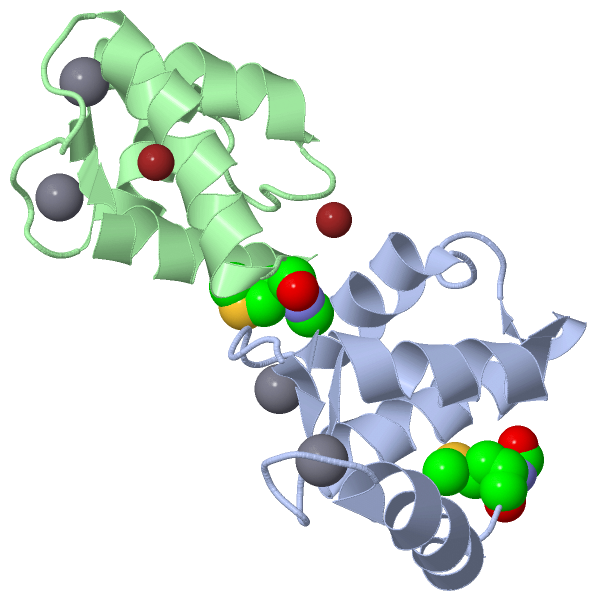
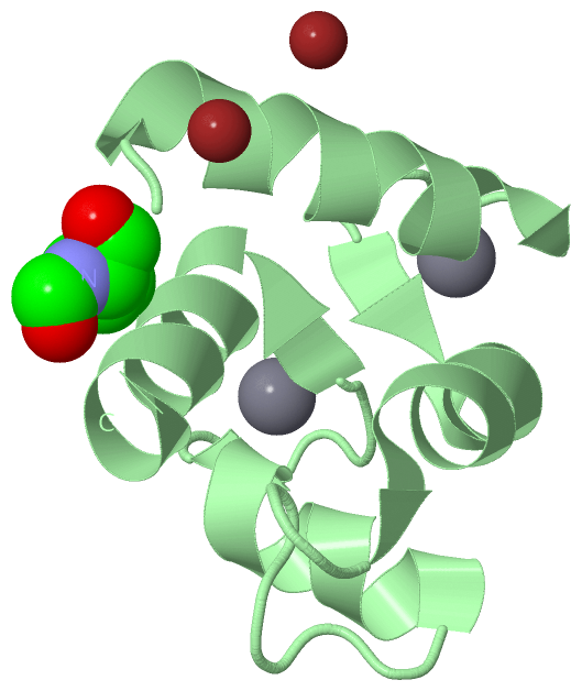
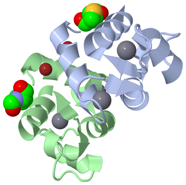
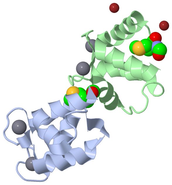
Asymmetric Unit (3, 8)
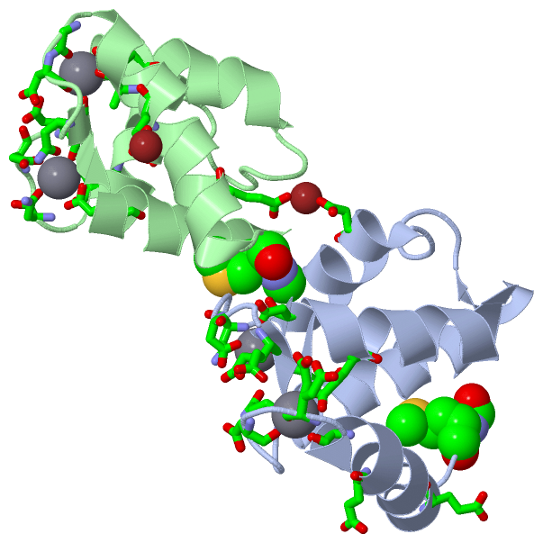
No.NameCountTypeFull Name
1CA4Ligand/IonCALCIUM ION
2FME2Mod. Amino AcidN-FORMYLMETHIONINE
3ZN2Ligand/IonZINC ION
Biological Unit 1 (1, 1)
No.NameCountTypeFull Name
1CA-1Ligand/IonCALCIUM ION
2FME1Mod. Amino AcidN-FORMYLMETHIONINE
3ZN-1Ligand/IonZINC ION
Biological Unit 2 (1, 1)
No.NameCountTypeFull Name
1CA-1Ligand/IonCALCIUM ION
2FME1Mod. Amino AcidN-FORMYLMETHIONINE
3ZN-1Ligand/IonZINC ION
Biological Unit 3 (1, 1)
No.NameCountTypeFull Name
1CA-1Ligand/IonCALCIUM ION
2FME1Mod. Amino AcidN-FORMYLMETHIONINE
3ZN-1Ligand/IonZINC ION
Biological Unit 4 (1, 1)
No.NameCountTypeFull Name
1CA-1Ligand/IonCALCIUM ION
2FME1Mod. Amino AcidN-FORMYLMETHIONINE
3ZN-1Ligand/IonZINC ION
Biological Unit 5 (1, 2)
No.NameCountTypeFull Name
1CA-1Ligand/IonCALCIUM ION
2FME2Mod. Amino AcidN-FORMYLMETHIONINE
3ZN-1Ligand/IonZINC ION

(-) Sites  (6, 6)

Asymmetric Unit (6, 6)
No.NameEvidenceResiduesDescription
1AC1SOFTWAREASP A:54 , ASN A:56 , ASP A:58 , GLU A:60 , GLU A:65 , HOH A:1017BINDING SITE FOR RESIDUE CA A 1001
2AC2SOFTWAREASP B:54 , ASN B:56 , ASP B:58 , GLU B:60 , GLU B:65 , HOH B:1016BINDING SITE FOR RESIDUE CA B 1002
3AC3SOFTWAREGLU A:11 , ASP A:47 , HOH A:1114 , GLU B:5BINDING SITE FOR RESIDUE ZN B 1003
4AC4SOFTWAREGLU A:5 , GLU B:11 , GLU B:48 , GLU B:51BINDING SITE FOR RESIDUE ZN B 1004
5AC5SOFTWAREALA A:14 , GLU A:17 , ASP A:19 , GLN A:22 , GLU A:27 , HOH A:1010BINDING SITE FOR RESIDUE CA A 1005
6AC6SOFTWAREALA B:14 , GLY B:18 , ASP B:19 , GLN B:22 , GLU B:27 , HOH B:1010BINDING SITE FOR RESIDUE CA B 1006

(-) SS Bonds  (0, 0)

(no "SS Bond" information available for 1QX2)

(-) Cis Peptide Bonds  (0, 0)

(no "Cis Peptide Bond" information available for 1QX2)

 Sequence-Structure Mapping

(-) SAPs(SNPs)/Variants  (0, 0)

(no "SAP(SNP)/Variant" information available for 1QX2)

(-) PROSITE Motifs  (3, 6)

Asymmetric Unit (3, 6)
 PROSITEUniProtKBPDB
No.IDACDescriptionIDLocationCountLocation
1EF_HAND_2PS50222 EF-hand calcium-binding domain profile.S100G_BOVIN45-79
 
  2A:41-75
B:41-74
2S100_CABPPS00303 S-100/ICaBP type calcium binding protein signature.S100G_BOVIN53-74
 
  2A:51-68
B:51-68
3EF_HAND_1PS00018 EF-hand calcium-binding domain.S100G_BOVIN58-70
 
  2A:54-66
B:54-66
Biological Unit 1 (3, 3)
 PROSITEUniProtKBPDB
No.IDACDescriptionIDLocationCountLocation
1EF_HAND_2PS50222 EF-hand calcium-binding domain profile.S100G_BOVIN45-79
 
  1A:41-75
-
2S100_CABPPS00303 S-100/ICaBP type calcium binding protein signature.S100G_BOVIN53-74
 
  1A:51-68
-
3EF_HAND_1PS00018 EF-hand calcium-binding domain.S100G_BOVIN58-70
 
  1A:54-66
-
Biological Unit 2 (3, 3)
 PROSITEUniProtKBPDB
No.IDACDescriptionIDLocationCountLocation
1EF_HAND_2PS50222 EF-hand calcium-binding domain profile.S100G_BOVIN45-79
 
  1-
B:41-74
2S100_CABPPS00303 S-100/ICaBP type calcium binding protein signature.S100G_BOVIN53-74
 
  1-
B:51-68
3EF_HAND_1PS00018 EF-hand calcium-binding domain.S100G_BOVIN58-70
 
  1-
B:54-66
Biological Unit 3 (3, 6)
 PROSITEUniProtKBPDB
No.IDACDescriptionIDLocationCountLocation
1EF_HAND_2PS50222 EF-hand calcium-binding domain profile.S100G_BOVIN45-79
 
  2A:41-75
B:41-74
2S100_CABPPS00303 S-100/ICaBP type calcium binding protein signature.S100G_BOVIN53-74
 
  2A:51-68
B:51-68
3EF_HAND_1PS00018 EF-hand calcium-binding domain.S100G_BOVIN58-70
 
  2A:54-66
B:54-66
Biological Unit 4 (3, 6)
 PROSITEUniProtKBPDB
No.IDACDescriptionIDLocationCountLocation
1EF_HAND_2PS50222 EF-hand calcium-binding domain profile.S100G_BOVIN45-79
 
  2A:41-75
B:41-74
2S100_CABPPS00303 S-100/ICaBP type calcium binding protein signature.S100G_BOVIN53-74
 
  2A:51-68
B:51-68
3EF_HAND_1PS00018 EF-hand calcium-binding domain.S100G_BOVIN58-70
 
  2A:54-66
B:54-66
Biological Unit 5 (3, 6)
 PROSITEUniProtKBPDB
No.IDACDescriptionIDLocationCountLocation
1EF_HAND_2PS50222 EF-hand calcium-binding domain profile.S100G_BOVIN45-79
 
  2A:41-75
B:41-74
2S100_CABPPS00303 S-100/ICaBP type calcium binding protein signature.S100G_BOVIN53-74
 
  2A:51-68
B:51-68
3EF_HAND_1PS00018 EF-hand calcium-binding domain.S100G_BOVIN58-70
 
  2A:54-66
B:54-66

(-) Exons   (2, 4)

Asymmetric Unit (2, 4)
 ENSEMBLUniProtKBPDB
No.Transcript IDExonExon IDGenome LocationLengthIDLocationLengthCountLocationLength
1.1ENSBTAT000000226301ENSBTAE00000325724Un.004.79:167452-16749746S100G_BOVIN-00--
1.2ENSBTAT000000226302ENSBTAE00000184836Un.004.79:168025-168168144S100G_BOVIN1-45452A:0-41
B:0-41
42
42
1.3ENSBTAT000000226303ENSBTAE00000184843Un.004.79:171280-171540261S100G_BOVIN46-79342A:42-75
B:42-74
34
33

(-) Sequences/Alignments

Asymmetric Unit
   Reformat: Number of residues per line =  ('0' or empty: single-line sequence representation)
  Number of residues per labelling interval =   
  UniProt sequence: complete  aligned part    
   Show mapping: SCOP domains CATH domains Pfam domains Secondary structure (by author)
SAPs(SNPs) PROSITE motifs Exons
(details for a mapped element are shown in a popup box when the mouse pointer rests over it)
Chain A from PDB  Type:PROTEIN  Length:76
 aligned with S100G_BOVIN | P02633 from UniProtKB/Swiss-Prot  Length:79

    Alignment length:76
                                    13        23        33        43        53        63        73      
           S100G_BOVIN    4 KKSPEELKGIFEKYAAKEGDPNQLSKEELKLLLQTEFPSLLKGPSTLDELFEELDKNGDGEVSFEEFQVLVKKISQ 79
               SCOP domains d1qx2a_ A: Calbindin D9K                                                     SCOP domains
               CATH domains -1qx2A00 A:1-75 EF-hand                                                      CATH domains
               Pfam domains ---------------------------------------------------------------------------- Pfam domains
         Sec.struct. author ..hhhhhhhhhhhhhh.......eehhhhhhhhhhhhhhh.....hhhhhhhhhh.....eehhhhhhhhhhhhh. Sec.struct. author
                 SAPs(SNPs) ---------------------------------------------------------------------------- SAPs(SNPs)
                PROSITE (1) -----------------------------------------EF_HAND_2  PDB: A:41-75             PROSITE (1)
                PROSITE (2) -------------------------------------------------S100_CABP  PDB: A:51-6----- PROSITE (2)
                PROSITE (3) ------------------------------------------------------EF_HAND_1    --------- PROSITE (3)
               Transcript 1 Exon 1.2  PDB: A:0-41 UniProt: 1-45       Exon 1.3  PDB: A:42-75             Transcript 1
                  1qx2 A  0 mKSPEEIKGAFEVFAAKEGDPNQISKEELKLVMQTLGPSLLKGMSTLDEMIEEVDKNGDGEVSFEEFLVMMKKISQ 75
                            |        9        19        29        39        49        59        69      
                            |                                                                           
                            0-FME                                                                       

Chain B from PDB  Type:PROTEIN  Length:75
 aligned with S100G_BOVIN | P02633 from UniProtKB/Swiss-Prot  Length:79

    Alignment length:75
                                    13        23        33        43        53        63        73     
           S100G_BOVIN    4 KKSPEELKGIFEKYAAKEGDPNQLSKEELKLLLQTEFPSLLKGPSTLDELFEELDKNGDGEVSFEEFQVLVKKIS 78
               SCOP domains d1qx2b_ B: Calbindin D9K                                                    SCOP domains
               CATH domains -1qx2B00 B:1-74 EF-hand                                                     CATH domains
               Pfam domains --------------------------------------------------------------------------- Pfam domains
         Sec.struct. author ..hhhhhhhhhhhhhhh.....eehhhhhhhhhhhhh........hhhhhhhhhh......eehhhhhhhhhhhh Sec.struct. author
                 SAPs(SNPs) --------------------------------------------------------------------------- SAPs(SNPs)
                PROSITE (1) -----------------------------------------EF_HAND_2  PDB: B:41-74            PROSITE (1)
                PROSITE (2) -------------------------------------------------S100_CABP  PDB: B:51-6---- PROSITE (2)
                PROSITE (3) ------------------------------------------------------EF_HAND_1    -------- PROSITE (3)
               Transcript 1 Exon 1.2  PDB: B:0-41 UniProt: 1-45       Exon 1.3  PDB: B:42-74            Transcript 1
                  1qx2 B  0 mKSPEEIKGAFEVFAAKEGDPNQISKEELKLVMQTLGPSLLKGMSTLDEMIEEVDKNGDGEVSFEEFLVMMKKIS 74
                            |        9        19        29        39        49        59        69     
                            0-FME                                                                      

   Legend:   → Mismatch (orange background)
  - → Gap (green background, '-', border residues have a numbering label)
    → Modified Residue (blue background, lower-case, 'x' indicates undefined single-letter code, labelled with number + name)
  x → Chemical Group (purple background, 'x', labelled with number + name, e.g. ACE or NH2)
  extra numbering lines below/above indicate numbering irregularities and modified residue names etc., number ends below/above '|'

 Classification and Annotation

(-) SCOP Domains  (1, 2)

Asymmetric Unit

(-) CATH Domains  (1, 2)

Asymmetric Unit

(-) Pfam Domains  (0, 0)

(no "Pfam Domain" information available for 1QX2)

(-) Gene Ontology  (5, 5)

Asymmetric Unit(hide GO term definitions)
Chain A,B   (S100G_BOVIN | P02633)
molecular function
    GO:0005509    calcium ion binding    Interacting selectively and non-covalently with calcium ions (Ca2+).
    GO:0046872    metal ion binding    Interacting selectively and non-covalently with any metal ion.
    GO:0005499    vitamin D binding    Interacting selectively and non-covalently with vitamin D, any of a group of related, fat-soluble compounds that are derived from delta-5,7 steroids and play a central role in calcium metabolism. Specific forms of vitamin D include calciferol (ergocalciferol; vitamin D2) and cholecalciferol (calciol; vitamin D3).
cellular component
    GO:0016324    apical plasma membrane    The region of the plasma membrane located at the apical end of the cell.
    GO:0016323    basolateral plasma membrane    The region of the plasma membrane that includes the basal end and sides of the cell. Often used in reference to animal polarized epithelial membranes, where the basal membrane is the part attached to the extracellular matrix, or in plant cells, where the basal membrane is defined with respect to the zygotic axis.

 Visualization

(-) Interactive Views

Asymmetric Unit
  Complete Structure
    Jena3D(integrated viewing of ligand, site, SAP, PROSITE, SCOP information)
    WebMol | AstexViewer[tm]@PDBe
(Java Applets, require no local installation except for Java; loading may be slow)
    STRAP
(Java WebStart application, automatic local installation, requires Java; full application with system access!)
    RasMol
(require local installation)
    Molscript (VRML)
(requires installation of a VRML viewer; select preferred view via VRML and generate a mono or stereo PDF format file)
 
  Ligands, Modified Residues, Ions
    CA  [ RasMol | Jena3D ]  +environment [ RasMol | Jena3D ]
    FME  [ RasMol | Jena3D ]  +environment [ RasMol | Jena3D ]
    ZN  [ RasMol | Jena3D ]  +environment [ RasMol | Jena3D ]
 
  Sites
    AC1  [ RasMol ]  +environment [ RasMol ]
    AC2  [ RasMol ]  +environment [ RasMol ]
    AC3  [ RasMol ]  +environment [ RasMol ]
    AC4  [ RasMol ]  +environment [ RasMol ]
    AC5  [ RasMol ]  +environment [ RasMol ]
    AC6  [ RasMol ]  +environment [ RasMol ]
 
  Cis Peptide Bonds
(no "Cis Peptide Bonds" information available for 1qx2)
 
Biological Units
  Complete Structure
    Biological Unit 1  [ Jena3D ]
    Biological Unit 2  [ Jena3D ]
    Biological Unit 3  [ Jena3D ]
    Biological Unit 4  [ Jena3D ]
    Biological Unit 5  [ Jena3D ]

(-) Still Images

Jmol
  protein: cartoon or spacefill or dots and stick; nucleic acid: cartoon and stick; ligands: spacefill; active site: stick
Molscript
  protein, nucleic acid: cartoon; ligands: spacefill; active site: ball and stick

 Databases and Analysis Tools

(-) Databases

Access by PDB/NDB ID
  1qx2
    Family and Domain InformationProDom | SYSTERS
    General Structural InformationGlycoscienceDB | MMDB | NDB | OCA | PDB | PDBe | PDBj | PDBsum | PDBWiki | PQS | PROTEOPEDIA
    Orientation in MembranesOPM
    Protein SurfaceSURFACE
    Secondary StructureDSSP (structure derived) | HSSP (homology derived)
    Structural GenomicsGeneCensus
    Structural NeighboursCE | VAST
    Structure ClassificationCATH | Dali | SCOP
    Validation and Original DataBMRB Data View | BMRB Restraints Grid | EDS | PROCHECK | RECOORD | WHAT_CHECK
 
Access by UniProt ID/Accession number
  S100G_BOVIN | P02633
    Comparative Protein Structure ModelsModBase
    Genomic InformationEnsembl
    Protein-protein InteractionDIP
    Sequence, Family and Domain InformationInterPro | Pfam | SMART | UniProtKB/SwissProt
 
Access by Enzyme Classificator   (EC Number)
  (no 'Enzyme Classificator' available)
    General Enzyme InformationBRENDA | EC-PDB | Enzyme | IntEnz
    PathwayKEGG | MetaCyc
 
Access by Disease Identifier   (MIM ID)
  (no 'MIM ID' available)
    Disease InformationOMIM
 
Access by GenAge ID
  (no 'GenAge ID' available)
    Age Related InformationGenAge

(-) Analysis Tools

Access by PDB/NDB ID
    Domain InformationXDom
    Interatomic Contacts of Structural UnitsCSU
    Ligand-protein ContactsLPC
    Protein CavitiescastP
    Sequence and Secondary StructurePDBCartoon
    Structure AlignmentSTRAP(Java WebStart application, automatic local installation, requires Java; full application with system access!)
    Structure and Sequence BrowserSTING
 
Access by UniProt ID/Accession number
  S100G_BOVIN | P02633
    Protein Disorder PredictionDisEMBL | FoldIndex | GLOBPLOT (for more information see DisProt)

 Related Entries

(-) Entries Sharing at Least One Protein Chain (UniProt ID)

UniProtKB/Swiss-Prot
        S100G_BOVIN | P026331b1g 1boc 1bod 1cdn 1clb 1d1o 1ht9 1ig5 1igv 1kcy 1kqv 1ksm 1n65 1rt0 2bca 2bcb 2maz 3icb 4icb

(-) Related Entries Specified in the PDB File

(no "Related Entries Specified in the PDB File" available for 1QX2)