|
|
|
|
 Description
|
|
Compounds
|
||||||||||||||||||||||||||||||||||||||||
Chains, Units
Summary Information (see also Sequences/Alignments below) |
Ligands, Modified Residues, Ions (0, 0)| (no "Ligand,Modified Residues,Ions" information available for 1CLB) |
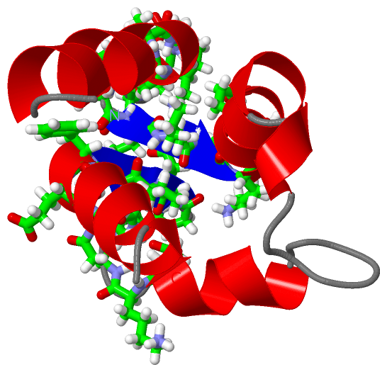
Sites (2, 2)
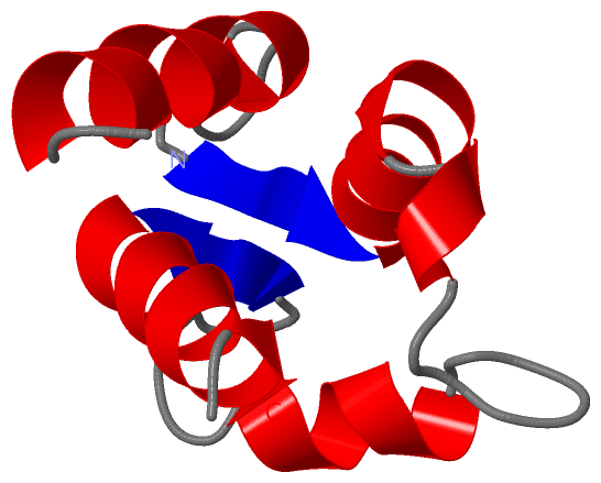
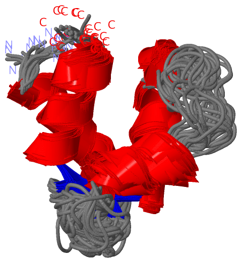
NMR Structure (2, 2)
|
SS Bonds (0, 0)| (no "SS Bond" information available for 1CLB) |
Cis Peptide Bonds (0, 0)| (no "Cis Peptide Bond" information available for 1CLB) |
SAPs(SNPs)/Variants (0, 0)| (no "SAP(SNP)/Variant" information available for 1CLB) |
PROSITE Motifs (3, 3)
NMR Structure (3, 3)
|
||||||||||||||||||||||||||||||||||||||||
Exons (2, 2)
NMR Structure (2, 2)
|
||||||||||||||||||||||||||||||||||||||||||||||||||||||||||||
Sequences/Alignments
NMR StructureChain A from PDB Type:PROTEIN Length:75 aligned with S100G_BOVIN | P02633 from UniProtKB/Swiss-Prot Length:79 Alignment length:75 14 24 34 44 54 64 74 S100G_BOVIN 5 KSPEELKGIFEKYAAKEGDPNQLSKEELKLLLQTEFPSLLKGPSTLDELFEELDKNGDGEVSFEEFQVLVKKISQ 79 SCOP domains d1clba_ A: Calbindin D9K SCOP domains CATH domains 1clbA00 A:1-75 EF-hand CATH domains Pfam domains --------------------------------------------------------------------------- Pfam domains SAPs(SNPs) --------------------------------------------------------------------------- SAPs(SNPs) PROSITE (1) ----------------------------------------EF_HAND_2 PDB: A:41-75 PROSITE (1) PROSITE (2) ------------------------------------------------S100_CABP PDB: A:49-7----- PROSITE (2) PROSITE (3) -----------------------------------------------------EF_HAND_1 --------- PROSITE (3) Transcript 1 Exon 1.2 PDB: A:1-41 UniProt: 1-45 Exon 1.3 PDB: A:42-75 Transcript 1 1clb A 1 KSPEELKGIFEKYAAKEGDPNQLSKEELKLLLQTEFPSLLKGGSTLDELFEELDKNGDGEVSFEEFQVLVKKISQ 75 10 20 30 40 50 60 70
|
||||||||||||||||||||
SCOP Domains (1, 1)
NMR Structure

|
CATH Domains (1, 1)
NMR Structure
|
Pfam Domains (0, 0)| (no "Pfam Domain" information available for 1CLB) |
Gene Ontology (5, 5)|
NMR Structure(hide GO term definitions) Chain A (S100G_BOVIN | P02633)
|
||||||||||||||||||||||||||||||||||||||||||
Interactive Views
|
||||||||||||||||||||||||||||||||||||||||||||||||||||||||||||||||||||||||||||||||||||||||||||||||||||||||||||||||||||||||||||
Still Images
|
||||||||||||||||||||||||||||||||||||||||||
Databases
|
||||||||||||||||||||||||||||||||||||||||||||||||||||||||||||||||||||||||||||||||||||||||||||||||||||||||||||||||||||||||||||||||||||||||||||||||||||||||||||||||
Analysis Tools
|
|||||||||||||||||||||||||||||||||||||||||||||||||||||||||||||
Entries Sharing at Least One Protein Chain (UniProt ID)
Related Entries Specified in the PDB File
|
|