|
|
|
|
 Description
|
|
Compounds
|
||||||||||||||||||||||||||||||||||||||||||||||||
Chains, Units
Summary Information (see also Sequences/Alignments below) |
Ligands, Modified Residues, Ions (2, 18)| Asymmetric Unit (2, 18) Biological Unit 1 (0, 0) Biological Unit 2 (0, 0) Biological Unit 3 (0, 0) |
Sites (18, 18)
Asymmetric Unit (18, 18)
|
SS Bonds (0, 0)| (no "SS Bond" information available for 1ODB) |
Cis Peptide Bonds (0, 0)| (no "Cis Peptide Bond" information available for 1ODB) |
SAPs(SNPs)/Variants (0, 0)| (no "SAP(SNP)/Variant" information available for 1ODB) |
PROSITE Motifs (2, 12)
Asymmetric Unit (2, 12)
|
||||||||||||||||||||||||||||||||||||||||||||||||||||||||||||||||||||||||||||||||||||||||||||||||||||||||||||||||||||||||||||||||
Exons (2, 12)
Asymmetric Unit (2, 12)
|
||||||||||||||||||||||||||||||||||||||||||||||||||||||||||||
Sequences/Alignments
Asymmetric UnitChain A from PDB Type:PROTEIN Length:91 aligned with S10AC_HUMAN | P80511 from UniProtKB/Swiss-Prot Length:92 Alignment length:91 10 20 30 40 50 60 70 80 90 S10AC_HUMAN 1 MTKLEEHLEGIVNIFHQYSVRKGHFDTLSKGELKQLLTKELANTIKNIKDKAVIDEIFQGLDANQDEQVDFQEFISLVAIALKAAHYHTHK 91 SCOP domains d1odba_ A: Calcyclin (S100) SCOP domains CATH domains 1odbA00 A:0-90 EF-hand CATH domains Pfam domains ------------------------------------------------------------------------------------------- Pfam domains SAPs(SNPs) ------------------------------------------------------------------------------------------- SAPs(SNPs) PROSITE (1) ------------------------------------------------EF_HAND_2 PDB: A:48-83 ------- PROSITE (1) PROSITE (2) --------------------------------------------------------S100_CABP PDB: A:56-7------------- PROSITE (2) Transcript 1 Exon 1.2 PDB: A:0-45 UniProt: 1-46 Exon 1.4a PDB: A:46-90 UniProt: 47-92 Transcript 1 1odb A 0 STKLEEHLEGIVNIFHQYSVRKGHFDTLSKGELKQLLTKELANTIKNIKDKAVIDEIFQGLDANQDEQVDFQEFISLVAIALKAAHYHTHK 90 9 19 29 39 49 59 69 79 89 Chain B from PDB Type:PROTEIN Length:91 aligned with S10AC_HUMAN | P80511 from UniProtKB/Swiss-Prot Length:92 Alignment length:91 10 20 30 40 50 60 70 80 90 S10AC_HUMAN 1 MTKLEEHLEGIVNIFHQYSVRKGHFDTLSKGELKQLLTKELANTIKNIKDKAVIDEIFQGLDANQDEQVDFQEFISLVAIALKAAHYHTHK 91 SCOP domains d1odbb_ B: Calcyclin (S100) SCOP domains CATH domains 1odbB00 B:0-90 EF-hand CATH domains Pfam domains ------------------------------------------------------------------------------------------- Pfam domains SAPs(SNPs) ------------------------------------------------------------------------------------------- SAPs(SNPs) PROSITE (1) ------------------------------------------------EF_HAND_2 PDB: B:48-83 ------- PROSITE (1) PROSITE (2) --------------------------------------------------------S100_CABP PDB: B:56-7------------- PROSITE (2) Transcript 1 Exon 1.2 PDB: B:0-45 UniProt: 1-46 Exon 1.4a PDB: B:46-90 UniProt: 47-92 Transcript 1 1odb B 0 STKLEEHLEGIVNIFHQYSVRKGHFDTLSKGELKQLLTKELANTIKNIKDKAVIDEIFQGLDANQDEQVDFQEFISLVAIALKAAHYHTHK 90 9 19 29 39 49 59 69 79 89 Chain C from PDB Type:PROTEIN Length:91 aligned with S10AC_HUMAN | P80511 from UniProtKB/Swiss-Prot Length:92 Alignment length:91 10 20 30 40 50 60 70 80 90 S10AC_HUMAN 1 MTKLEEHLEGIVNIFHQYSVRKGHFDTLSKGELKQLLTKELANTIKNIKDKAVIDEIFQGLDANQDEQVDFQEFISLVAIALKAAHYHTHK 91 SCOP domains d1odbc_ C: Calcyclin (S100) SCOP domains CATH domains 1odbC00 C:0-90 EF-hand CATH domains Pfam domains ------------------------------------------------------------------------------------------- Pfam domains SAPs(SNPs) ------------------------------------------------------------------------------------------- SAPs(SNPs) PROSITE (1) ------------------------------------------------EF_HAND_2 PDB: C:48-83 ------- PROSITE (1) PROSITE (2) --------------------------------------------------------S100_CABP PDB: C:56-7------------- PROSITE (2) Transcript 1 Exon 1.2 PDB: C:0-45 UniProt: 1-46 Exon 1.4a PDB: C:46-90 UniProt: 47-92 Transcript 1 1odb C 0 STKLEEHLEGIVNIFHQYSVRKGHFDTLSKGELKQLLTKELANTIKNIKDKAVIDEIFQGLDANQDEQVDFQEFISLVAIALKAAHYHTHK 90 9 19 29 39 49 59 69 79 89 Chain D from PDB Type:PROTEIN Length:91 aligned with S10AC_HUMAN | P80511 from UniProtKB/Swiss-Prot Length:92 Alignment length:91 10 20 30 40 50 60 70 80 90 S10AC_HUMAN 1 MTKLEEHLEGIVNIFHQYSVRKGHFDTLSKGELKQLLTKELANTIKNIKDKAVIDEIFQGLDANQDEQVDFQEFISLVAIALKAAHYHTHK 91 SCOP domains d1odbd_ D: Calcyclin (S100) SCOP domains CATH domains 1odbD00 D:0-90 EF-hand CATH domains Pfam domains ------------------------------------------------------------------------------------------- Pfam domains SAPs(SNPs) ------------------------------------------------------------------------------------------- SAPs(SNPs) PROSITE (1) ------------------------------------------------EF_HAND_2 PDB: D:48-83 ------- PROSITE (1) PROSITE (2) --------------------------------------------------------S100_CABP PDB: D:56-7------------- PROSITE (2) Transcript 1 Exon 1.2 PDB: D:0-45 UniProt: 1-46 Exon 1.4a PDB: D:46-90 UniProt: 47-92 Transcript 1 1odb D 0 STKLEEHLEGIVNIFHQYSVRKGHFDTLSKGELKQLLTKELANTIKNIKDKAVIDEIFQGLDANQDEQVDFQEFISLVAIALKAAHYHTHK 90 9 19 29 39 49 59 69 79 89 Chain E from PDB Type:PROTEIN Length:91 aligned with S10AC_HUMAN | P80511 from UniProtKB/Swiss-Prot Length:92 Alignment length:91 10 20 30 40 50 60 70 80 90 S10AC_HUMAN 1 MTKLEEHLEGIVNIFHQYSVRKGHFDTLSKGELKQLLTKELANTIKNIKDKAVIDEIFQGLDANQDEQVDFQEFISLVAIALKAAHYHTHK 91 SCOP domains d1odbe_ E: Calcyclin (S100) SCOP domains CATH domains 1odbE00 E:0-90 EF-hand CATH domains Pfam domains ------------------------------------------------------------------------------------------- Pfam domains SAPs(SNPs) ------------------------------------------------------------------------------------------- SAPs(SNPs) PROSITE (1) ------------------------------------------------EF_HAND_2 PDB: E:48-83 ------- PROSITE (1) PROSITE (2) --------------------------------------------------------S100_CABP PDB: E:56-7------------- PROSITE (2) Transcript 1 Exon 1.2 PDB: E:0-45 UniProt: 1-46 Exon 1.4a PDB: E:46-90 UniProt: 47-92 Transcript 1 1odb E 0 STKLEEHLEGIVNIFHQYSVRKGHFDTLSKGELKQLLTKELANTIKNIKDKAVIDEIFQGLDANQDEQVDFQEFISLVAIALKAAHYHTHK 90 9 19 29 39 49 59 69 79 89 Chain F from PDB Type:PROTEIN Length:90 aligned with S10AC_HUMAN | P80511 from UniProtKB/Swiss-Prot Length:92 Alignment length:90 10 20 30 40 50 60 70 80 90 S10AC_HUMAN 1 MTKLEEHLEGIVNIFHQYSVRKGHFDTLSKGELKQLLTKELANTIKNIKDKAVIDEIFQGLDANQDEQVDFQEFISLVAIALKAAHYHTH 90 SCOP domains d1odbf_ F: Calcyclin (S100) SCOP domains CATH domains 1odbF00 F:0-89 EF-hand CATH domains Pfam domains (1) ---S_100-1odbF01 F:3-46 ------------------------------------------- Pfam domains (1) Pfam domains (2) ---S_100-1odbF02 F:3-46 ------------------------------------------- Pfam domains (2) Pfam domains (3) ---S_100-1odbF03 F:3-46 ------------------------------------------- Pfam domains (3) Pfam domains (4) ---S_100-1odbF04 F:3-46 ------------------------------------------- Pfam domains (4) Pfam domains (5) ---S_100-1odbF05 F:3-46 ------------------------------------------- Pfam domains (5) Pfam domains (6) ---S_100-1odbF06 F:3-46 ------------------------------------------- Pfam domains (6) SAPs(SNPs) ------------------------------------------------------------------------------------------ SAPs(SNPs) PROSITE (1) ------------------------------------------------EF_HAND_2 PDB: F:48-83 ------ PROSITE (1) PROSITE (2) --------------------------------------------------------S100_CABP PDB: F:56-7------------ PROSITE (2) Transcript 1 Exon 1.2 PDB: F:0-45 UniProt: 1-46 Exon 1.4a PDB: F:46-89 UniProt: 47-92 Transcript 1 1odb F 0 STKLEEHLEGIVNIFHQYSVRKGHFDTLSKGELKQLLTKELANTIKNIKDKAVIDEIFQGLDANQDEQVDFQEFISLVAIALKAAHYHTH 89 9 19 29 39 49 59 69 79 89
|
||||||||||||||||||||
SCOP Domains (1, 6)
Asymmetric Unit
 |
CATH Domains (1, 6)
Asymmetric Unit
|
Pfam Domains (1, 6)
Asymmetric Unit
|
Gene Ontology (28, 28)|
Asymmetric Unit(hide GO term definitions) Chain A,B,C,D,E,F (S10AC_HUMAN | P80511)
|
||||||||||||||||||||||||||||||||||||||||||||||||||||||||||||||||||||||||||||||||||||||||||||||||||||||||||||||||||||||||||||||||||||||||||||||||||||||||||||||||||||||||||||||||||||||||||
Interactive Views
|
||||||||||||||||||||||||||||||||||||||||||||||||||||||||||||||||||||||||||||||||||||||||||||||||||||||||||||||||||||||||||||||||||||||||||||||||||||||||||||||||||||||||||||||||||||||||||||||||||||||||||||||||||||||||||||||||||||||||||||||||||||||||||||||||||||||||||||||||
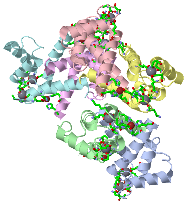
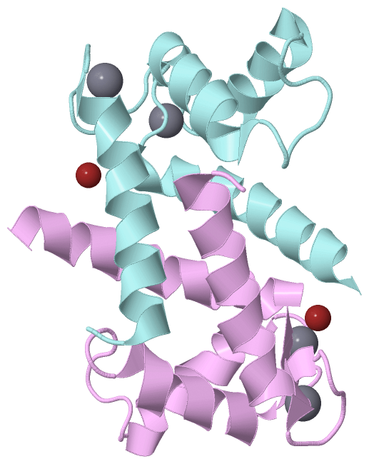
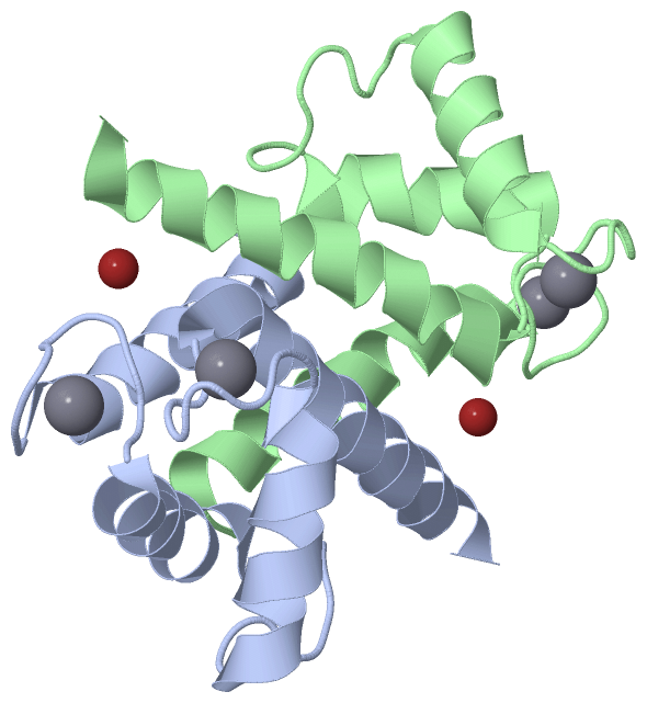
Still Images
|
||||||||||||||||
Databases
|
||||||||||||||||||||||||||||||||||||||||||||||||||||||||||||||||||||||||||||||||||||||||||||||||||||||||||||||||||||||||||||||||||||||||||||||||||||||||||||||||
Analysis Tools
|
|||||||||||||||||||||||||||||||||||||||||||||||||||||||||||||
Entries Sharing at Least One Protein Chain (UniProt ID)
Related Entries Specified in the PDB File
|
|