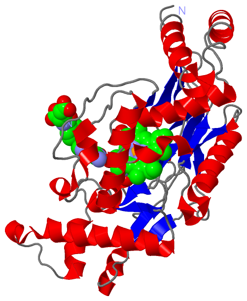
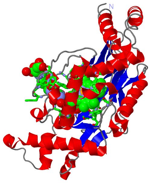
Show PDB file:   
         Plain Text   HTML   (compressed file size)
QuickSearch:   
by PDB,NDB,UniProt,PROSITE Code or Search Term(s)  
(-)Biol.Unit 1 - manually
(-)Asym.Unit - manually
(-)Asymmetric Unit
(-)Asym. Unit - sites
(-)Biological Unit 1
(-)Biol. Unit 1 - sites
collapse expand < >
Image Biol.Unit 1 - manually
Biol.Unit 1 - manually  (Jmol Viewer)
Image Asym.Unit - manually
Asym.Unit - manually  (Jmol Viewer)
Image Asymmetric Unit
Asymmetric Unit  (Jmol Viewer)
Image Asym. Unit - sites
Asym. Unit - sites  (Jmol Viewer)
Image Biological Unit 1
Biological Unit 1  (Jmol Viewer)
Image Biol. Unit 1 - sites
Biol. Unit 1 - sites  (Jmol Viewer)

(-) Description

Title :  STRUCTURE OF NITRIC OXIDE SYNTHASE HEME PROTEIN FROM BACILLUS SUBTILIS WITH N-HYDROXY-ARGININE AND TETRAHYDROFOLATE BOUND
 
Authors :  K. Pant, A. M. Bilwes, S. Adak, D. J. Stuehr, B. R. Crane
Date :  23 Jul 02  (Deposition) - 30 Oct 02  (Release) - 24 Feb 09  (Revision)
Method :  X-RAY DIFFRACTION
Resolution :  2.14
Chains :  Asym. Unit :  A
Biol. Unit 1:  A  (2x)
Keywords :  Oxygenase, Tetrahydrofolate, Pterin, Bacteria, Heme, Hydroxy Arginine, Oxidoreductase (Keyword Search: [Gene Ontology, PubMed, Web (Google))
 
Reference :  K. Pant, A. M. Bilwes, S. Adak, D. J. Stuehr, B. R. Crane
Structure Of A Nitric Oxide Synthase Heme Protein From Bacillus Subtilis.
Biochemistry V. 41 11071 2002
PubMed-ID: 12220171  |  Reference-DOI: 10.1021/BI0263715
(for further references see the PDB file header)

(-) Compounds

Molecule 1 - NITRIC OXIDE SYNTHASE
    ChainsA
    EC Number1.14.13.39
    EngineeredYES
    Expression SystemESCHERICHIA COLI BL21(DE3)
    Expression System PlasmidPET15B
    Expression System StrainBL-21 (DE3)
    Expression System Taxid469008
    Expression System Vector TypePLASMID
    Organism ScientificBACILLUS SUBTILIS
    Organism Taxid1423
    SynonymNITRIC-OXIDE SYNTHETASE, NO SYNTHASE, ENDOTHELIUM- DERIVED RELAXATION FACTOR-FORMING ENZYME, ENDOTHELIUM- DERIVED RELAXINGFACTOR SYNTHASE

 Structural Features

(-) Chains, Units

  1
Asymmetric Unit A
Biological Unit 1 (2x)A

Summary Information (see also Sequences/Alignments below)

(-) Ligands, Modified Residues, Ions  (3, 3)

Asymmetric Unit (3, 3)
No.NameCountTypeFull Name
1HAR1Ligand/IonN-OMEGA-HYDROXY-L-ARGININE
2HEM1Ligand/IonPROTOPORPHYRIN IX CONTAINING FE
3THG1Ligand/Ion(6S)-5,6,7,8-TETRAHYDROFOLATE
Biological Unit 1 (3, 6)
No.NameCountTypeFull Name
1HAR2Ligand/IonN-OMEGA-HYDROXY-L-ARGININE
2HEM2Ligand/IonPROTOPORPHYRIN IX CONTAINING FE
3THG2Ligand/Ion(6S)-5,6,7,8-TETRAHYDROFOLATE

(-) Sites  (3, 3)

Asymmetric Unit (3, 3)
No.NameEvidenceResiduesDescription
1AC1SOFTWARETRP A:56 , SER A:59 , ARG A:61 , CYS A:62 , PHE A:231 , ASN A:232 , GLY A:233 , TRP A:234 , GLU A:239 , TRP A:325 , TYR A:351 , TYR A:353 , HAR A:909 , HOH A:957 , HOH A:975 , HOH A:991 , HOH A:1029 , THG A:1999BINDING SITE FOR RESIDUE HEM A 901
2AC2SOFTWAREGLN A:125 , ARG A:128 , TYR A:209 , ILE A:214 , GLY A:233 , TRP A:234 , TYR A:235 , GLU A:239 , ASN A:244 , HEM A:901 , HOH A:945 , HOH A:1015 , HOH A:1198BINDING SITE FOR RESIDUE HAR A 909
3AC3SOFTWAREARG A:243 , TRP A:323 , THR A:324 , TRP A:325 , PHE A:338 , ARG A:340 , TYR A:342 , HEM A:901 , HOH A:1149 , HOH A:1182 , HOH A:1187 , HOH A:1194BINDING SITE FOR RESIDUE THG A 1999

(-) SS Bonds  (0, 0)

(no "SS Bond" information available for 1M7Z)

(-) Cis Peptide Bonds  (1, 1)

Asymmetric Unit
No.Residues
1Lys A:348 -Pro A:349

 Sequence-Structure Mapping

(-) SAPs(SNPs)/Variants  (0, 0)

(no "SAP(SNP)/Variant" information available for 1M7Z)

(-) PROSITE Motifs  (1, 1)

Asymmetric Unit (1, 1)
 PROSITEUniProtKBPDB
No.IDACDescriptionIDLocationCountLocation
1NOSPS60001 Nitric oxide synthase (NOS) signature.NOSO_BACSU65-72  1A:61-68
Biological Unit 1 (1, 2)
 PROSITEUniProtKBPDB
No.IDACDescriptionIDLocationCountLocation
1NOSPS60001 Nitric oxide synthase (NOS) signature.NOSO_BACSU65-72  2A:61-68

(-) Exons   (0, 0)

(no "Exon" information available for 1M7Z)

(-) Sequences/Alignments

Asymmetric Unit
   Reformat: Number of residues per line =  ('0' or empty: single-line sequence representation)
  Number of residues per labelling interval =   
  UniProt sequence: complete  aligned part    
   Show mapping: SCOP domains CATH domains Pfam domains Secondary structure (by author)
SAPs(SNPs) PROSITE motifs Exons
(details for a mapped element are shown in a popup box when the mouse pointer rests over it)
Chain A from PDB  Type:PROTEIN  Length:361
 aligned with NOSO_BACSU | O34453 from UniProtKB/Swiss-Prot  Length:363

    Alignment length:363
                                    10        20        30        40        50        60        70        80        90       100       110       120       130       140       150       160       170       180       190       200       210       220       230       240       250       260       270       280       290       300       310       320       330       340       350       360   
           NOSO_BACSU     1 MEEKEILWNEAKAFIAACYQELGKEEEVKDRLADIKSEIDLTGSYVHTKEELEHGAKMAWRNSNRCIGRLFWNSLNVIDRRDVRTKEEVRDALFHHIETATNNGKIRPTITIFPPEEKGEKQVEIWNHQLIRYAGYESDGERIGDPASCSLTAACEELGWRGERTDFDLLPLIFRMKGDEQPVWYELPRSLVIEVPITHPDIEAFSDLELKWYGVPIISDMKLEVGGIHYNAAPFNGWYMGTEIGARNLADEKRYDKLKKVASVIGIAADYNTDLWKDQALVELNKAVLHSYKKQGVSIVDHHTAASQFKRFEEQEEEAGRKLTGDWTWLIPPISPAATHIFHRSYDNSIVKPNYFYQDKPYE 363
               SCOP domains d1m7za_ A: Nitric oxide (NO) synthase oxygenase domain                                                                                                                                                                                                                                                                                                                      SCOP domains
               CATH domains 1m7zA01 A:-3-77,A:104-114,A:218-232,A:302-326                                    1m7zA02                   1m7zA01    ---1m7zA02 A:78-103  ,A:118-189,A:353-357                                  1m7zA03                -----1m7zA01        --1m7zA03 A:190-212,A:235-300                                       -1m7zA01                  --------------------------1m7zA-- CATH domains
               Pfam domains ----NO_synthase-1m7zA01 A:1-359                                                                                                                                                                                                                                                                                                                                             Pfam domains
         Sec.struct. author .....hhhhhhhhhhhhhh.hhh......hhhhhhhhhhhhh.....hhhhhhhhhhhhhhh.....hhhhhhhheeee.....hhhhhhhhhhhhhhhhhhhhh...eeee..........eee............--.....hhhhhhhhhhhhh..............eeeee......eee........eee.....hhhhhhhh.eee...ee...eeee..eee....ee..eehhhhhh..........hhhhhhhhh.....hhhhhhhhhhhhhhhhhhhhhhhhh...eehhhhhhhhhhhhhhhhhhh......hhhhhh...hhhhh..............eee....... Sec.struct. author
                 SAPs(SNPs) --------------------------------------------------------------------------------------------------------------------------------------------------------------------------------------------------------------------------------------------------------------------------------------------------------------------------------------------------------------------------- SAPs(SNPs)
                    PROSITE ----------------------------------------------------------------NOS     --------------------------------------------------------------------------------------------------------------------------------------------------------------------------------------------------------------------------------------------------------------------------------------------------- PROSITE
                 Transcript --------------------------------------------------------------------------------------------------------------------------------------------------------------------------------------------------------------------------------------------------------------------------------------------------------------------------------------------------------------------------- Transcript
                 1m7z A  -3 GSHMEILWNEAKAFIAECYQELGKEEEVKDRLDSIKSEIDRTGSYVHTKEELEHGAKMAWRNSNRCIGRLFWNSLNVIDRRDVRTKEDVRDALFHHIETATNNGKIRPSITIFPPEEKGEKQVEIWNHQLIRYAGYE--GERIGDPASRSLTAACEQLGWRGERTDFDLLPLIFRMRGDEQPVWYELPRSLVIEVPITHPDIEAFSDLELKWYGVPIISDMKLEVGGIHYNAAPFNGWYMGTEIGARNLADEKRYDKLKKVASVIGISTNYNTDLWKDQALVELNKAVLYSYKKQGVSIVDHHTAASQFKRFEEQEEEAGRKLTGDWTWLIPPISPAATHIFHRSYDNSIVKPNYFYQDKPYE 359
                                     6        16        26        36        46        56        66        76        86        96       106       116       126      |136       146       156       166       176       186       196       206       216       226       236       246       256       266       276       286       296       306       316       326       336       346       356   
                                                                                                                                                                  133  |                                                                                                                                                                                                                               
                                                                                                                                                                     136                                                                                                                                                                                                                               

   Legend:   → Mismatch (orange background)
  - → Gap (green background, '-', border residues have a numbering label)
    → Modified Residue (blue background, lower-case, 'x' indicates undefined single-letter code, labelled with number + name)
  x → Chemical Group (purple background, 'x', labelled with number + name, e.g. ACE or NH2)
  extra numbering lines below/above indicate numbering irregularities and modified residue names etc., number ends below/above '|'

 Classification and Annotation

(-) SCOP Domains  (1, 1)

Asymmetric Unit

(-) CATH Domains  (3, 3)

Asymmetric Unit
(-)
Class: Alpha Beta (26913)

(-) Pfam Domains  (1, 1)

Asymmetric Unit

(-) Gene Ontology  (7, 7)

Asymmetric Unit(hide GO term definitions)
Chain A   (NOSO_BACSU | O34453)
molecular function
    GO:0020037    heme binding    Interacting selectively and non-covalently with heme, any compound of iron complexed in a porphyrin (tetrapyrrole) ring.
    GO:0046872    metal ion binding    Interacting selectively and non-covalently with any metal ion.
    GO:0004517    nitric-oxide synthase activity    Catalysis of the reaction: L-arginine + n NADPH + n H+ + m O2 = citrulline + nitric oxide + n NADP+.
    GO:0016491    oxidoreductase activity    Catalysis of an oxidation-reduction (redox) reaction, a reversible chemical reaction in which the oxidation state of an atom or atoms within a molecule is altered. One substrate acts as a hydrogen or electron donor and becomes oxidized, while the other acts as hydrogen or electron acceptor and becomes reduced.
biological process
    GO:0006809    nitric oxide biosynthetic process    The chemical reactions and pathways resulting in the formation of nitric oxide, nitrogen monoxide (NO), a colorless gas only slightly soluble in water.
    GO:0055114    oxidation-reduction process    A metabolic process that results in the removal or addition of one or more electrons to or from a substance, with or without the concomitant removal or addition of a proton or protons.
cellular component
    GO:0005575    cellular_component    The part of a cell, extracellular environment or virus in which a gene product is located. A gene product may be located in one or more parts of a cell and its location may be as specific as a particular macromolecular complex, that is, a stable, persistent association of macromolecules that function together.

 Visualization

(-) Interactive Views

Asymmetric Unit
  Complete Structure
    Jena3D(integrated viewing of ligand, site, SAP, PROSITE, SCOP information)
    WebMol | AstexViewer[tm]@PDBe
(Java Applets, require no local installation except for Java; loading may be slow)
    STRAP
(Java WebStart application, automatic local installation, requires Java; full application with system access!)
    RasMol
(require local installation)
    Molscript (VRML)
(requires installation of a VRML viewer; select preferred view via VRML and generate a mono or stereo PDF format file)
 
  Ligands, Modified Residues, Ions
    HAR  [ RasMol | Jena3D ]  +environment [ RasMol | Jena3D ]
    HEM  [ RasMol | Jena3D ]  +environment [ RasMol | Jena3D ]
    THG  [ RasMol | Jena3D ]  +environment [ RasMol | Jena3D ]
 
  Sites
    AC1  [ RasMol ]  +environment [ RasMol ]
    AC2  [ RasMol ]  +environment [ RasMol ]
    AC3  [ RasMol ]  +environment [ RasMol ]
 
  Cis Peptide Bonds
    Lys A:348 - Pro A:349   [ RasMol ]  
 
Biological Unit
  Complete Structure
    Biological Unit 1  [ Jena3D ]

(-) Still Images

Jmol
  protein: cartoon or spacefill or dots and stick; nucleic acid: cartoon and stick; ligands: spacefill; active site: stick
Molscript
  protein, nucleic acid: cartoon; ligands: spacefill; active site: ball and stick

 Databases and Analysis Tools

(-) Databases

Access by PDB/NDB ID
  1m7z
    Family and Domain InformationProDom | SYSTERS
    General Structural InformationGlycoscienceDB | MMDB | NDB | OCA | PDB | PDBe | PDBj | PDBsum | PDBWiki | PQS | PROTEOPEDIA
    Orientation in MembranesOPM
    Protein SurfaceSURFACE
    Secondary StructureDSSP (structure derived) | HSSP (homology derived)
    Structural GenomicsGeneCensus
    Structural NeighboursCE | VAST
    Structure ClassificationCATH | Dali | SCOP
    Validation and Original DataBMRB Data View | BMRB Restraints Grid | EDS | PROCHECK | RECOORD | WHAT_CHECK
 
Access by UniProt ID/Accession number
  NOSO_BACSU | O34453
    Comparative Protein Structure ModelsModBase
    Genomic InformationEnsembl
    Protein-protein InteractionDIP
    Sequence, Family and Domain InformationInterPro | Pfam | SMART | UniProtKB/SwissProt
 
Access by Enzyme Classificator   (EC Number)
  1.14.13.39
    General Enzyme InformationBRENDA | EC-PDB | Enzyme | IntEnz
    PathwayKEGG | MetaCyc
 
Access by Disease Identifier   (MIM ID)
  (no 'MIM ID' available)
    Disease InformationOMIM
 
Access by GenAge ID
  (no 'GenAge ID' available)
    Age Related InformationGenAge

(-) Analysis Tools

Access by PDB/NDB ID
    Domain InformationXDom
    Interatomic Contacts of Structural UnitsCSU
    Ligand-protein ContactsLPC
    Protein CavitiescastP
    Sequence and Secondary StructurePDBCartoon
    Structure AlignmentSTRAP(Java WebStart application, automatic local installation, requires Java; full application with system access!)
    Structure and Sequence BrowserSTING
 
Access by UniProt ID/Accession number
  NOSO_BACSU | O34453
    Protein Disorder PredictionDisEMBL | FoldIndex | GLOBPLOT (for more information see DisProt)

 Related Entries

(-) Entries Sharing at Least One Protein Chain (UniProt ID)

UniProtKB/Swiss-Prot
        NOSO_BACSU | O344531m7v 2amo 2an0 2an2 2fbz 2fc1 2fc2 4d3i 4d3j 4d3k 4d3m 4d3n 4d3o 4d3t 4d3u 4d3v 4d7h 4d7i 4d7j 4lwa 4lwb 4ug5 4ug6 4ug7 4ug8 4ug9 4uga 4ugb 4ugc 4ugd 4uge 4ugf 4ugg 4ugh 4ugi 4ugj 4ugk 4ugl 4ugm 4ugn 4ugo 4ugp 4ugq 4ugr 4ugs 4ugt 4ugu 4ugv 4ugw 4ugx 4ugy 4uqr 4uqs 5g65 5g66 5g67 5g68 5g69 5g6a 5g6b 5g6c 5g6d 5g6e 5g6f 5g6g 5g6h 5g6i 5g6j 5g6k 5g6l 5g6m 5g6n 5g6o 5g6p 5g6q

(-) Related Entries Specified in the PDB File

1dww MURINE INDUCIBLE NITRIC OXIDE SYNTHASE OXYGENASE DIMER N- HYDROXYARGININE AND DIHYDROBIOPTERIN
1m7v STRUCTURE OF A NITRIC OXIDE SYNTHASE HEME PROTEIN FROM BACILLUS SUBTILIS WITH TETRAHYDROFOLATE AND ARGININE BOUND