Show PDB file:   
         Plain Text   HTML   (compressed file size)
QuickSearch:   
by PDB,NDB,UniProt,PROSITE Code or Search Term(s)  
(-)Asym./Biol. Unit
(-)Asym./Biol. Unit - sites
collapse expand < >
Image Asym./Biol. Unit
Asym./Biol. Unit  (Jmol Viewer)
Image Asym./Biol. Unit - sites
Asym./Biol. Unit - sites  (Jmol Viewer)

(-) Description

Title :  CRYSTAL STRUCTURE OF MCL-1 IN COMPLEX WITH A BIM COMPETITIVE INHIBITOR
 
Authors :  R. A. Judge, A. J. Souers
Date :  21 Apr 17  (Deposition) - 03 May 17  (Release) - 03 May 17  (Revision)
Method :  X-RAY DIFFRACTION
Resolution :  2.31
Chains :  Asym./Biol. Unit :  A,B
Keywords :  Mcl-1, Signaling Protein-Inhibitor Complex (Keyword Search: [Gene Ontology, PubMed, Web (Google))
 
Reference :  M. Bruncko, L. Wang, G. S. Sheppard, D. C. Phillips, S. K. Tahir, J. Xue, S. Erickson, S. Fidanze, E. Fry, L. Hasvold, G. J. Jenkins, S. Jin, R. A. Judge, P. J. Kovar, D. Madar, P. Nimmer, C. Park, A. M. Petros, S. H. Rosenberg, M. L. Smith, X. Song, C. Sun, Z. F. Tao, X. Wang, Y. Xiao, H. Zhang, C. Tse, J. D. Leverson, S. W. Elmore, A. J. Souers
Structure-Guided Design Of A Series Of Mcl-1 Inhibitors Wit High Affinity And Selectivity.
J. Med. Chem. V. 58 2180 2015
PubMed-ID: 25679114  |  Reference-DOI: 10.1021/JM501258M

(-) Compounds

Molecule 1 - INDUCED MYELOID LEUKEMIA CELL DIFFERENTIATION PROTEIN MCL- 1
    ChainsA, B
    EngineeredYES
    Expression SystemESCHERICHIA COLI
    Expression System Taxid562
    GeneMCL1, BCL2L3
    Organism CommonHUMAN
    Organism ScientificHOMO SAPIENS
    Organism Taxid9606
    SynonymBCL-2-LIKE PROTEIN 3,BCL2-L-3,BCL-2-RELATED PROTEIN EAT/MCL1,MCL1/EAT

 Structural Features

(-) Chains, Units

  12
Asymmetric/Biological Unit AB

Summary Information (see also Sequences/Alignments below)

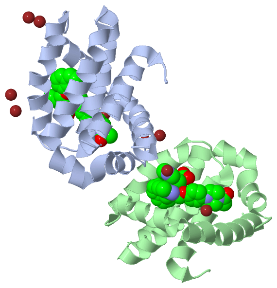
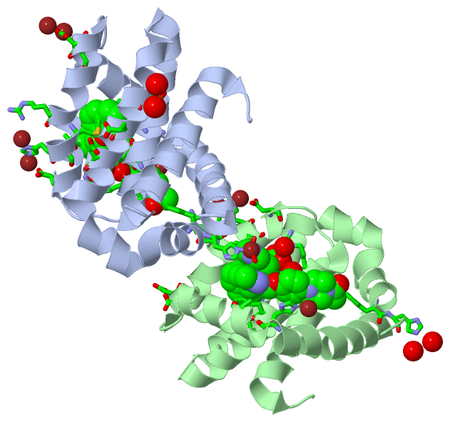
(-) Ligands, Modified Residues, Ions  (2, 12)

Asymmetric/Biological Unit (2, 12)
No.NameCountTypeFull Name
19EA2Ligand/Ion7-(3-{[4-(4-ACETYLPIPERAZIN-1-YL)PHENOXY]METHYL}-1,5-DIMETHYL-1H-PYRAZOL-4-YL)-3-{3-[(NAPHTHALEN-1-YL)OXY]PROPYL}-1-[(PYRIDIN-3-YL)METHYL]-1H-INDOLE-2-CARBOXYLIC ACID
2ZN10Ligand/IonZINC ION

(-) Sites  (12, 12)

Asymmetric Unit (12, 12)
No.NameEvidenceResiduesDescription
01AC1SOFTWAREHIS A:224 , ALA A:227 , MET A:250 , VAL A:253 , GLY A:262 , ARG A:263 , THR A:266 , LEU A:267 , PHE A:270 , PHE A:319 , ZN A:403 , HOH A:506 , HOH A:509 , HOH A:512 , HIS B:320binding site for residue 9EA A 401
02AC2SOFTWAREHIS A:224 , GLU A:288 , HOH A:506 , HOH A:529binding site for residue ZN A 402
03AC3SOFTWARE9EA A:401 , HIS B:320 , HOH B:537 , HOH B:550binding site for residue ZN A 403
04AC4SOFTWAREHIS A:252 , ASP A:256 , HOH A:537binding site for residue ZN A 404
05AC5SOFTWAREARG A:248 , HIS A:252 , HOH A:515 , GLU B:292 , ASP B:296binding site for residue ZN A 405
06AC6SOFTWAREASP A:242 , ASP B:241binding site for residue ZN A 406
07AC7SOFTWAREASP A:241 , ASP B:242binding site for residue ZN A 407
08AC8SOFTWAREHIS A:320 , HOH A:540 , 9EA B:401 , HOH B:541binding site for residue ZN A 408
09AC9SOFTWAREHIS A:320 , ZN A:408 , HIS B:224 , ALA B:227 , MET B:250 , VAL B:253 , GLY B:262 , ARG B:263 , THR B:266 , LEU B:267 , PHE B:270 , PHE B:319 , HOH B:502 , HOH B:518 , HOH B:538 , HOH B:541binding site for residue 9EA B 401
10AD1SOFTWAREHIS B:224 , GLU B:288 , HOH B:518 , HOH B:543binding site for residue ZN B 402
11AD2SOFTWAREGLU A:292 , ASP A:296 , ARG B:248 , HIS B:252 , HOH B:532binding site for residue ZN B 403
12AD3SOFTWAREGLU A:322 , HIS B:252 , ASP B:256 , HOH B:533binding site for residue ZN B 404

(-) SS Bonds  (0, 0)

(no "SS Bond" information available for 5VKC)

(-) Cis Peptide Bonds  (0, 0)

(no "Cis Peptide Bond" information available for 5VKC)

 Sequence-Structure Mapping

(-) SAPs(SNPs)/Variants  (0, 0)

(no "SAP(SNP)/Variant" information available for 5VKC)

(-) PROSITE Motifs  (0, 0)

(no "PROSITE Motif" information available for 5VKC)

(-) Exons   (0, 0)

(no "Exon" information available for 5VKC)

(-) Sequences/Alignments

Asymmetric/Biological Unit
   Reformat: Number of residues per line =  ('0' or empty: single-line sequence representation)
  Number of residues per labelling interval =   
  UniProt sequence: complete  aligned part    
   Show mapping: SCOP domains CATH domains Pfam domains Secondary structure (by author)
SAPs(SNPs) PROSITE motifs Exons
(details for a mapped element are shown in a popup box when the mouse pointer rests over it)
Chain A from PDB  Type:PROTEIN  Length:144
                                                                                                                                                                                
               SCOP domains ------------------------------------------------------------------------------------------------------------------------------------------------ SCOP domains
               CATH domains ------------------------------------------------------------------------------------------------------------------------------------------------ CATH domains
               Pfam domains ------------------------------------------------------------------------------------------------------------------------------------------------ Pfam domains
         Sec.struct. author .hhhhhhhhhhhhhhhhhhhh...hhhhhhhhhhhhhhhhhhhhhhhhhhhhhhhhhh..hhhhhhhhhhhhhhhhhh...hhhhhhhhhhhhhhhhhhhhhh.hhhhhhhhhhhhhhhhhhhhhhhhhhhhhhhhhhhhh... Sec.struct. author
                 SAPs(SNPs) ------------------------------------------------------------------------------------------------------------------------------------------------ SAPs(SNPs)
                    PROSITE ------------------------------------------------------------------------------------------------------------------------------------------------ PROSITE
                 Transcript ------------------------------------------------------------------------------------------------------------------------------------------------ Transcript
                 5vkc A 172 MDLYRQSLEIISRYLREQATGAKSGATSRKALETLRRVGDGVQRNHETAFQGMLRKLDIKNEDDVKSLSRVMIHVFSDGVTNWGRIVTLISFGAFVAKHLKTINQESCIEPLAESITDVLVRTKRDWLVKQRGWDGFVEFFHVE 322
                                   181       191  ||   208       218       228       238       248       258       268       278       288       298       308       318    
                                                194|                                                                                                                        
                                                 202                                                                                                                        

Chain B from PDB  Type:PROTEIN  Length:144
                                                                                                                                                                                
               SCOP domains ------------------------------------------------------------------------------------------------------------------------------------------------ SCOP domains
               CATH domains ------------------------------------------------------------------------------------------------------------------------------------------------ CATH domains
               Pfam domains ------------------------------------------------------------------------------------------------------------------------------------------------ Pfam domains
         Sec.struct. author .hhhhhhhhhhhhhhhhhhhh...hhhhhhhhhhhhhhhhhhhhhhhhhhhhhhhhhh..hhhhhhhhhhhhhhhhhh...hhhhhhhhhhhhhhhhhhhhhh.hhhhhhhhhhhhhhhhhhhhhhhhhhhhhhhhhhhhh... Sec.struct. author
                 SAPs(SNPs) ------------------------------------------------------------------------------------------------------------------------------------------------ SAPs(SNPs)
                    PROSITE ------------------------------------------------------------------------------------------------------------------------------------------------ PROSITE
                 Transcript ------------------------------------------------------------------------------------------------------------------------------------------------ Transcript
                 5vkc B 172 MDLYRQSLEIISRYLREQATGAKSGATSRKALETLRRVGDGVQRNHETAFQGMLRKLDIKNEDDVKSLSRVMIHVFSDGVTNWGRIVTLISFGAFVAKHLKTINQESCIEPLAESITDVLVRTKRDWLVKQRGWDGFVEFFHVE 322
                                   181       191  ||   208       218       228       238       248       258       268       278       288       298       308       318    
                                                194|                                                                                                                        
                                                 202                                                                                                                        

   Legend:   → Mismatch (orange background)
  - → Gap (green background, '-', border residues have a numbering label)
    → Modified Residue (blue background, lower-case, 'x' indicates undefined single-letter code, labelled with number + name)
  x → Chemical Group (purple background, 'x', labelled with number + name, e.g. ACE or NH2)
  extra numbering lines below/above indicate numbering irregularities and modified residue names etc., number ends below/above '|'

 Classification and Annotation

(-) SCOP Domains  (0, 0)

(no "SCOP Domain" information available for 5VKC)

(-) CATH Domains  (0, 0)

(no "CATH Domain" information available for 5VKC)

(-) Pfam Domains  (0, 0)

(no "Pfam Domain" information available for 5VKC)

(-) Gene Ontology  (32, 32)

Asymmetric/Biological Unit(hide GO term definitions)

 Visualization

(-) Interactive Views

Asymmetric/Biological Unit
  Complete Structure
    Jena3D(integrated viewing of ligand, site, SAP, PROSITE, SCOP information)
    WebMol | AstexViewer[tm]@PDBe
(Java Applets, require no local installation except for Java; loading may be slow)
    STRAP
(Java WebStart application, automatic local installation, requires Java; full application with system access!)
    RasMol
(require local installation)
    Molscript (VRML)
(requires installation of a VRML viewer; select preferred view via VRML and generate a mono or stereo PDF format file)
 
  Ligands, Modified Residues, Ions
    9EA  [ RasMol | Jena3D ]  +environment [ RasMol | Jena3D ]
    ZN  [ RasMol | Jena3D ]  +environment [ RasMol | Jena3D ]
 
  Sites
    AC1  [ RasMol ]  +environment [ RasMol ]
    AC2  [ RasMol ]  +environment [ RasMol ]
    AC3  [ RasMol ]  +environment [ RasMol ]
    AC4  [ RasMol ]  +environment [ RasMol ]
    AC5  [ RasMol ]  +environment [ RasMol ]
    AC6  [ RasMol ]  +environment [ RasMol ]
    AC7  [ RasMol ]  +environment [ RasMol ]
    AC8  [ RasMol ]  +environment [ RasMol ]
    AC9  [ RasMol ]  +environment [ RasMol ]
    AD1  [ RasMol ]  +environment [ RasMol ]
    AD2  [ RasMol ]  +environment [ RasMol ]
    AD3  [ RasMol ]  +environment [ RasMol ]
 
  Cis Peptide Bonds
(no "Cis Peptide Bonds" information available for 5vkc)
 

(-) Still Images

Jmol
  protein: cartoon or spacefill or dots and stick; nucleic acid: cartoon and stick; ligands: spacefill; active site: stick
Molscript
  protein, nucleic acid: cartoon; ligands: spacefill; active site: ball and stick

 Databases and Analysis Tools

(-) Databases

Access by PDB/NDB ID
  5vkc
    Family and Domain InformationProDom | SYSTERS
    General Structural InformationGlycoscienceDB | MMDB | NDB | OCA | PDB | PDBe | PDBj | PDBsum | PDBWiki | PQS | PROTEOPEDIA
    Orientation in MembranesOPM
    Protein SurfaceSURFACE
    Secondary StructureDSSP (structure derived) | HSSP (homology derived)
    Structural GenomicsGeneCensus
    Structural NeighboursCE | VAST
    Structure ClassificationCATH | Dali | SCOP
    Validation and Original DataBMRB Data View | BMRB Restraints Grid | EDS | PROCHECK | RECOORD | WHAT_CHECK
 
Access by UniProt ID/Accession number
  MCL1_HUMAN | Q07820
    Comparative Protein Structure ModelsModBase
    Genomic InformationEnsembl
    Protein-protein InteractionDIP
    Sequence, Family and Domain InformationInterPro | Pfam | SMART | UniProtKB/SwissProt
 
Access by Enzyme Classificator   (EC Number)
  (no 'Enzyme Classificator' available)
    General Enzyme InformationBRENDA | EC-PDB | Enzyme | IntEnz
    PathwayKEGG | MetaCyc
 
Access by Disease Identifier   (MIM ID)
  (no 'MIM ID' available)
    Disease InformationOMIM
 
Access by GenAge ID
  (no 'GenAge ID' available)
    Age Related InformationGenAge

(-) Analysis Tools

Access by PDB/NDB ID
    Domain InformationXDom
    Interatomic Contacts of Structural UnitsCSU
    Ligand-protein ContactsLPC
    Protein CavitiescastP
    Sequence and Secondary StructurePDBCartoon
    Structure AlignmentSTRAP(Java WebStart application, automatic local installation, requires Java; full application with system access!)
    Structure and Sequence BrowserSTING
 
Access by UniProt ID/Accession number
  MCL1_HUMAN | Q07820
    Protein Disorder PredictionDisEMBL | FoldIndex | GLOBPLOT (for more information see DisProt)

 Related Entries

(-) Entries Sharing at Least One Protein Chain (UniProt ID)

UniProtKB/Swiss-Prot
        MCL1_HUMAN | Q078202kbw 2mhs 2nl9 2nla 2pqk 3d7v 3io9 3kj0 3kj1 3kj2 3kz0 3mk8 3pk1 3twu 3wix 3wiy 4bpi 4bpj 4hw2 4hw3 4hw4 4oq5 4oq6 4wgi 4wmr 4wms 4wmt 4wmu 4wmv 4wmw 4wmx 4zbf 4zbi 5c3f 5c6h 5fc4 5fdo 5fdr 5iez 5if4 5jsb 5ku9 5lof 5mes 5mev 5uum

(-) Related Entries Specified in the PDB File

(no "Related Entries Specified in the PDB File" available for 5VKC)