Show PDB file:   
         Plain Text   HTML   (compressed file size)
QuickSearch:   
by PDB,NDB,UniProt,PROSITE Code or Search Term(s)  
(-)Asymmetric Unit
(-)Asym. Unit - sites
(-)Biological Unit 1
(-)Biol. Unit 1 - sites
(-)Biological Unit 2
collapse expand < >
Image Asymmetric Unit
Asymmetric Unit  (Jmol Viewer)
Image Asym. Unit - sites
Asym. Unit - sites  (Jmol Viewer)
Image Biological Unit 1
Biological Unit 1  (Jmol Viewer)
Image Biol. Unit 1 - sites
Biol. Unit 1 - sites  (Jmol Viewer)
Image Biological Unit 2
Biological Unit 2  (Jmol Viewer)

(-) Description

Title :  CRYSTAL STRUCTURE OF A SINGLE CHAIN MONELLIN MUTANT: Q28K/C41S/Y65R-MNEI
 
Authors :  A. Pica, A. Merlino
Date :  19 Jun 16  (Deposition) - 05 Oct 16  (Release) - 05 Oct 16  (Revision)
Method :  X-RAY DIFFRACTION
Resolution :  1.70
Chains :  Asym. Unit :  A,B
Biol. Unit 1:  A  (1x)
Biol. Unit 2:  B  (1x)
Keywords :  Plant Protein, Sweet Protein (Keyword Search: [Gene Ontology, PubMed, Web (Google))
 
Reference :  S. Leone, A. Pica, A. Merlino, F. Sannino, P. A. Temussi, D. Picone
Sweeter And Stronger: Enhancing Sweetness And Stability Of The Single Chain Monellin Mnei Through Molecular Design.
Sci Rep V. 6 34045 2016
PubMed-ID: 27658853  |  Reference-DOI: 10.1038/SREP34045

(-) Compounds

Molecule 1 - MONELLIN CHAIN B,MONELLIN CHAIN A
    ChainsA, B
    EngineeredYES
    Expression SystemESCHERICHIA COLI 'BL21-GOLD(DE3)PLYSS AG'
    Expression System Taxid866768
    Organism CommonSERENDIPITY BERRY
    Organism ScientificDIOSCOREOPHYLLUM CUMMINSII
    Organism Taxid3457
    SynonymMONELLIN CHAIN II,MONELLIN CHAIN I

 Structural Features

(-) Chains, Units

  12
Asymmetric Unit AB
Biological Unit 1 (1x)A 
Biological Unit 2 (1x) B

Summary Information (see also Sequences/Alignments below)

(-) Ligands, Modified Residues, Ions  (1, 4)

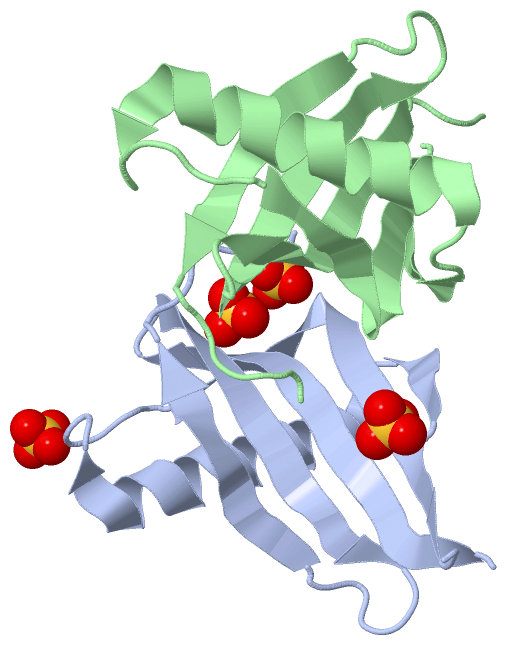
Asymmetric Unit (1, 4)
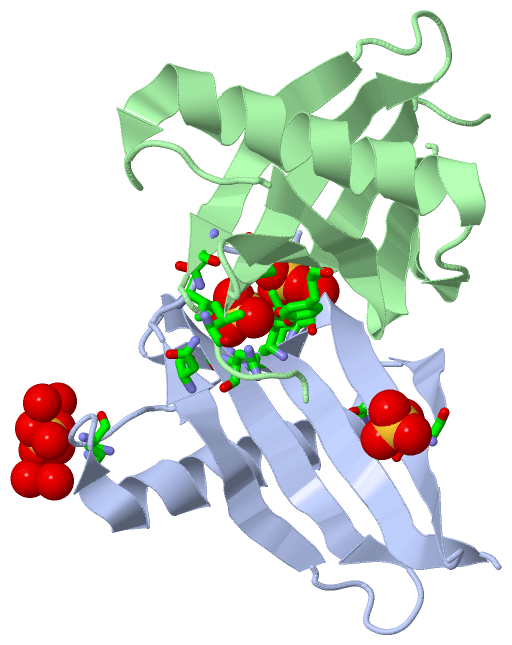
No.NameCountTypeFull Name
1SO44Ligand/IonSULFATE ION
Biological Unit 1 (1, 4)
No.NameCountTypeFull Name
1SO44Ligand/IonSULFATE ION
Biological Unit 2 (0, 0)
No.NameCountTypeFull Name
1SO4-1Ligand/IonSULFATE ION

(-) Sites  (4, 4)

Asymmetric Unit (4, 4)
No.NameEvidenceResiduesDescription
1AC1SOFTWARETYR A:63 , ARG A:72 , ASN A:90 , VAL A:93 , TYR B:63 , ARG B:72 , ASN B:90 , VAL B:93binding site for residue SO4 A 101
2AC2SOFTWAREGLY A:9 , PRO A:10 , PHE A:11 , ARG A:31 , HOH A:207 , HOH A:230 , HOH A:231 , HOH A:235 , HOH A:252binding site for residue SO4 A 102
3AC3SOFTWARELYS A:36 , ILE A:38 , ARG A:65 , HOH A:201binding site for residue SO4 A 103
4AC4SOFTWAREGLY A:1 , LYS A:44binding site for residue SO4 A 104

(-) SS Bonds  (0, 0)

(no "SS Bond" information available for 5LC6)

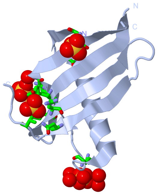
(-) Cis Peptide Bonds  (4, 4)
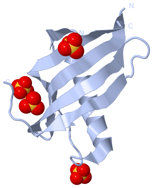
Asymmetric Unit
No.Residues
1Arg A:39 -Pro A:40
2Gly A:91 -Pro A:92
3Arg B:39 -Pro B:40
4Gly B:91 -Pro B:92

 Sequence-Structure Mapping

(-) SAPs(SNPs)/Variants  (0, 0)

(no "SAP(SNP)/Variant" information available for 5LC6)

(-) PROSITE Motifs  (0, 0)

(no "PROSITE Motif" information available for 5LC6)

(-) Exons   (0, 0)

(no "Exon" information available for 5LC6)

(-) Sequences/Alignments

Asymmetric Unit
   Reformat: Number of residues per line =  ('0' or empty: single-line sequence representation)
  Number of residues per labelling interval =   
  UniProt sequence: complete  aligned part    
   Show mapping: SCOP domains CATH domains Pfam domains Secondary structure (by author)
SAPs(SNPs) PROSITE motifs Exons
(details for a mapped element are shown in a popup box when the mouse pointer rests over it)
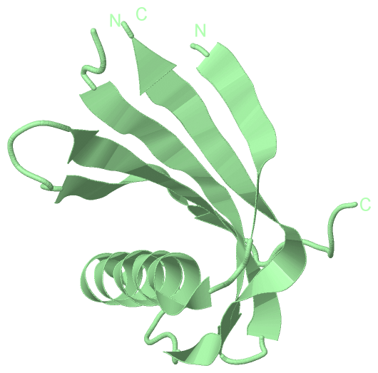
Chain A from PDB  Type:PROTEIN  Length:92
                                                                                                                           
               SCOP domains -------------------------------------------------------------------------------------------- SCOP domains
               CATH domains -------------------------------------------------------------------------------------------- CATH domains
               Pfam domains -------------------------------------------------------------------------------------------- Pfam domains
         Sec.struct. author .eeeee..hhhhhhhhhhhhhhhhhhh......eeeeeeeeeeeeee...eeeeeeeeeeee..eeeeeeeeee.....eeeeeee...... Sec.struct. author
                 SAPs(SNPs) -------------------------------------------------------------------------------------------- SAPs(SNPs)
                    PROSITE -------------------------------------------------------------------------------------------- PROSITE
                 Transcript -------------------------------------------------------------------------------------------- Transcript
                  5lc6 A  1 GEWEIIDIGPFTQNLGKFAVDEENKIGKYGRLTFNKVIRPSMKKTIYENEIKGYEYQLYVRASDKLFRADISEDYKTRGRKLLRFNGPVPPP 96
                                    10        20        30        40        54        64        74        84        94  
                                                                           49|                                          
                                                                            54                                          

Chain B from PDB  Type:PROTEIN  Length:90
                                                                                                                         
               SCOP domains ------------------------------------------------------------------------------------------ SCOP domains
               CATH domains ------------------------------------------------------------------------------------------ CATH domains
               Pfam domains ------------------------------------------------------------------------------------------ Pfam domains
         Sec.struct. author .eeeee..hhhhhhhhhhhhhhhhhhh......eeeeeeeeeeeee....eeeeeeeeee..eeeeeeeeee.....eeeeeee...... Sec.struct. author
                 SAPs(SNPs) ------------------------------------------------------------------------------------------ SAPs(SNPs)
                    PROSITE ------------------------------------------------------------------------------------------ PROSITE
                 Transcript ------------------------------------------------------------------------------------------ Transcript
                  5lc6 B  1 GEWEIIDIGPFTQNLGKFAVDEENKIGKYGRLTFNKVIRPSMKKTIYEIKGYEYQLYVRASDKLFRADISEDYKTRGRKLLRFNGPVPPP 96
                                    10        20        30        40      ||56        66        76        86        96
                                                                         47|                                          
                                                                          54                                          

   Legend:   → Mismatch (orange background)
  - → Gap (green background, '-', border residues have a numbering label)
    → Modified Residue (blue background, lower-case, 'x' indicates undefined single-letter code, labelled with number + name)
  x → Chemical Group (purple background, 'x', labelled with number + name, e.g. ACE or NH2)
  extra numbering lines below/above indicate numbering irregularities and modified residue names etc., number ends below/above '|'

 Classification and Annotation

(-) SCOP Domains  (0, 0)

(no "SCOP Domain" information available for 5LC6)

(-) CATH Domains  (0, 0)

(no "CATH Domain" information available for 5LC6)

(-) Pfam Domains  (0, 0)

(no "Pfam Domain" information available for 5LC6)

(-) Gene Ontology  (0, 0)

Asymmetric Unit(hide GO term definitions)
    (no "Gene Ontology" information available for 5LC6)

 Visualization

(-) Interactive Views

Asymmetric Unit
  Complete Structure
    Jena3D(integrated viewing of ligand, site, SAP, PROSITE, SCOP information)
    WebMol | AstexViewer[tm]@PDBe
(Java Applets, require no local installation except for Java; loading may be slow)
    STRAP
(Java WebStart application, automatic local installation, requires Java; full application with system access!)
    RasMol
(require local installation)
    Molscript (VRML)
(requires installation of a VRML viewer; select preferred view via VRML and generate a mono or stereo PDF format file)
 
  Ligands, Modified Residues, Ions
    SO4  [ RasMol | Jena3D ]  +environment [ RasMol | Jena3D ]
 
  Sites
    AC1  [ RasMol ]  +environment [ RasMol ]
    AC2  [ RasMol ]  +environment [ RasMol ]
    AC3  [ RasMol ]  +environment [ RasMol ]
    AC4  [ RasMol ]  +environment [ RasMol ]
 
  Cis Peptide Bonds
    Arg A:39 - Pro A:40   [ RasMol ]  
    Arg B:39 - Pro B:40   [ RasMol ]  
    Gly A:91 - Pro A:92   [ RasMol ]  
    Gly B:91 - Pro B:92   [ RasMol ]  
 
Biological Units
  Complete Structure
    Biological Unit 1  [ Jena3D ]
    Biological Unit 2  [ Jena3D ]

(-) Still Images

Jmol
  protein: cartoon or spacefill or dots and stick; nucleic acid: cartoon and stick; ligands: spacefill; active site: stick
Molscript
  protein, nucleic acid: cartoon; ligands: spacefill; active site: ball and stick

 Databases and Analysis Tools

(-) Databases

Access by PDB/NDB ID
  5lc6
    Family and Domain InformationProDom | SYSTERS
    General Structural InformationGlycoscienceDB | MMDB | NDB | OCA | PDB | PDBe | PDBj | PDBsum | PDBWiki | PQS | PROTEOPEDIA
    Orientation in MembranesOPM
    Protein SurfaceSURFACE
    Secondary StructureDSSP (structure derived) | HSSP (homology derived)
    Structural GenomicsGeneCensus
    Structural NeighboursCE | VAST
    Structure ClassificationCATH | Dali | SCOP
    Validation and Original DataBMRB Data View | BMRB Restraints Grid | EDS | PROCHECK | RECOORD | WHAT_CHECK
 
Access by UniProt ID/Accession number
  MONA_DIOCU | P02881
    Comparative Protein Structure ModelsModBase
    Genomic InformationEnsembl
    Protein-protein InteractionDIP
    Sequence, Family and Domain InformationInterPro | Pfam | SMART | UniProtKB/SwissProt
  MONB_DIOCU | P02882
    Comparative Protein Structure ModelsModBase
    Genomic InformationEnsembl
    Protein-protein InteractionDIP
    Sequence, Family and Domain InformationInterPro | Pfam | SMART | UniProtKB/SwissProt
 
Access by Enzyme Classificator   (EC Number)
  (no 'Enzyme Classificator' available)
    General Enzyme InformationBRENDA | EC-PDB | Enzyme | IntEnz
    PathwayKEGG | MetaCyc
 
Access by Disease Identifier   (MIM ID)
  (no 'MIM ID' available)
    Disease InformationOMIM
 
Access by GenAge ID
  (no 'GenAge ID' available)
    Age Related InformationGenAge

(-) Analysis Tools

Access by PDB/NDB ID
    Domain InformationXDom
    Interatomic Contacts of Structural UnitsCSU
    Ligand-protein ContactsLPC
    Protein CavitiescastP
    Sequence and Secondary StructurePDBCartoon
    Structure AlignmentSTRAP(Java WebStart application, automatic local installation, requires Java; full application with system access!)
    Structure and Sequence BrowserSTING
 
Access by UniProt ID/Accession number
  MONA_DIOCU | P02881
    Protein Disorder PredictionDisEMBL | FoldIndex | GLOBPLOT (for more information see DisProt)
  MONB_DIOCU | P02882
    Protein Disorder PredictionDisEMBL | FoldIndex | GLOBPLOT (for more information see DisProt)

 Related Entries

(-) Entries Sharing at Least One Protein Chain (UniProt ID)

UniProtKB/Swiss-Prot
        MONA_DIOCU | P028811fa3 1fuw 1iv7 1iv9 1krl 1m9g 1mnl 2gpk 2o9u 3mon 3pxm 3pyj 3q2p 4mon 5lc7
        MONB_DIOCU | P028821fa3 1fuw 1iv7 1iv9 1krl 1m9g 1mnl 1mol 2o9u 3mon 3pxm 3pyj 3q2p 4mon 5lc7

(-) Related Entries Specified in the PDB File

(no "Related Entries Specified in the PDB File" available for 5LC6)