Show PDB file:   
         Plain Text   HTML   (compressed file size)
QuickSearch:   
by PDB,NDB,UniProt,PROSITE Code or Search Term(s)  
(-)Asymmetric Unit
(-)Asym. Unit - sites
(-)Biological Unit 1
(-)Biol. Unit 1 - sites
(-)Biological Unit 2
collapse expand < >
Image Asymmetric Unit
Asymmetric Unit  (Jmol Viewer)
Image Asym. Unit - sites
Asym. Unit - sites  (Jmol Viewer)
Image Biological Unit 1
Biological Unit 1  (Jmol Viewer)
Image Biol. Unit 1 - sites
Biol. Unit 1 - sites  (Jmol Viewer)
Image Biological Unit 2
Biological Unit 2  (Jmol Viewer)

(-) Description

Title :  CRYSTAL STRUCTURE OF THE E. COLI TYPE 1 PILUS SUBUNIT FIMG (ENGINEERED VARIANT WITH SUBSTITUTION Q134E; N-TERMINAL FIMG RESIDUES 1-12 TRUNCATED) IN COMPLEX WITH THE DONOR STRAND PEPTIDE DSF_T4R-T6R-D13N
 
Authors :  C. Giese, J. Eras, A. Kern, M. A. Scharer, G. Capitani, R. Glockshuber
Date :  11 Mar 16  (Deposition) - 06 Jul 16  (Release) - 10 Aug 16  (Revision)
Method :  X-RAY DIFFRACTION
Resolution :  1.50
Chains :  Asym. Unit :  A,B,F,G
Biol. Unit 1:  F,G  (1x)
Biol. Unit 2:  A,B  (1x)
Keywords :  Complex, Protein, Fimgt, Cell Adhesion (Keyword Search: [Gene Ontology, PubMed, Web (Google))
 
Reference :  C. Giese, J. Eras, A. Kern, M. A. Scharer, G. Capitani, R. Glockshuber
Accelerating The Association Of The Most Stable Protein-Ligand Complex By More Than Two Orders Of Magnitude
Angew. Chem. Int. Ed. Engl. V. 55 9350 2016
PubMed-ID: 27351462  |  Reference-DOI: 10.1002/ANIE.201603652

(-) Compounds

Molecule 1 - PROTEIN FIMG
    ChainsG, A
    EngineeredYES
    Expression SystemESCHERICHIA COLI
    Expression System Taxid562
    FragmentUNP RESIDUES 36-167
    GeneFIMG, B4319, JW4282
    Organism ScientificESCHERICHIA COLI K-12
    Organism Taxid83333
 
Molecule 2 - PROTEIN FIMF
    ChainsF, B
    EngineeredYES
    FragmentUNP RESIDUES 23-37
    Organism ScientificESCHERICHIA COLI K-12
    Organism Taxid83333
    SyntheticYES

 Structural Features

(-) Chains, Units

  1234
Asymmetric Unit ABFG
Biological Unit 1 (1x)  FG
Biological Unit 2 (1x)AB  

Summary Information (see also Sequences/Alignments below)

(-) Ligands, Modified Residues, Ions  (1, 4)

Asymmetric Unit (1, 4)
No.NameCountTypeFull Name
1CO4Ligand/IonCOBALT (II) ION
Biological Unit 1 (0, 0)
No.NameCountTypeFull Name
1CO-1Ligand/IonCOBALT (II) ION
Biological Unit 2 (0, 0)
No.NameCountTypeFull Name
1CO-1Ligand/IonCOBALT (II) ION

(-) Sites  (4, 4)

Asymmetric Unit (4, 4)
No.NameEvidenceResiduesDescription
1AC1SOFTWAREHOH A:304 , HIS G:45 , HOH G:465binding site for residue CO G 201
2AC2SOFTWAREHIS A:114 , HOH A:424 , HOH A:429 , ASP G:26 , HOH G:317 , HOH G:444binding site for residue CO G 202
3AC3SOFTWAREASP A:26 , HOH A:340 , HOH A:432 , HIS G:114 , HOH G:426 , HOH G:430binding site for residue CO G 203
4AC4SOFTWARELEU A:27 , HIS A:45 , HOH A:302 , HOH A:367 , HOH A:422 , HOH A:469 , HOH G:308binding site for residue CO A 201

(-) SS Bonds  (2, 2)

Asymmetric Unit
No.Residues
1A:16 -A:54
2G:16 -G:54

(-) Cis Peptide Bonds  (0, 0)

(no "Cis Peptide Bond" information available for 5IQM)

 Sequence-Structure Mapping

(-) SAPs(SNPs)/Variants  (0, 0)

(no "SAP(SNP)/Variant" information available for 5IQM)

(-) PROSITE Motifs  (0, 0)

(no "PROSITE Motif" information available for 5IQM)

(-) Exons   (0, 0)

(no "Exon" information available for 5IQM)

(-) Sequences/Alignments

Asymmetric Unit
   Reformat: Number of residues per line =  ('0' or empty: single-line sequence representation)
  Number of residues per labelling interval =   
  UniProt sequence: complete  aligned part    
   Show mapping: SCOP domains CATH domains Pfam domains Secondary structure (by author)
SAPs(SNPs) PROSITE motifs Exons
(details for a mapped element are shown in a popup box when the mouse pointer rests over it)
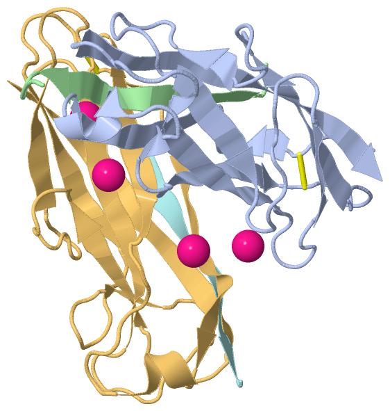
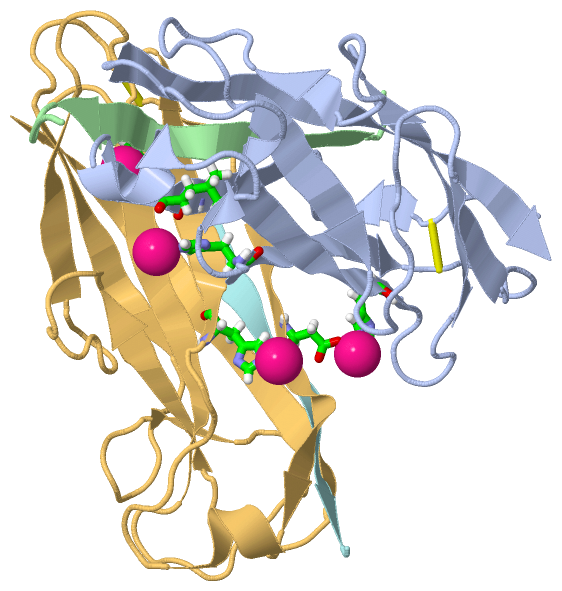
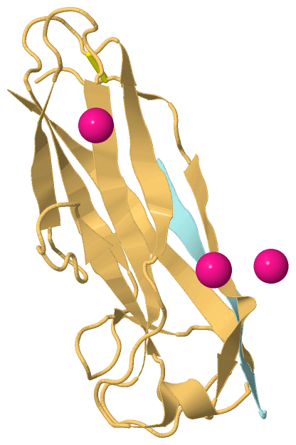
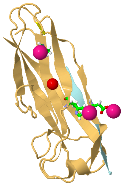
Chain A from PDB  Type:PROTEIN  Length:132
                                                                                                                                                                    
               SCOP domains ------------------------------------------------------------------------------------------------------------------------------------ SCOP domains
               CATH domains ------------------------------------------------------------------------------------------------------------------------------------ CATH domains
               Pfam domains ------------------------------------------------------------------------------------------------------------------------------------ Pfam domains
         Sec.struct. author ....ee...eeeeeeeeee.hhhh...ee...eeeeeeee.......eeeeeee........ee........eeeee...........eeeee.......eeeeeeeeeee.......eeeeeeeeeeeeee Sec.struct. author
                 SAPs(SNPs) ------------------------------------------------------------------------------------------------------------------------------------ SAPs(SNPs)
                    PROSITE ------------------------------------------------------------------------------------------------------------------------------------ PROSITE
                 Transcript ------------------------------------------------------------------------------------------------------------------------------------ Transcript
                 5iqm A  13 AKPCTVSTTNATVDLGDLYSFSLMSAGAASAWHDVALELTNCPVGTSRVTASFSGAADSTGYYKNQGTAQNIQLELQDDSGNTLNTGATKTVQVDDSSQSAHFPLQVRALTVNGGATQGTIEAVISITYTYS 144
                                    22        32        42        52        62        72        82        92       102       112       122       132       142  

Chain B from PDB  Type:PROTEIN  Length:15
                                               
               SCOP domains --------------- SCOP domains
               CATH domains --------------- CATH domains
               Pfam domains --------------- Pfam domains
         Sec.struct. author ..eeeeeeeeee... Sec.struct. author
                 SAPs(SNPs) --------------- SAPs(SNPs)
                    PROSITE --------------- PROSITE
                 Transcript --------------- Transcript
                 5iqm B   1 ADSRIRIRGYVRNNG  15
                                    10     

Chain F from PDB  Type:PROTEIN  Length:13
                                             
               SCOP domains ------------- SCOP domains
               CATH domains ------------- CATH domains
               Pfam domains ------------- Pfam domains
         Sec.struct. author ..eeeeeeeeee. Sec.struct. author
                 SAPs(SNPs) ------------- SAPs(SNPs)
                    PROSITE ------------- PROSITE
                 Transcript ------------- Transcript
                 5iqm F   1 ADSRIRIRGYVRN  13
                                    10   

Chain G from PDB  Type:PROTEIN  Length:132
                                                                                                                                                                    
               SCOP domains ------------------------------------------------------------------------------------------------------------------------------------ SCOP domains
               CATH domains ------------------------------------------------------------------------------------------------------------------------------------ CATH domains
               Pfam domains ------------------------------------------------------------------------------------------------------------------------------------ Pfam domains
         Sec.struct. author ....ee...eeeeeeeeee.hhhh........eeeeeeee.......eeeeeee........ee........eeeee...........eeeee.......eeeeeeeeee........eeeeeeeeeeeeee Sec.struct. author
                 SAPs(SNPs) ------------------------------------------------------------------------------------------------------------------------------------ SAPs(SNPs)
                    PROSITE ------------------------------------------------------------------------------------------------------------------------------------ PROSITE
                 Transcript ------------------------------------------------------------------------------------------------------------------------------------ Transcript
                 5iqm G  13 AKPCTVSTTNATVDLGDLYSFSLMSAGAASAWHDVALELTNCPVGTSRVTASFSGAADSTGYYKNQGTAQNIQLELQDDSGNTLNTGATKTVQVDDSSQSAHFPLQVRALTVNGGATQGTIEAVISITYTYS 144
                                    22        32        42        52        62        72        82        92       102       112       122       132       142  

   Legend:   → Mismatch (orange background)
  - → Gap (green background, '-', border residues have a numbering label)
    → Modified Residue (blue background, lower-case, 'x' indicates undefined single-letter code, labelled with number + name)
  x → Chemical Group (purple background, 'x', labelled with number + name, e.g. ACE or NH2)
  extra numbering lines below/above indicate numbering irregularities and modified residue names etc., number ends below/above '|'

 Classification and Annotation

(-) SCOP Domains  (0, 0)

(no "SCOP Domain" information available for 5IQM)

(-) CATH Domains  (0, 0)

(no "CATH Domain" information available for 5IQM)

(-) Pfam Domains  (0, 0)

(no "Pfam Domain" information available for 5IQM)

(-) Gene Ontology  (2, 4)

Asymmetric Unit(hide GO term definitions)

 Visualization

(-) Interactive Views

Asymmetric Unit
  Complete Structure
    Jena3D(integrated viewing of ligand, site, SAP, PROSITE, SCOP information)
    WebMol | AstexViewer[tm]@PDBe
(Java Applets, require no local installation except for Java; loading may be slow)
    STRAP
(Java WebStart application, automatic local installation, requires Java; full application with system access!)
    RasMol
(require local installation)
    Molscript (VRML)
(requires installation of a VRML viewer; select preferred view via VRML and generate a mono or stereo PDF format file)
 
  Ligands, Modified Residues, Ions
    CO  [ RasMol | Jena3D ]  +environment [ RasMol | Jena3D ]
 
  Sites
    AC1  [ RasMol ]  +environment [ RasMol ]
    AC2  [ RasMol ]  +environment [ RasMol ]
    AC3  [ RasMol ]  +environment [ RasMol ]
    AC4  [ RasMol ]  +environment [ RasMol ]
 
  Cis Peptide Bonds
(no "Cis Peptide Bonds" information available for 5iqm)
 
Biological Units
  Complete Structure
    Biological Unit 1  [ Jena3D ]
    Biological Unit 2  [ Jena3D ]

(-) Still Images

Jmol
  protein: cartoon or spacefill or dots and stick; nucleic acid: cartoon and stick; ligands: spacefill; active site: stick
Molscript
  protein, nucleic acid: cartoon; ligands: spacefill; active site: ball and stick

 Databases and Analysis Tools

(-) Databases

Access by PDB/NDB ID
  5iqm
    Family and Domain InformationProDom | SYSTERS
    General Structural InformationGlycoscienceDB | MMDB | NDB | OCA | PDB | PDBe | PDBj | PDBsum | PDBWiki | PQS | PROTEOPEDIA
    Orientation in MembranesOPM
    Protein SurfaceSURFACE
    Secondary StructureDSSP (structure derived) | HSSP (homology derived)
    Structural GenomicsGeneCensus
    Structural NeighboursCE | VAST
    Structure ClassificationCATH | Dali | SCOP
    Validation and Original DataBMRB Data View | BMRB Restraints Grid | EDS | PROCHECK | RECOORD | WHAT_CHECK
 
Access by UniProt ID/Accession number
  FIMF_ECOLI | P08189
    Comparative Protein Structure ModelsModBase
    Genomic InformationEnsembl
    Protein-protein InteractionDIP
    Sequence, Family and Domain InformationInterPro | Pfam | SMART | UniProtKB/SwissProt
  FIMG_ECOLI | P08190
    Comparative Protein Structure ModelsModBase
    Genomic InformationEnsembl
    Protein-protein InteractionDIP
    Sequence, Family and Domain InformationInterPro | Pfam | SMART | UniProtKB/SwissProt
 
Access by Enzyme Classificator   (EC Number)
  (no 'Enzyme Classificator' available)
    General Enzyme InformationBRENDA | EC-PDB | Enzyme | IntEnz
    PathwayKEGG | MetaCyc
 
Access by Disease Identifier   (MIM ID)
  (no 'MIM ID' available)
    Disease InformationOMIM
 
Access by GenAge ID
  (no 'GenAge ID' available)
    Age Related InformationGenAge

(-) Analysis Tools

Access by PDB/NDB ID
    Domain InformationXDom
    Interatomic Contacts of Structural UnitsCSU
    Ligand-protein ContactsLPC
    Protein CavitiescastP
    Sequence and Secondary StructurePDBCartoon
    Structure AlignmentSTRAP(Java WebStart application, automatic local installation, requires Java; full application with system access!)
    Structure and Sequence BrowserSTING
 
Access by UniProt ID/Accession number
  FIMF_ECOLI | P08189
    Protein Disorder PredictionDisEMBL | FoldIndex | GLOBPLOT (for more information see DisProt)
  FIMG_ECOLI | P08190
    Protein Disorder PredictionDisEMBL | FoldIndex | GLOBPLOT (for more information see DisProt)

 Related Entries

(-) Entries Sharing at Least One Protein Chain (UniProt ID)

UniProtKB/Swiss-Prot
        FIMF_ECOLI | P081892jmr 3bfq 3bfw 3bwu 3jwn 4j3o 4xob 5iqn 5iqo
        FIMG_ECOLI | P081903bfq 3bfw 3jwn 4j3o 4xo9 4xoa 4xod 5iqn 5iqo

(-) Related Entries Specified in the PDB File

(no "Related Entries Specified in the PDB File" available for 5IQM)