Show PDB file:   
         Plain Text   HTML   (compressed file size)
QuickSearch:   
by PDB,NDB,UniProt,PROSITE Code or Search Term(s)  
(-)Asym./Biol. Unit
(-)Asym./Biol. Unit - sites
collapse expand < >
Image Asym./Biol. Unit
Asym./Biol. Unit  (Jmol Viewer)
Image Asym./Biol. Unit - sites
Asym./Biol. Unit - sites  (Jmol Viewer)

(-) Description

Title :  ULTRA HIGH RESOLUTION CRYSTAL STRUCTURE OF THE APO- FORM OF SECOND BROMODOMAIN OF BRD2.
 
Authors :  S. K. Tripathi, B. Padmanabhan
Date :  22 Feb 16  (Deposition) - 22 Jun 16  (Release) - 22 Jun 16  (Revision)
Method :  X-RAY DIFFRACTION
Resolution :  0.94
Chains :  Asym./Biol. Unit :  A
Keywords :  Bet-Family, Acetyl-Lysine Binding, Transcription (Keyword Search: [Gene Ontology, PubMed, Web (Google))
 
Reference :  S. Tripathi, S. Mathur, P. Deshmukh, R. Manjula, B. Padmanabhan
A Novel Phenanthridionone Based Scaffold As A Potential Inhibitor Of The Brd2 Bromodomain: Crystal Structure Of The Complex
Plos One V. 11 56344 2016
PubMed-ID: 27243809  |  Reference-DOI: 10.1371/JOURNAL.PONE.0156344

(-) Compounds

Molecule 1 - BROMODOMAIN-CONTAINING PROTEIN 2
    ChainsA
    EngineeredYES
    Expression SystemESCHERICHIA COLI BL21
    Expression System Taxid511693
    FragmentUNP RESIDUES 348-455
    GeneBRD2, KIAA9001, RING3
    Organism CommonHUMAN
    Organism ScientificHOMO SAPIENS
    Organism Taxid9606
    SynonymO27.1.1,REALLY INTERESTING NEW GENE 3 PROTEIN

 Structural Features

(-) Chains, Units

  1
Asymmetric/Biological Unit A

Summary Information (see also Sequences/Alignments below)

(-) Ligands, Modified Residues, Ions  (3, 8)

Asymmetric/Biological Unit (3, 8)
No.NameCountTypeFull Name
1CL1Ligand/IonCHLORIDE ION
2GOL5Ligand/IonGLYCEROL
3PGE2Ligand/IonTRIETHYLENE GLYCOL

(-) Sites  (8, 8)

Asymmetric Unit (8, 8)
No.NameEvidenceResiduesDescription
1AC1SOFTWAREGLN A:412 , ARG A:419binding site for residue CL A 501
2AC2SOFTWARETRP A:370 , PRO A:371 , MET A:438binding site for residue GOL A 502
3AC3SOFTWARESER A:397 , THR A:398 , ARG A:401 , HOH A:712binding site for residue GOL A 503
4AC4SOFTWARELEU A:383 , ASN A:429 , HOH A:604 , HOH A:614 , HOH A:617binding site for residue GOL A 504
5AC5SOFTWARETYR A:368 , LYS A:402 , ASP A:407 , MET A:438 , LYS A:441 , HOH A:609 , HOH A:627 , HOH A:681 , HOH A:715binding site for residue GOL A 505
6AC6SOFTWAREHIS A:387 , ILE A:390 , LYS A:391 , PRO A:393 , HOH A:621 , HOH A:673binding site for residue GOL A 506
7AC7SOFTWAREARG A:409 , ASP A:410 , PRO A:431 , ARG A:440 , ASP A:444 , VAL A:445 , PHE A:448 , HOH A:619 , HOH A:652 , HOH A:665 , HOH A:708 , HOH A:735binding site for residue PGE A 507
8AC8SOFTWAREPRO A:347 , LEU A:350 , LYS A:351 , ASN A:354 , HOH A:683binding site for residue PGE A 508

(-) SS Bonds  (0, 0)

(no "SS Bond" information available for 5IBN)

(-) Cis Peptide Bonds  (0, 0)

(no "Cis Peptide Bond" information available for 5IBN)

 Sequence-Structure Mapping

(-) SAPs(SNPs)/Variants  (0, 0)

(no "SAP(SNP)/Variant" information available for 5IBN)

(-) PROSITE Motifs  (0, 0)

(no "PROSITE Motif" information available for 5IBN)

(-) Exons   (0, 0)

(no "Exon" information available for 5IBN)

(-) Sequences/Alignments

Asymmetric/Biological Unit
   Reformat: Number of residues per line =  ('0' or empty: single-line sequence representation)
  Number of residues per labelling interval =   
  UniProt sequence: complete  aligned part    
   Show mapping: SCOP domains CATH domains Pfam domains Secondary structure (by author)
SAPs(SNPs) PROSITE motifs Exons
(details for a mapped element are shown in a popup box when the mouse pointer rests over it)
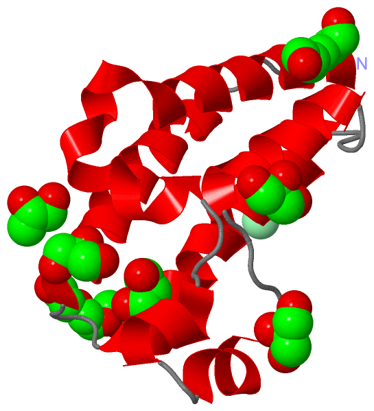
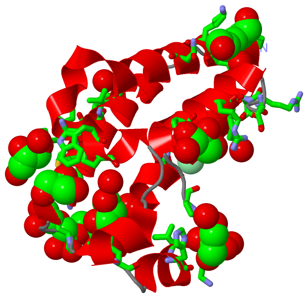
Chain A from PDB  Type:PROTEIN  Length:111
                                                                                                                                               
               SCOP domains --------------------------------------------------------------------------------------------------------------- SCOP domains
               CATH domains --------------------------------------------------------------------------------------------------------------- CATH domains
               Pfam domains --------------------------------------------------------------------------------------------------------------- Pfam domains
         Sec.struct. author hhhhhhhhhhhhhhhhhhhhhhhhhhhhh...hhhhhh..hhhhhh....hhhhhhhhhhh....hhhhhhhhhhhhhhhhhhhh...hhhhhhhhhhhhhhhhhhh.... Sec.struct. author
                 SAPs(SNPs) --------------------------------------------------------------------------------------------------------------- SAPs(SNPs)
                    PROSITE --------------------------------------------------------------------------------------------------------------- PROSITE
                 Transcript --------------------------------------------------------------------------------------------------------------- Transcript
                 5ibn A 345 ANPEQLKHCNGILKELLSKKHAAYAWPFYKPVDASALGLHDYHDIIKHPMDLSTVKRKMENRDYRDAQEFAADVRLMFSNCYKYNPPDHDVVAMARKLQDVFEFRYAKMPD 455
                                   354       364       374       384       394       404       414       424       434       444       454 

   Legend:   → Mismatch (orange background)
  - → Gap (green background, '-', border residues have a numbering label)
    → Modified Residue (blue background, lower-case, 'x' indicates undefined single-letter code, labelled with number + name)
  x → Chemical Group (purple background, 'x', labelled with number + name, e.g. ACE or NH2)
  extra numbering lines below/above indicate numbering irregularities and modified residue names etc., number ends below/above '|'

 Classification and Annotation

(-) SCOP Domains  (0, 0)

(no "SCOP Domain" information available for 5IBN)

(-) CATH Domains  (0, 0)

(no "CATH Domain" information available for 5IBN)

(-) Pfam Domains  (0, 0)

(no "Pfam Domain" information available for 5IBN)

(-) Gene Ontology  (10, 10)

Asymmetric/Biological Unit(hide GO term definitions)

 Visualization

(-) Interactive Views

Asymmetric/Biological Unit
  Complete Structure
    Jena3D(integrated viewing of ligand, site, SAP, PROSITE, SCOP information)
    WebMol | AstexViewer[tm]@PDBe
(Java Applets, require no local installation except for Java; loading may be slow)
    STRAP
(Java WebStart application, automatic local installation, requires Java; full application with system access!)
    RasMol
(require local installation)
    Molscript (VRML)
(requires installation of a VRML viewer; select preferred view via VRML and generate a mono or stereo PDF format file)
 
  Ligands, Modified Residues, Ions
    CL  [ RasMol | Jena3D ]  +environment [ RasMol | Jena3D ]
    GOL  [ RasMol | Jena3D ]  +environment [ RasMol | Jena3D ]
    PGE  [ RasMol | Jena3D ]  +environment [ RasMol | Jena3D ]
 
  Sites
    AC1  [ RasMol ]  +environment [ RasMol ]
    AC2  [ RasMol ]  +environment [ RasMol ]
    AC3  [ RasMol ]  +environment [ RasMol ]
    AC4  [ RasMol ]  +environment [ RasMol ]
    AC5  [ RasMol ]  +environment [ RasMol ]
    AC6  [ RasMol ]  +environment [ RasMol ]
    AC7  [ RasMol ]  +environment [ RasMol ]
    AC8  [ RasMol ]  +environment [ RasMol ]
 
  Cis Peptide Bonds
(no "Cis Peptide Bonds" information available for 5ibn)
 

(-) Still Images

Jmol
  protein: cartoon or spacefill or dots and stick; nucleic acid: cartoon and stick; ligands: spacefill; active site: stick
Molscript
  protein, nucleic acid: cartoon; ligands: spacefill; active site: ball and stick

 Databases and Analysis Tools

(-) Databases

Access by PDB/NDB ID
  5ibn
    Family and Domain InformationProDom | SYSTERS
    General Structural InformationGlycoscienceDB | MMDB | NDB | OCA | PDB | PDBe | PDBj | PDBsum | PDBWiki | PQS | PROTEOPEDIA
    Orientation in MembranesOPM
    Protein SurfaceSURFACE
    Secondary StructureDSSP (structure derived) | HSSP (homology derived)
    Structural GenomicsGeneCensus
    Structural NeighboursCE | VAST
    Structure ClassificationCATH | Dali | SCOP
    Validation and Original DataBMRB Data View | BMRB Restraints Grid | EDS | PROCHECK | RECOORD | WHAT_CHECK
 
Access by UniProt ID/Accession number
  BRD2_HUMAN | P25440
    Comparative Protein Structure ModelsModBase
    Genomic InformationEnsembl
    Protein-protein InteractionDIP
    Sequence, Family and Domain InformationInterPro | Pfam | SMART | UniProtKB/SwissProt
 
Access by Enzyme Classificator   (EC Number)
  (no 'Enzyme Classificator' available)
    General Enzyme InformationBRENDA | EC-PDB | Enzyme | IntEnz
    PathwayKEGG | MetaCyc
 
Access by Disease Identifier   (MIM ID)
  (no 'MIM ID' available)
    Disease InformationOMIM
 
Access by GenAge ID
  (no 'GenAge ID' available)
    Age Related InformationGenAge

(-) Analysis Tools

Access by PDB/NDB ID
    Domain InformationXDom
    Interatomic Contacts of Structural UnitsCSU
    Ligand-protein ContactsLPC
    Protein CavitiescastP
    Sequence and Secondary StructurePDBCartoon
    Structure AlignmentSTRAP(Java WebStart application, automatic local installation, requires Java; full application with system access!)
    Structure and Sequence BrowserSTING
 
Access by UniProt ID/Accession number
  BRD2_HUMAN | P25440
    Protein Disorder PredictionDisEMBL | FoldIndex | GLOBPLOT (for more information see DisProt)

 Related Entries

(-) Entries Sharing at Least One Protein Chain (UniProt ID)

UniProtKB/Swiss-Prot
        BRD2_HUMAN | P254401x0j 2dvq 2dvr 2dvs 2dvv 2e3k 2g4a 2ydw 2yek 3aqa 3oni 4a9e 4a9f 4a9h 4a9i 4a9j 4a9m 4a9n 4a9o 4akn 4alg 4alh 4j1p 4mr5 4mr6 4qeu 4qev 4qew 4uyf 4uyg 4uyh 5bt5 5dfb 5dfc 5dfd 5dw1 5ek9 5hel 5hem 5hen 5hfq 5ig6 5u5s 5uew

(-) Related Entries Specified in the PDB File

(no "Related Entries Specified in the PDB File" available for 5IBN)