|
|
|
|
 Description
|
|
Compounds
|
||||||||||||||||||||||||||||||||||||||||||||||||||||||||
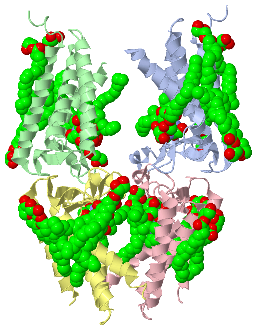
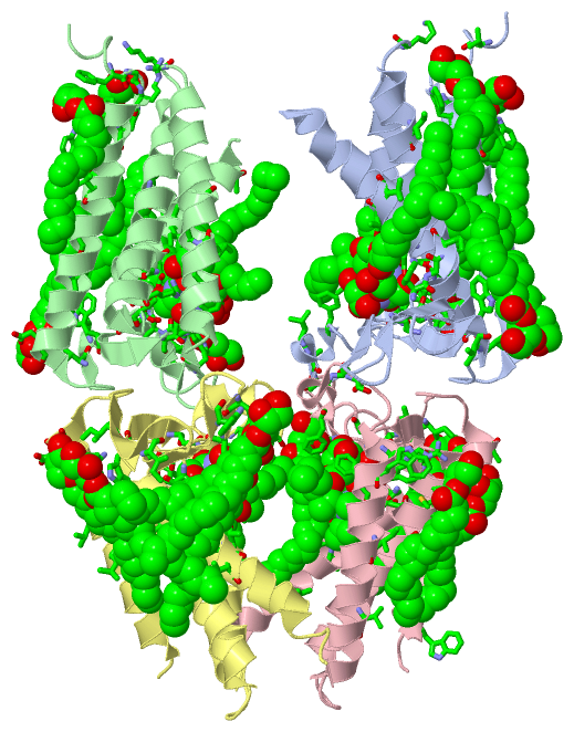
Chains, Units
Summary Information (see also Sequences/Alignments below) |
Ligands, Modified Residues, Ions (6, 44)
Asymmetric Unit (6, 44)
|
Sites (28, 28)
Asymmetric Unit (28, 28)
|
SS Bonds (0, 0)| (no "SS Bond" information available for 5DIR) |
Cis Peptide Bonds (0, 0)| (no "Cis Peptide Bond" information available for 5DIR) |
SAPs(SNPs)/Variants (0, 0)| (no "SAP(SNP)/Variant" information available for 5DIR) |
PROSITE Motifs (0, 0)| (no "PROSITE Motif" information available for 5DIR) |
Exons (0, 0)| (no "Exon" information available for 5DIR) |
Sequences/Alignments
Asymmetric Unit
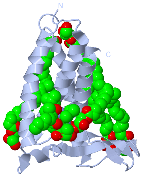
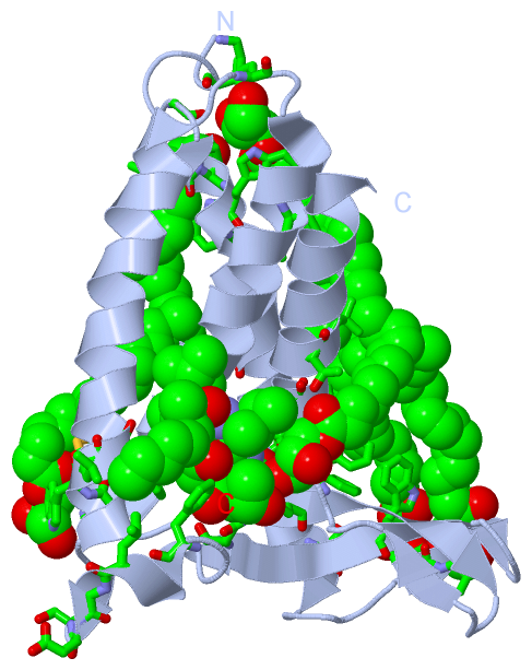
Chain A from PDB Type:PROTEIN Length:157
SCOP domains ------------------------------------------------------------------------------------------------------------------------------------------------------------- SCOP domains
CATH domains ------------------------------------------------------------------------------------------------------------------------------------------------------------- CATH domains
Pfam domains ------------------------------------------------------------------------------------------------------------------------------------------------------------- Pfam domains
SAPs(SNPs) ------------------------------------------------------------------------------------------------------------------------------------------------------------- SAPs(SNPs)
PROSITE ------------------------------------------------------------------------------------------------------------------------------------------------------------- PROSITE
Transcript ------------------------------------------------------------------------------------------------------------------------------------------------------------- Transcript
5dir A 2 PDVDRFGRLPWLWITVLVFVLDQVSKAFFQAELSMYQQIVVIPDLFSWTLAYNTGAAFSFLADSSGWQRWLFALIAIVVSASLVVWLKRLKKGETWLAIALALVLGGALGNLYDRMVLGHVVDFILVHWQNRWYFPAFNLADSAITVGAVMLALDMF 158
11 21 31 41 51 61 71 81 91 101 111 121 131 141 151
Chain B from PDB Type:PROTEIN Length:149
SCOP domains ----------------------------------------------------------------------------------------------------------------------------------------------------- SCOP domains
CATH domains ----------------------------------------------------------------------------------------------------------------------------------------------------- CATH domains
Pfam domains ----------------------------------------------------------------------------------------------------------------------------------------------------- Pfam domains
SAPs(SNPs) ----------------------------------------------------------------------------------------------------------------------------------------------------- SAPs(SNPs)
PROSITE ----------------------------------------------------------------------------------------------------------------------------------------------------- PROSITE
Transcript ----------------------------------------------------------------------------------------------------------------------------------------------------- Transcript
5dir B 2 PDVDRFGRLPWLWITVLVFVLDQVSKAFFQAELSMYQQIVVIPDLFSWTLAYNTGAAFSGWQRWLFALIAIVVSASLVVWLKRLKKGETWLAIALALVLGGALGNLYDRMVLGHVVDFILVHWQNRWYFPAFNLADSAITVGAVMLALD 156
11 21 31 41 51 67 77 87 97 107 117 127 137 147
60|
67
Chain C from PDB Type:PROTEIN Length:149
SCOP domains ----------------------------------------------------------------------------------------------------------------------------------------------------- SCOP domains
CATH domains ----------------------------------------------------------------------------------------------------------------------------------------------------- CATH domains
Pfam domains ----------------------------------------------------------------------------------------------------------------------------------------------------- Pfam domains
SAPs(SNPs) ----------------------------------------------------------------------------------------------------------------------------------------------------- SAPs(SNPs)
PROSITE ----------------------------------------------------------------------------------------------------------------------------------------------------- PROSITE
Transcript ----------------------------------------------------------------------------------------------------------------------------------------------------- Transcript
5dir C 11 PWLWITVLVFVLDQVSKAFFQAELSMYQQIVVIPDLFSWTLAYNTGAAFSFLADSSGWQRWLFALIAIVVSASLVVWLKRLKKGETWLAIALALVLGGALGNLYDRMVLGHVVDFILVHWQNRWYFPAFNLADSAITVGAVMLALDMFR 159
20 30 40 50 60 70 80 90 100 110 120 130 140 150
Chain D from PDB Type:PROTEIN Length:149
SCOP domains ----------------------------------------------------------------------------------------------------------------------------------------------------- SCOP domains
CATH domains ----------------------------------------------------------------------------------------------------------------------------------------------------- CATH domains
Pfam domains ----------------------------------------------------------------------------------------------------------------------------------------------------- Pfam domains
SAPs(SNPs) ----------------------------------------------------------------------------------------------------------------------------------------------------- SAPs(SNPs)
PROSITE ----------------------------------------------------------------------------------------------------------------------------------------------------- PROSITE
Transcript ----------------------------------------------------------------------------------------------------------------------------------------------------- Transcript
5dir D 11 PWLWITVLVFVLDQVSKAFFQAELSMYQQIVVIPDLFSWTLAYNTGAAFSFLADSSGWQRWLFALIAIVVSASLVVWLKRLKKGETWLAIALALVLGGALGNLYDRMVLGHVVDFILVHWQNRWYFPAFNLADSAITVGAVMLALDMFR 159
20 30 40 50 60 70 80 90 100 110 120 130 140 150
|
||||||||||||||||||||
SCOP Domains (0, 0)| (no "SCOP Domain" information available for 5DIR) |
CATH Domains (0, 0)| (no "CATH Domain" information available for 5DIR) |
Pfam Domains (0, 0)| (no "Pfam Domain" information available for 5DIR) |
Gene Ontology (7, 7)|
Asymmetric Unit(hide GO term definitions) |
Interactive Views
|
|||||||||||||||||||||||||||||||||||||||||||||||||||||||||||||||||||||||||||||||||||||||||||||||||||||||||||||||||||||||||||||||||||||||||||||||||||||||||||||||||||||||||||||||||||||||||||||||||||||||||||||||||||||||||||||||||||||||||||||||||||||||||||||||||||||||||||||||||||||||||||||||||||||||||||||||||||||||||||||||||||||||||||||||||||||||||||||||||||||||||||||||||||||||
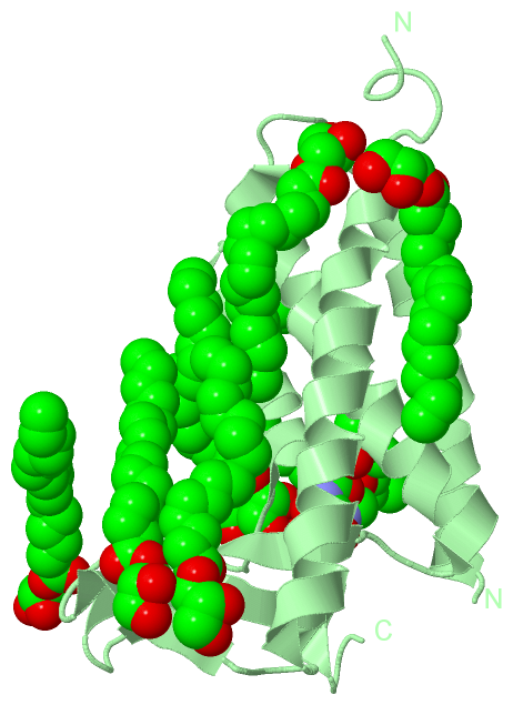
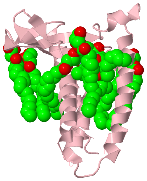
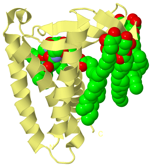
Still Images
|
||||||||||||||||
Databases
|
||||||||||||||||||||||||||||||||||||||||||||||||||||||||||||||||||||||||||||||||||||||||||||||||||||||||||||||||||||||||||||||||||||||||||||||||||||||||||||||||
Analysis Tools
|
|||||||||||||||||||||||||||||||||||||||||||||||||||||||||||||
Entries Sharing at Least One Protein Chain (UniProt ID)
Related Entries Specified in the PDB File
|
|