Show PDB file:   
         Plain Text   HTML   (compressed file size)
QuickSearch:   
by PDB,NDB,UniProt,PROSITE Code or Search Term(s)  
(-)Asymmetric Unit
(-)Asym. Unit - sites
(-)Biological Unit 1
(-)Biol. Unit 1 - sites
(-)Biological Unit 2
(-)Biological Unit 3
(-)Biological Unit 4
collapse expand < >
Image Asymmetric Unit
Asymmetric Unit  (Jmol Viewer)
Image Asym. Unit - sites
Asym. Unit - sites  (Jmol Viewer)
Image Biological Unit 1
Biological Unit 1  (Jmol Viewer)
Image Biol. Unit 1 - sites
Biol. Unit 1 - sites  (Jmol Viewer)
Image Biological Unit 2
Biological Unit 2  (Jmol Viewer)
Image Biological Unit 3
Biological Unit 3  (Jmol Viewer)
Image Biological Unit 4
Biological Unit 4  (Jmol Viewer)

(-) Description

Title :  CRYSTAL STRUCTURE OF THE SECOND BROMODOMAIN OF HUMAN BRD3 IN COMPLEX WITH COMPOUND
 
Authors :  M. Welin, R. Kimbung, C. Diehl, M. Hakansson, D. T. Logan, B. Walse
Date :  03 Jul 15  (Deposition) - 16 Mar 16  (Release) - 28 Sep 16  (Revision)
Method :  X-RAY DIFFRACTION
Resolution :  1.90
Chains :  Asym. Unit :  A,B,C,D
Biol. Unit 1:  A  (1x)
Biol. Unit 2:  B  (1x)
Biol. Unit 3:  C  (1x)
Biol. Unit 4:  D  (1x)
Keywords :  Dna Binding Protein, Brd3, Bromodomain Containing Protein 3, Ring3-Like Protein, Brd3 Domain 2 (Keyword Search: [Gene Ontology, PubMed, Web (Google))
 
Reference :  L. M. Nilsson, L. C. Green, S. V. Muralidharan, D. Demir, M. Welin, J. Bhadury, D. T. Logan, B. Walse, J. A. Nilsson
Cancer Differentiating Agent Hexamethylene Bisacetamide Inhibits Bet Bromodomain Proteins.
Cancer Res. V. 76 2376 2016
PubMed-ID: 26941288  |  Reference-DOI: 10.1158/0008-5472.CAN-15-2721

(-) Compounds

Molecule 1 - BROMODOMAIN-CONTAINING PROTEIN 3
    ChainsA, B, C, D
    EngineeredYES
    Expression SystemESCHERICHIA COLI
    Expression System PlasmidPNIC28-BSA4
    Expression System StrainBL21(DE3)
    Expression System Taxid469008
    Expression System VariantT1R PRARE2
    Expression System Vector TypePLASMID
    FragmentBROMO 2 DOMAIN, RESIDUES 306-416
    Organism CommonHUMAN
    Organism ScientificHOMO SAPIENS
    Organism Taxid9606
    SynonymRING3-LIKE PROTEIN

 Structural Features

(-) Chains, Units

  1234
Asymmetric Unit ABCD
Biological Unit 1 (1x)A   
Biological Unit 2 (1x) B  
Biological Unit 3 (1x)  C 
Biological Unit 4 (1x)   D

Summary Information (see also Sequences/Alignments below)

(-) Ligands, Modified Residues, Ions  (2, 9)

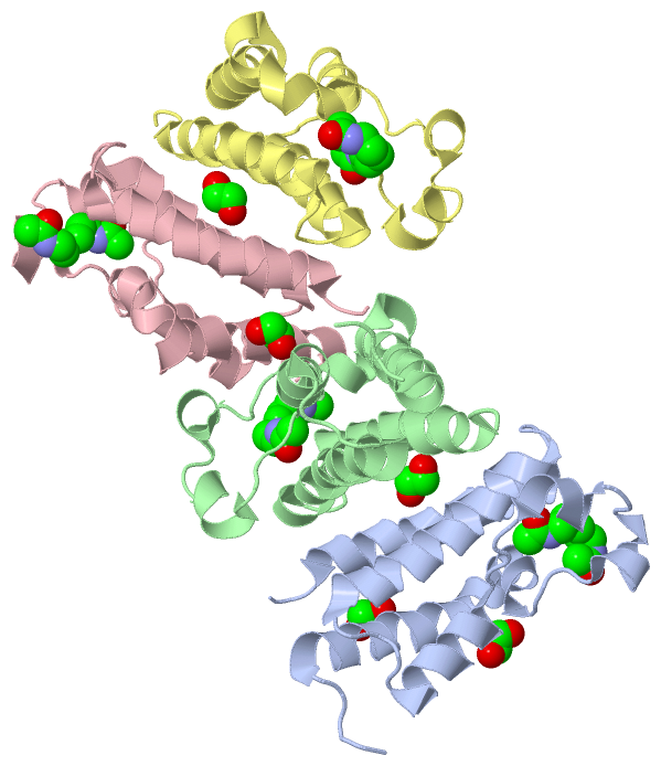
Asymmetric Unit (2, 9)
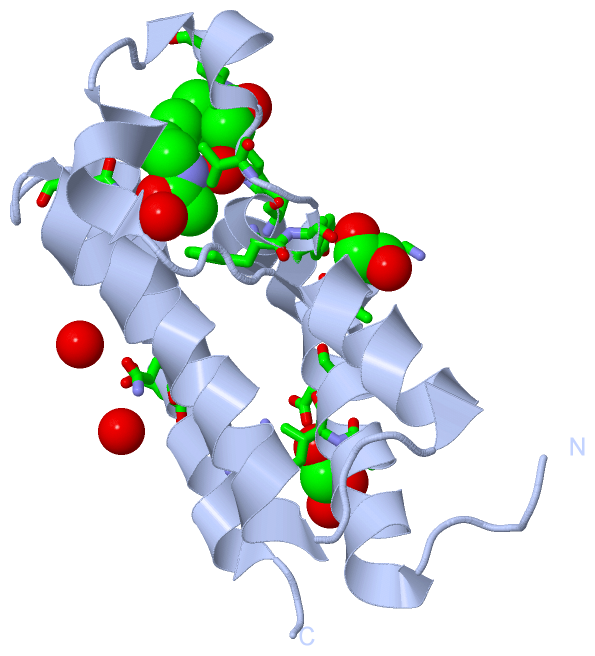
No.NameCountTypeFull Name
15D44Ligand/IonN-(6-ACETAMIDOHEXYL)ACETAMIDE
2EDO5Ligand/Ion1,2-ETHANEDIOL
Biological Unit 1 (2, 3)
No.NameCountTypeFull Name
15D41Ligand/IonN-(6-ACETAMIDOHEXYL)ACETAMIDE
2EDO2Ligand/Ion1,2-ETHANEDIOL
Biological Unit 2 (2, 2)
No.NameCountTypeFull Name
15D41Ligand/IonN-(6-ACETAMIDOHEXYL)ACETAMIDE
2EDO1Ligand/Ion1,2-ETHANEDIOL
Biological Unit 3 (2, 2)
No.NameCountTypeFull Name
15D41Ligand/IonN-(6-ACETAMIDOHEXYL)ACETAMIDE
2EDO1Ligand/Ion1,2-ETHANEDIOL
Biological Unit 4 (2, 2)
No.NameCountTypeFull Name
15D41Ligand/IonN-(6-ACETAMIDOHEXYL)ACETAMIDE
2EDO1Ligand/Ion1,2-ETHANEDIOL

(-) Sites  (9, 9)

Asymmetric Unit (9, 9)
No.NameEvidenceResiduesDescription
1AC1SOFTWARETRP A:332 , PHE A:334 , VAL A:338 , LEU A:343 , ASN A:391 , HOH A:2029 , HOH A:2044 , HOH B:2038 , HOH B:2043BINDING SITE FOR RESIDUE 5D4 A 500
2AC2SOFTWARESER A:317 , ILE A:318 , GLU A:321 , ARG A:411 , HOH A:2106 , TRP D:332BINDING SITE FOR RESIDUE EDO A 501
3AC3SOFTWARELEU A:323 , LYS A:325 , ALA A:328 , TYR A:335BINDING SITE FOR RESIDUE EDO A 502
4AC4SOFTWAREVAL B:338 , LEU B:345 , ASN B:391 , HIS B:395 , GLU B:396 , MET B:400 , HOH B:2028 , HOH B:2045 , ARG C:313BINDING SITE FOR RESIDUE 5D4 B 500
5AC5SOFTWAREGLN A:405 , ASP A:406 , HOH A:2078 , HOH A:2090 , GLN B:405 , ASP B:406BINDING SITE FOR RESIDUE EDO B 501
6AC6SOFTWAREPHE C:334 , ASN C:391 , HIS C:395 , HOH C:2020 , HOH C:2029BINDING SITE FOR RESIDUE 5D4 C 500
7AC7SOFTWARETRP B:332 , HOH B:2025 , TYR C:314 , SER C:317 , ILE C:318 , GLU C:321 , ARG C:411BINDING SITE FOR RESIDUE EDO C 501
8AC8SOFTWARETRP D:332 , PHE D:334 , VAL D:338 , ASN D:391 , HIS D:395 , HOH D:2025 , HOH D:2036BINDING SITE FOR RESIDUE 5D4 D 500
9AC9SOFTWAREGLN C:405 , ASP C:406 , HOH C:2072 , GLN D:405 , ASP D:406BINDING SITE FOR RESIDUE EDO D 501

(-) SS Bonds  (0, 0)

(no "SS Bond" information available for 5A7C)

(-) Cis Peptide Bonds  (0, 0)

(no "Cis Peptide Bond" information available for 5A7C)

 Sequence-Structure Mapping

(-) SAPs(SNPs)/Variants  (0, 0)

(no "SAP(SNP)/Variant" information available for 5A7C)

(-) PROSITE Motifs  (0, 0)

(no "PROSITE Motif" information available for 5A7C)

(-) Exons   (0, 0)

(no "Exon" information available for 5A7C)

(-) Sequences/Alignments

Asymmetric Unit
   Reformat: Number of residues per line =  ('0' or empty: single-line sequence representation)
  Number of residues per labelling interval =   
  UniProt sequence: complete  aligned part    
   Show mapping: SCOP domains CATH domains Pfam domains Secondary structure (by author)
SAPs(SNPs) PROSITE motifs Exons
(details for a mapped element are shown in a popup box when the mouse pointer rests over it)
Chain A from PDB  Type:PROTEIN  Length:112
                                                                                                                                                
               SCOP domains ---------------------------------------------------------------------------------------------------------------- SCOP domains
               CATH domains ---------------------------------------------------------------------------------------------------------------- CATH domains
               Pfam domains ---------------------------------------------------------------------------------------------------------------- Pfam domains
         Sec.struct. author ....hhhhhhhhhhhhhhhhhhhhhhhhhhh...hhhhhh..hhhhhh....hhhhhhhhhhh....hhhhhhhhhhhhhhhhhhhh...hhhhhhhhhhhhhhhhhhhh.. Sec.struct. author
                 SAPs(SNPs) ---------------------------------------------------------------------------------------------------------------- SAPs(SNPs)
                    PROSITE ---------------------------------------------------------------------------------------------------------------- PROSITE
                 Transcript ---------------------------------------------------------------------------------------------------------------- Transcript
                 5a7c A 305 MGKLSEHLRYCDSILREMLSKKHAAYAWPFYKPVDAEALELHDYHDIIKHPMDLSTVKRKMDGREYPDAQGFAADVRLMFSNCYKYNPPDHEVVAMARKLQDVFEMRFAKMP 416
                                   314       324       334       344       354       364       374       384       394       404       414  

Chain B from PDB  Type:PROTEIN  Length:111
                                                                                                                                               
               SCOP domains --------------------------------------------------------------------------------------------------------------- SCOP domains
               CATH domains --------------------------------------------------------------------------------------------------------------- CATH domains
               Pfam domains --------------------------------------------------------------------------------------------------------------- Pfam domains
         Sec.struct. author ...hhhhhhhhhhhhhhhhhhhhhhhhhhh...hhhhhh..hhhhhh....hhhhhhhhhhh....hhhhhhhhhhhhhhhhhhhh...hhhhhhhhhhhhhhhhhhhh.. Sec.struct. author
                 SAPs(SNPs) --------------------------------------------------------------------------------------------------------------- SAPs(SNPs)
                    PROSITE --------------------------------------------------------------------------------------------------------------- PROSITE
                 Transcript --------------------------------------------------------------------------------------------------------------- Transcript
                 5a7c B 306 GKLSEHLRYCDSILREMLSKKHAAYAWPFYKPVDAEALELHDYHDIIKHPMDLSTVKRKMDGREYPDAQGFAADVRLMFSNCYKYNPPDHEVVAMARKLQDVFEMRFAKMP 416
                                   315       325       335       345       355       365       375       385       395       405       415 

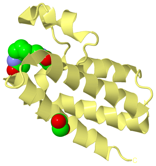
Chain C from PDB  Type:PROTEIN  Length:109
                                                                                                                                             
               SCOP domains ------------------------------------------------------------------------------------------------------------- SCOP domains
               CATH domains ------------------------------------------------------------------------------------------------------------- CATH domains
               Pfam domains ------------------------------------------------------------------------------------------------------------- Pfam domains
         Sec.struct. author .hhhhhhhhhhhhhhhhhhhhhhhhhhh...hhhhhh..hhhhhh....hhhhhhhhhhh....hhhhhhhhhhhhhhhhhhhh...hhhhhhhhhhhhhhhhhhhh.. Sec.struct. author
                 SAPs(SNPs) ------------------------------------------------------------------------------------------------------------- SAPs(SNPs)
                    PROSITE ------------------------------------------------------------------------------------------------------------- PROSITE
                 Transcript ------------------------------------------------------------------------------------------------------------- Transcript
                 5a7c C 308 LSEHLRYCDSILREMLSKKHAAYAWPFYKPVDAEALELHDYHDIIKHPMDLSTVKRKMDGREYPDAQGFAADVRLMFSNCYKYNPPDHEVVAMARKLQDVFEMRFAKMP 416
                                   317       327       337       347       357       367       377       387       397       407         

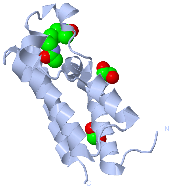
Chain D from PDB  Type:PROTEIN  Length:109
                                                                                                                                             
               SCOP domains ------------------------------------------------------------------------------------------------------------- SCOP domains
               CATH domains ------------------------------------------------------------------------------------------------------------- CATH domains
               Pfam domains ------------------------------------------------------------------------------------------------------------- Pfam domains
         Sec.struct. author .hhhhhhhhhhhhhhhhhhhhhhhhhhh...hhhhhh..hhhhhh....hhhhhhhhhhh....hhhhhhhhhhhhhhhhhhhh...hhhhhhhhhhhhhhhhhhhh.. Sec.struct. author
                 SAPs(SNPs) ------------------------------------------------------------------------------------------------------------- SAPs(SNPs)
                    PROSITE ------------------------------------------------------------------------------------------------------------- PROSITE
                 Transcript ------------------------------------------------------------------------------------------------------------- Transcript
                 5a7c D 308 LSEHLRYCDSILREMLSKKHAAYAWPFYKPVDAEALELHDYHDIIKHPMDLSTVKRKMDGREYPDAQGFAADVRLMFSNCYKYNPPDHEVVAMARKLQDVFEMRFAKMP 416
                                   317       327       337       347       357       367       377       387       397       407         

   Legend:   → Mismatch (orange background)
  - → Gap (green background, '-', border residues have a numbering label)
    → Modified Residue (blue background, lower-case, 'x' indicates undefined single-letter code, labelled with number + name)
  x → Chemical Group (purple background, 'x', labelled with number + name, e.g. ACE or NH2)
  extra numbering lines below/above indicate numbering irregularities and modified residue names etc., number ends below/above '|'

 Classification and Annotation

(-) SCOP Domains  (0, 0)

(no "SCOP Domain" information available for 5A7C)

(-) CATH Domains  (0, 0)

(no "CATH Domain" information available for 5A7C)

(-) Pfam Domains  (0, 0)

(no "Pfam Domain" information available for 5A7C)

(-) Gene Ontology  (7, 7)

Asymmetric Unit(hide GO term definitions)

 Visualization

(-) Interactive Views

Asymmetric Unit
  Complete Structure
    Jena3D(integrated viewing of ligand, site, SAP, PROSITE, SCOP information)
    WebMol | AstexViewer[tm]@PDBe
(Java Applets, require no local installation except for Java; loading may be slow)
    STRAP
(Java WebStart application, automatic local installation, requires Java; full application with system access!)
    RasMol
(require local installation)
    Molscript (VRML)
(requires installation of a VRML viewer; select preferred view via VRML and generate a mono or stereo PDF format file)
 
  Ligands, Modified Residues, Ions
    5D4  [ RasMol | Jena3D ]  +environment [ RasMol | Jena3D ]
    EDO  [ RasMol | Jena3D ]  +environment [ RasMol | Jena3D ]
 
  Sites
    AC1  [ RasMol ]  +environment [ RasMol ]
    AC2  [ RasMol ]  +environment [ RasMol ]
    AC3  [ RasMol ]  +environment [ RasMol ]
    AC4  [ RasMol ]  +environment [ RasMol ]
    AC5  [ RasMol ]  +environment [ RasMol ]
    AC6  [ RasMol ]  +environment [ RasMol ]
    AC7  [ RasMol ]  +environment [ RasMol ]
    AC8  [ RasMol ]  +environment [ RasMol ]
    AC9  [ RasMol ]  +environment [ RasMol ]
 
  Cis Peptide Bonds
(no "Cis Peptide Bonds" information available for 5a7c)
 
Biological Units
  Complete Structure
    Biological Unit 1  [ Jena3D ]
    Biological Unit 2  [ Jena3D ]
    Biological Unit 3  [ Jena3D ]
    Biological Unit 4  [ Jena3D ]

(-) Still Images

Jmol
  protein: cartoon or spacefill or dots and stick; nucleic acid: cartoon and stick; ligands: spacefill; active site: stick
Molscript
  protein, nucleic acid: cartoon; ligands: spacefill; active site: ball and stick

 Databases and Analysis Tools

(-) Databases

Access by PDB/NDB ID
  5a7c
    Family and Domain InformationProDom | SYSTERS
    General Structural InformationGlycoscienceDB | MMDB | NDB | OCA | PDB | PDBe | PDBj | PDBsum | PDBWiki | PQS | PROTEOPEDIA
    Orientation in MembranesOPM
    Protein SurfaceSURFACE
    Secondary StructureDSSP (structure derived) | HSSP (homology derived)
    Structural GenomicsGeneCensus
    Structural NeighboursCE | VAST
    Structure ClassificationCATH | Dali | SCOP
    Validation and Original DataBMRB Data View | BMRB Restraints Grid | EDS | PROCHECK | RECOORD | WHAT_CHECK
 
Access by UniProt ID/Accession number
  BRD3_HUMAN | Q15059
    Comparative Protein Structure ModelsModBase
    Genomic InformationEnsembl
    Protein-protein InteractionDIP
    Sequence, Family and Domain InformationInterPro | Pfam | SMART | UniProtKB/SwissProt
 
Access by Enzyme Classificator   (EC Number)
  (no 'Enzyme Classificator' available)
    General Enzyme InformationBRENDA | EC-PDB | Enzyme | IntEnz
    PathwayKEGG | MetaCyc
 
Access by Disease Identifier   (MIM ID)
  (no 'MIM ID' available)
    Disease InformationOMIM
 
Access by GenAge ID
  (no 'GenAge ID' available)
    Age Related InformationGenAge

(-) Analysis Tools

Access by PDB/NDB ID
    Domain InformationXDom
    Interatomic Contacts of Structural UnitsCSU
    Ligand-protein ContactsLPC
    Protein CavitiescastP
    Sequence and Secondary StructurePDBCartoon
    Structure AlignmentSTRAP(Java WebStart application, automatic local installation, requires Java; full application with system access!)
    Structure and Sequence BrowserSTING
 
Access by UniProt ID/Accession number
  BRD3_HUMAN | Q15059
    Protein Disorder PredictionDisEMBL | FoldIndex | GLOBPLOT (for more information see DisProt)

 Related Entries

(-) Entries Sharing at Least One Protein Chain (UniProt ID)

UniProtKB/Swiss-Prot
        BRD3_HUMAN | Q150592e7n 2nxb 2oo1 2yw5 3s91 3s92 5hfr 5hjc

(-) Related Entries Specified in the PDB File

5a6n CRYSTAL STRUCTURE OF HUMAN DEATH ASSOCIATED PROTEIN KINASE 3 (DAPK3) IN COMPLEX WITH COMPOUND 2
5a6o CRYSTAL STRUCTURE OF THE APO FORM OF THE UNPHOSPHORYLATED HUMAN DEATH ASSOCIATED PROTEIN KINASE 3 (DAPK3)