Show PDB file:   
         Plain Text   HTML   (compressed file size)
QuickSearch:   
by PDB,NDB,UniProt,PROSITE Code or Search Term(s)  
(-)Asym./Biol. Unit
collapse expand < >
Image Asym./Biol. Unit
Asym./Biol. Unit  (Jmol Viewer)

(-) Description

Title :  CRYSTAL STRUCTURE OF PASTA DOMAINS 3 AND 4 OF MYCOBACTERIUM TUBERCULOSIS PROTEIN KINASE B
 
Authors :  D. M. Prigozhin, Tb Structural Genomics Consortium (Tbsgc)
Date :  29 Sep 15  (Deposition) - 14 Sep 16  (Release) - 09 Nov 16  (Revision)
Method :  X-RAY DIFFRACTION
Resolution :  2.00
Chains :  Asym./Biol. Unit :  A
Keywords :  Kinase, Extracellular Sensor Domain, Peptidoglycan Binding, Structural Genomics, Tb Structural Genomics Consortium, Tbsgc, Transferase (Keyword Search: [Gene Ontology, PubMed, Web (Google))
 
Reference :  D. M. Prigozhin, K. G. Papavinasasundaram, C. E. Baer, K. C. Murphy, A. Moskaleva, T. Y. Chen, T. Alber, C. M. Sassetti
Structural And Genetic Analyses Of The Mycobacterium Tuberculosis Protein Kinase B Sensor Domain Identify A Potential Ligand-Binding Site.
J. Biol. Chem. V. 291 22961 2016
PubMed-ID: 27601474  |  Reference-DOI: 10.1074/JBC.M116.731760

(-) Compounds

Molecule 1 - SERINE/THREONINE-PROTEIN KINASE PKNB
    ChainsA
    EC Number2.7.11.1
    EngineeredYES
    Expression SystemESCHERICHIA COLI
    Expression System Taxid562
    FragmentUNP RESIDUES 491-626
    GenePKNB, RV0014C, MTCY10H4.14C
    Organism ScientificMYCOBACTERIUM TUBERCULOSIS (STRAIN ATCC 25618 / H37RV)
    Organism Taxid83332
    StrainATCC 25618 / H37RV

 Structural Features

(-) Chains, Units

  1
Asymmetric/Biological Unit A

Summary Information (see also Sequences/Alignments below)

(-) Ligands, Modified Residues, Ions  (0, 0)

(no "Ligand,Modified Residues,Ions" information available for 5E0Z)

(-) Sites  (0, 0)

(no "Site" information available for 5E0Z)

(-) SS Bonds  (0, 0)

(no "SS Bond" information available for 5E0Z)

(-) Cis Peptide Bonds  (2, 2)

Asymmetric/Biological Unit
No.Residues
1Asn A:538 -Pro A:539
2Asn A:607 -Pro A:608

 Sequence-Structure Mapping

(-) SAPs(SNPs)/Variants  (0, 0)

(no "SAP(SNP)/Variant" information available for 5E0Z)

(-) PROSITE Motifs  (0, 0)

(no "PROSITE Motif" information available for 5E0Z)

(-) Exons   (0, 0)

(no "Exon" information available for 5E0Z)

(-) Sequences/Alignments

Asymmetric/Biological Unit
   Reformat: Number of residues per line =  ('0' or empty: single-line sequence representation)
  Number of residues per labelling interval =   
  UniProt sequence: complete  aligned part    
   Show mapping: SCOP domains CATH domains Pfam domains Secondary structure (by author)
SAPs(SNPs) PROSITE motifs Exons
(details for a mapped element are shown in a popup box when the mouse pointer rests over it)
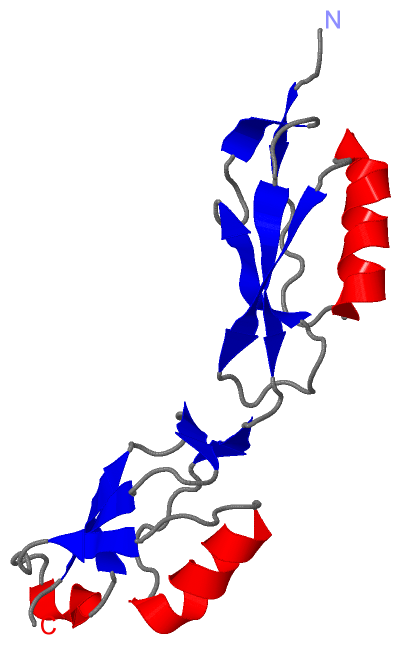
Chain A from PDB  Type:PROTEIN  Length:136
                                                                                                                                                                        
               SCOP domains ---------------------------------------------------------------------------------------------------------------------------------------- SCOP domains
               CATH domains ---------------------------------------------------------------------------------------------------------------------------------------- CATH domains
               Pfam domains ---------------------------------------------------------------------------------------------------------------------------------------- Pfam domains
         Sec.struct. author ...eee.......hhhhhhhhhhhhh...eeeeee........eeeee.....eee....eeeeee...eee.......hhhhhhhhhhh......eee......hhhhh..eeeee.....eee....eeeee.. Sec.struct. author
                 SAPs(SNPs) ---------------------------------------------------------------------------------------------------------------------------------------- SAPs(SNPs)
                    PROSITE ---------------------------------------------------------------------------------------------------------------------------------------- PROSITE
                 Transcript ---------------------------------------------------------------------------------------------------------------------------------------- Transcript
                 5e0z A 491 GPATKDIPDVAGQTVDVAQKNLNVYGFTKFSQASVDSPRPAGEVTGTNPPAGTTVPVDSVIELQVSKGNQFVMPDLSGMFWVDAEPRLRALGWTGMLDKGADVDAGGSQHNRVVYQNPPAGTGVNRDGIITLRFGQ 626
                                   500       510       520       530       540       550       560       570       580       590       600       610       620      

   Legend:   → Mismatch (orange background)
  - → Gap (green background, '-', border residues have a numbering label)
    → Modified Residue (blue background, lower-case, 'x' indicates undefined single-letter code, labelled with number + name)
  x → Chemical Group (purple background, 'x', labelled with number + name, e.g. ACE or NH2)
  extra numbering lines below/above indicate numbering irregularities and modified residue names etc., number ends below/above '|'

 Classification and Annotation

(-) SCOP Domains  (0, 0)

(no "SCOP Domain" information available for 5E0Z)

(-) CATH Domains  (0, 0)

(no "CATH Domain" information available for 5E0Z)

(-) Pfam Domains  (0, 0)

(no "Pfam Domain" information available for 5E0Z)

(-) Gene Ontology  (27, 27)

Asymmetric/Biological Unit(hide GO term definitions)

 Visualization

(-) Interactive Views

Asymmetric/Biological Unit
  Complete Structure
    Jena3D(integrated viewing of ligand, site, SAP, PROSITE, SCOP information)
    WebMol | AstexViewer[tm]@PDBe
(Java Applets, require no local installation except for Java; loading may be slow)
    STRAP
(Java WebStart application, automatic local installation, requires Java; full application with system access!)
    RasMol
(require local installation)
    Molscript (VRML)
(requires installation of a VRML viewer; select preferred view via VRML and generate a mono or stereo PDF format file)
 
  Ligands, Modified Residues, Ions
(no "Ligands, Modified Residues, Ions" information available for 5e0z)
 
  Sites
(no "Sites" information available for 5e0z)
 
  Cis Peptide Bonds
    Asn A:538 - Pro A:539   [ RasMol ]  
    Asn A:607 - Pro A:608   [ RasMol ]  
 

(-) Still Images

Jmol
  protein: cartoon or spacefill or dots and stick; nucleic acid: cartoon and stick; ligands: spacefill; active site: stick
Molscript
  protein, nucleic acid: cartoon; ligands: spacefill; active site: ball and stick

 Databases and Analysis Tools

(-) Databases

Access by PDB/NDB ID
  5e0z
    Family and Domain InformationProDom | SYSTERS
    General Structural InformationGlycoscienceDB | MMDB | NDB | OCA | PDB | PDBe | PDBj | PDBsum | PDBWiki | PQS | PROTEOPEDIA
    Orientation in MembranesOPM
    Protein SurfaceSURFACE
    Secondary StructureDSSP (structure derived) | HSSP (homology derived)
    Structural GenomicsGeneCensus
    Structural NeighboursCE | VAST
    Structure ClassificationCATH | Dali | SCOP
    Validation and Original DataBMRB Data View | BMRB Restraints Grid | EDS | PROCHECK | RECOORD | WHAT_CHECK
 
Access by UniProt ID/Accession number
  PKNB_MYCTU | P9WI81
    Comparative Protein Structure ModelsModBase
    Genomic InformationEnsembl
    Protein-protein InteractionDIP
    Sequence, Family and Domain InformationInterPro | Pfam | SMART | UniProtKB/SwissProt
 
Access by Enzyme Classificator   (EC Number)
  2.7.11.1
    General Enzyme InformationBRENDA | EC-PDB | Enzyme | IntEnz
    PathwayKEGG | MetaCyc
 
Access by Disease Identifier   (MIM ID)
  (no 'MIM ID' available)
    Disease InformationOMIM
 
Access by GenAge ID
  (no 'GenAge ID' available)
    Age Related InformationGenAge

(-) Analysis Tools

Access by PDB/NDB ID
    Domain InformationXDom
    Interatomic Contacts of Structural UnitsCSU
    Ligand-protein ContactsLPC
    Protein CavitiescastP
    Sequence and Secondary StructurePDBCartoon
    Structure AlignmentSTRAP(Java WebStart application, automatic local installation, requires Java; full application with system access!)
    Structure and Sequence BrowserSTING
 
Access by UniProt ID/Accession number
  PKNB_MYCTU | P9WI81
    Protein Disorder PredictionDisEMBL | FoldIndex | GLOBPLOT (for more information see DisProt)

 Related Entries

(-) Entries Sharing at Least One Protein Chain (UniProt ID)

UniProtKB/Swiss-Prot
        PKNB_MYCTU | P9WI811mru 1o6y 2fum 2kud 2kue 2kuf 2kui 3f61 3f69 5e0y 5e10 5e12

(-) Related Entries Specified in the PDB File

3ouv 5e0y 5e10 5e12